. 2019 Jan 7;15(1):e1007514.
doi: 10.1371/journal.ppat.1007514. eCollection 2019 Jan.

EBNA3C facilitates RASSF1A downregulation through ubiquitin-mediated degradation and promoter hypermethylation to drive B-cell proliferation

Affiliations

EBNA3C facilitates RASSF1A downregulation through ubiquitin-mediated degradation and promoter hypermethylation to drive B-cell proliferation

Shengwei Zhang et al. PLoS Pathog. .

Abstract

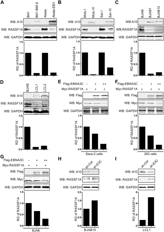
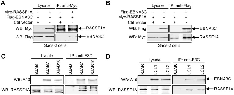
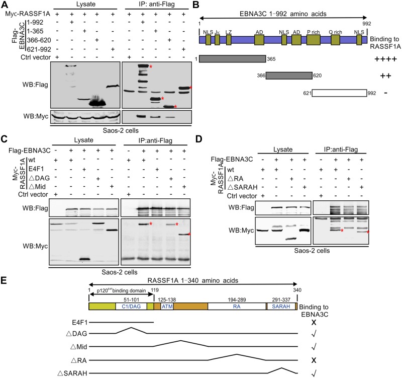
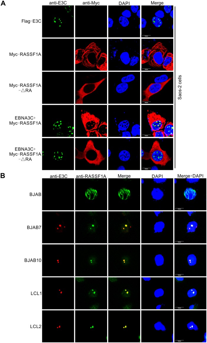
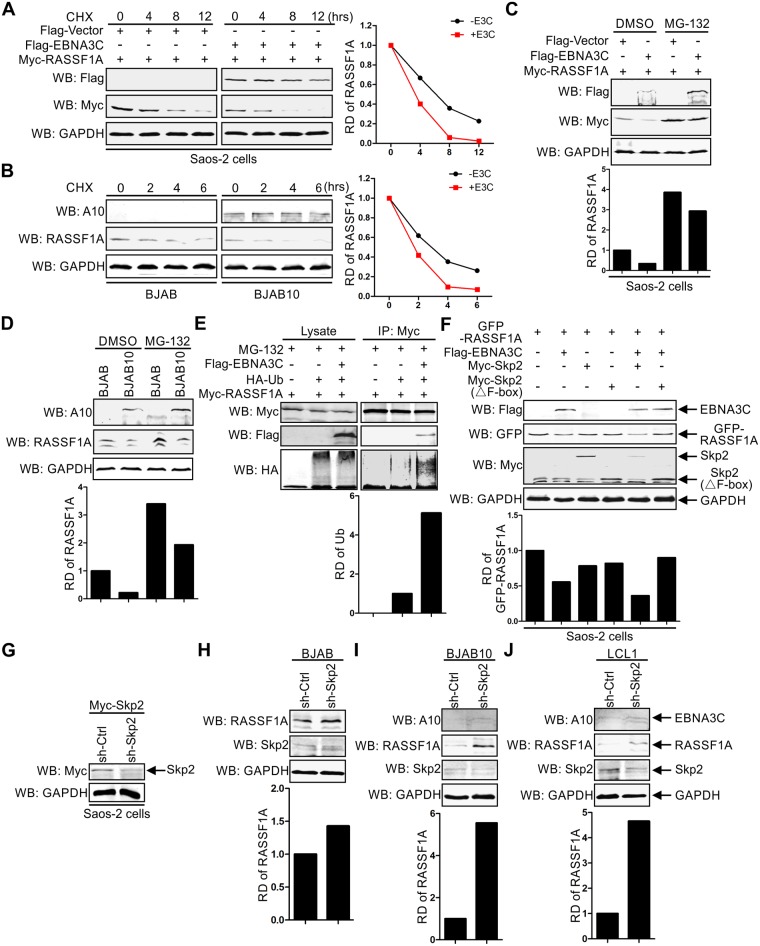
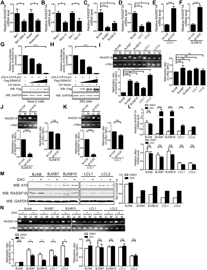
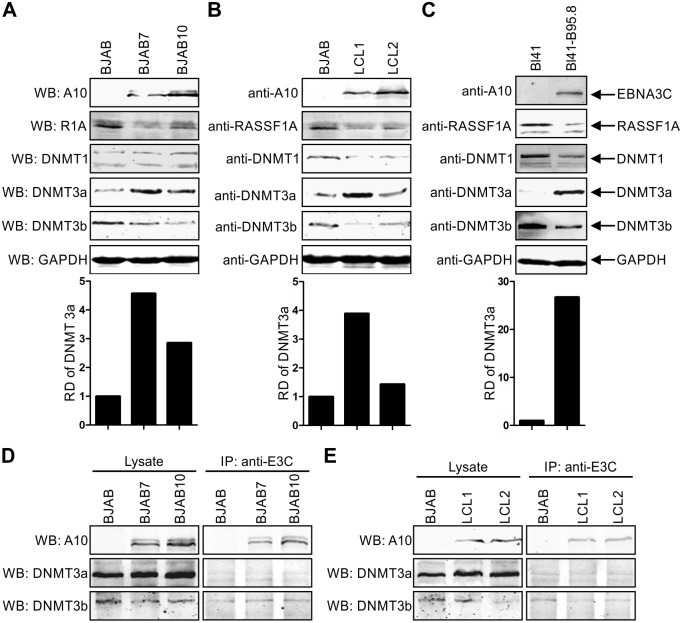
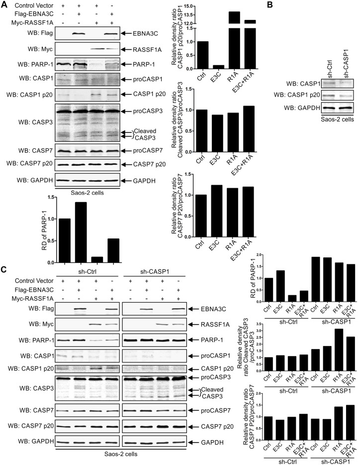
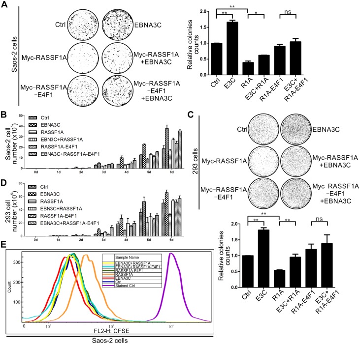
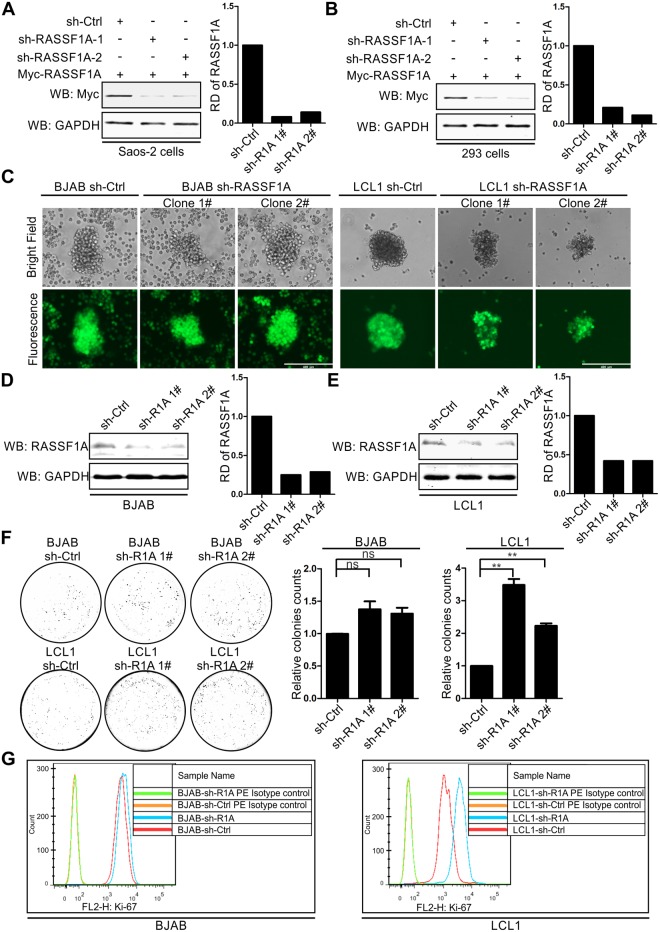
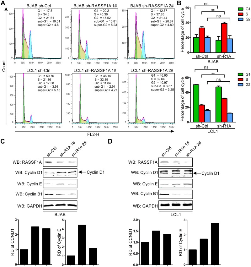
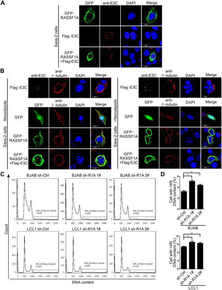
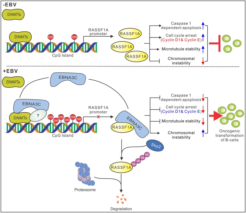
EBV latent antigen 3C (EBNA3C) is essential for EBV-induced primary B-cell transformation. Infection by EBV induces hypermethylation of a number of tumor suppressor genes, which contributes to the development of human cancers. The Ras association domain family isoform 1A (RASSF1A) is a cellular tumor suppressor, which regulates a broad range of cellular functions, including apoptosis, cell-cycle arrest, mitotic arrest, and migration. However, the expression of RASSF1A is lost in many human cancers by epigenetic silencing. In the present study, we showed that EBNA3C promoted B-cell transformation by specifically suppressing the expression of RASSF1A. EBNA3C directly interacted with RASSF1A and induced RASSF1A degradation via the ubiquitin-proteasome-dependent pathway. SCFSkp2, an E3-ubiquitin ligase, was recruited by EBNA3C to enhance RASSF1A degradation. Moreover, EBNA3C decreased the transcriptional activity of RASSF1A promoter by enhancing its methylation through EBNA3C-mediated modulation of DNMTs expression. EBNA3C also inhibited RASSF1A-mediated cell apoptosis, disrupted RASSF1A-mediated microtubule and chromosomal stability, and promoted cell proliferation by upregulating Cyclin D1 and Cyclin E expression. Our data provides new details, which sheds light on additional mechanisms by which EBNA3C can induce B-cell transformation. This will also facilitate the development of novel therapeutic approaches through targeting of the RASSF1A pathway.


Conflict of interest statement

The authors have declared that no competing interests exist.

Figures

Fig 1
Fig 1. EBNA3C specifically down-regulated RASSF1A protein expression.
(A-B) RASSF1A expression was decreased in EBV positive cell lines. 15 million A) Burkitt’s lymphoma cell BL41 and Akata B) Mutu I and Sav I cells and corresponding EBV positive A) BL41-B95.8 and Akata-EBV B) Mutu III and Sav III cells were harvested and lysed with RIPA buffer. The expression levels of RASSF1A were detected by western blot. The relative density (RD) of RASSF1A was quantitated and shown. (C-D) RASSF1A expression was repressed in EBNA3C stably expressing BJAB cells, and EBV-transformed LCLs. The expression of RASSF1A in C) BJAB7 and BJAB10 and D) LCL1 and LCL2 cells were detected as described for A. (E-G) EBNA3C decreased RASSF1A expression specifically. E) Saos-2, F) 293 and G) BJAB cells were transfected with Myc-tagged RASSF1A alone or co-transfected with an increasing amount of Flag-tagged EBNA3C. Western blots were performed to detect the expression of RASSF1A as described for A. (H-I) Knocking down EBNA3C rescued the expression level of RASSF1A in BJAB10 and LCL cells. H) 10 million BJAB10 cells were transfected with sh-EBNA3C or scramble control (sh-Ctrl) by electroporation. I) 15 million lentivirus transduced stable EBNA3C knocked-down LCL1 cells and LCL1-sh-Ctrl were harvested. 48h post-transfection, the cells were harvested and lysed for western blot. The relative density (RD) of RASSF1A was quantitated and shown.
Fig 2
Fig 2. EBNA3C associates with RASSF1A.
(A-B) 10 million Saos-2 cells were transfected with Myc-tagged RASSF1A alone or together with Flag-tagged EBNA3C. 48h post-transfection, the cells were harvested and lysed for immunoprecipitation with 1 μg A) anti-Myc or B) anti-Flag antibody. The inputs and immunoprecipitated samples fractionated and specific signals were detected by western blot by using antibodies against Flag and Myc. (C-D) EBNA3C associated with endogenous RASSF1A. 60 million C) BJAB, BJAB7, BJAB10, D) LCL1 and LCL2 were collected and lysed for immunoprecipitation with 1 μg anti-EBNA3C antibody. Western blot was used to detect specific signal in the inputs and immunoprecipitated samples.
Fig 3
Fig 3. The N-terminus of EBNA3C and C-terminus of RASSF1A are required for the interaction between EBNA3C and RASSF1A.
(A) The N terminus of EBNA3C is critical for EBNA3C and RASSF1A interaction. 10 million Saos-2 cells were transfected with Myc-tagged RASSF1A alone or together with Flag-tagged full-length EBNA3C or EBNA3C truncated mutants. 48h post-transfection, the cells were harvested and lysed for immunoprecipitation with 1 μg anti-Flag antibody. The input and immunoprecipitated samples were detected by western blot as described for Fig 2B. (B) The schematic diagram summarizes the binding domains between different regions of EBNA3C and RASSF1A. Jκ, RBP-Jκ; LZ, leucine zipper domain; AD, acidic domains; P rich, Proline-rich; Q rich, glutamine-proline-rich. NLS, nuclear localization signal. -, no binding; ++, binding; ++++, strongly binding. (C-D) The C-terminus of RASSF1A is important for EBNA3C and RASSF1A interaction. 10 million Saos-2 cells were transfected with Myc-tagged RASSF1A truncated mutants and Flag-tagged full-length EBNA3C. 48h post-transfection, the cells were harvested and lysed for immunoprecipitation as described for A. (E) The schematic diagram summarizes the binding domains between different regions of RASSF1A and EBNA3C. E4F1, E1A-regulation transcription factor p120E4F; DAG, Conserved region 1 diacylglycerol-binding domain; ATM, ATM-kinase consensus phosphorylation domain; RA, Ras association domain; SARAH, Sav/RASSF/Hpo interaction domain; X, no binding; √, binding.
Fig 4
Fig 4. EBNA3C co-localizes with RASSF1A in nuclear compartments in human cells.
(A) EBNA3C co-localized with RASSF1A in Saos-2 cells. 0.1 million Saos-2 cells were plated on coverslips and transfected with Flag-tagged EBNA3C, Myc-tagged RASSF1A or RASSF1A-ΔRA separately or together by using jetPRIME transfection reagent. 24h post-transfection, cells were subjected to immunofluorescence assays. (B) EBNA3C co-localized with endogenous RASSF1A in B-cells. BJAB, BJAB7, BJAB10, LCL1, and LCL2 cells were plated on the slide and air-dried. The cells were fixed and subjected to immunofluorescence assays as described in materials and methods.
Fig 5
Fig 5. EBNA3C modulates RASSF1A expression in a ubiquitin-proteasome-dependent manner.
A-B) EBNA3C decreased RASSF1A stability. (A) 10 million Saos-2 cells were transfected with Myc-tagged RASSF1A and Flag-tagged vector or Flag-tagged EBNA3C. 24 h post-transfection, cells were treated with 40 μg/ml Cycloheximide. (B) 10 million BJAB and BJAB10 cells were treated with 40 μg/ml Cycloheximide. At the indicated time points, the cells were harvested and lysed for western blot. The relative density (RD) of RASSF1A was quantitated to measure RASSF1A rate of degradation. (C-D) EBNA3C decreased RASSF1A stability. C) 10 million Saos-2 cells were transfected with Myc-tagged RASSF1A alone or together with Flag-tagged vector or Flag-tagged EBNA3C. 24 h post-transfection, cells were incubated with 20 μM MG132 for another 16 hours. D) 10 million BJAB and BJAB10 cells were incubated with 20 μM MG132 for 16 hours. Then cells were harvested and RASSF1A expression was detected by western blot. (E) EBNA3C enhanced RASSF1A poly-ubiquitination. 10 million Saos-2 cells were transfected with the indicated constructs. 24 h post-transfection, cells were incubated with 20 μM MG132 for another 16 hours. Then the cells were harvested and subjected to immunoprecipitation using an antibody against Myc. The inputs and immunoprecipitated samples were detected by western blot. (F) Skp2 protein was recruited by EBNA3C to mediate RASSF1A degradation. GFP-tagged RASSF1A was co-transfected with plasmids encoding EBNA3C and/or Skp2 or Skp2 truncated mutant. 24 h post-transfection, cells were harvested and lysed for western blot. The expression of RASSF1A, EBNA3C, and Skp2 was detected with specific antibodies. (G) The effect of sh-Skp2 in reducing Skp2 expression was detected in Saos-2 cells. Myc-tagged Skp2 and sh-Ctrl, or sh-Skp2 were transfected into Saos-2 cells. 48 hours post-transfection, the cells were harvested and the expression of Skp2 was detected by western blot. (H-J) Silencing Skp2 rescued RASSF1A protein expression. Skp2 knocked down BJAB H), BJAB10 I), and LCL1 cells J) were constructed by lentiviruses and selected by puromycin. 3 days later, the cells were harvested and lysed for western blot analyses. The expression levels of RASSF1A, EBNA3C, Skp2, and GAPDH were detected. The relative density (RD) of RASSF1A was quantitated and shown.
Fig 6
Fig 6. EBNA3C down-regulated RASSF1A mRNA expression by enhancing RASSF1A promoter methylation.
(A-D) RASSF1A mRNA expression was decreased in A-B) EBV positive cell lines and C-D) BJAB7, BJAB10, LCL1, and LCL2 cells. 5 million B-cells were collected and total RNA extracted via Trizol reagent. cDNAs were generated by reverse transcriptase kit and RASSF1A mRNA expression level was detected by real-time PCR. The mRNA expression of GAPDH was set as a control. Each sample was determined in triplicate. (E-F) Knock-down EBNA3C rescued RASSF1A mRNA expression. The mRNA expression of RASSF1A in EBNA3C knock-downed LCL1 cell lines or in sh-EBNA3C transfected BJAB10 cells was determined as described for A-D. (G-H) EBNA3C decreased the transcriptional activity of the RASSF1A promoter. 0.5 million Saos-2 and 293 cells were transfected with pGL4.2-R1A-promoter, pRL-TK and an increasing amount of Flag-tagged EBNA3C. Total amounts of plasmids were kept constant by co-transfecting with the vector. 36 h post-transfection, cells were harvested and lysed for dual-luciferase reporter assay according to the manufacturer’s instructions and the expression of Flag-tagged EBNA3C was detected by western blot. All assays were repeated at least three times for reproducibility. (I) The methylation levels of the RASSF1A promoter was specifically increased in BJAB7, BJAB10, LCL1, and LCL2 cells. 5 million BJAB, BJAB7, BJAB10, LCL1, and LCL2 cells were harvested and genomic DNA was extracted as described in materials and methods. 1 μg genomic DNA was used for bisulfite conversion and purification via the EZ DNA Methylation Gold Kit. 100 ng and 50 ng bisulfite-modified DNA was used in 15 μl methylation-specific PCR (MSP) reaction to amplify methylated (M) and unmethylated (U) DNA fragments. The relative density of methylated (M) and unmethylated (U) DNA fragments was measured using Image J software. The ratio of methylated (M) DNA fragments to unmethylated (U) DNA fragments of RASSF1A or c-Myc promoter in BJAB cells was set as the basic level separately. The methylation ratio was calculated by comparing the ratio of methylated (M) DNA fragments to unmethylated (U) DNA fragments of RASSF1A or c-Myc promoter in BJAB7, BJAB10, LCL1 and LCL2 cells to that in BJAB cells. Mean values and standard deviation of two independent experiment were presented. (J-K) Knockdown of EBNA3C decreased the methylation level of RASSF1A promoter. The methylation ratio of the RASSF1A and c-Myc promoter in LCL1-sh-EBN3C and sh-EBNA3C transfected BJAB10 cells were determined as described above. (L-N) RASSF1A mRNA, protein expression levels and methylation status were specifically increased in DAC-treated B cells. 5 million BJAB, BJAB7, BJAB10, LCL1, and LCL2 cells were treated with 5 μM DAC and the medium were refreshed every day. 5 days later, the cells were harvested and aliquoted into three fractions. The mRNA L) and protein M) expression levels of RASSF1A in DAC-treated B cells were detected by real-time PCR and western blot. The mRNA expression level of c-Myc and protein expression level of EBNA3C were determined simultaneously. N) The methylation status of RASSF1A and c-Myc were detected by methylation specific PCR.
Fig 7
Fig 7. EBNA3C modulated DNMT levels.
(A-B) The expression of DNMTs were detected in B cells. 15 million A) BJAB, BJAB7, BJAB10, B) LCL1, LCL2, C) BL41, and BL41-B95.8 were collected and lysed with RIPA buffer. The expression of DNMT1, DNMT3a, and DNMT3b was monitored by western blot with specific antibodies. The relative density (RD) of DNMT3a was quantitated and presented. (D-E) The interaction between EBNA3C and endogenous DNMT3a and DNMT3b were detected in B cells. 60 million D) BJAB, BJAB7, BJAB10, E) LCL1 and LCL2 were collected and lysed for immunoprecipitation with 1 μg anti-EBNA3C antibody. Western blot was used to detect specific signal in the input and immunoprecipitated samples.
Fig 8
Fig 8. EBNA3C inhibits RASSF1A-mediated cell apoptosis.
(A) 10 million Saos-2 cells were transfected with control vector, plasmids encoding EBNA3C, RASSF1A. 48 h post-transfection, cells were harvested and the expression of PARP-1, full-length and cleaved caspase-1, caspase 3 and caspase 7 were detected by western blot. The relative density (RD) of PARP-1 and cleaved caspase products to full-length caspase proteins were quantitated and presented. (B) Short hairpin RNA targeted caspase-1 (sh-CASP1) decreased caspase-1 expression in Saos-2 cells. sh-CASP1 was constructed and transfected into Saos-2 cells. 48 hours post-transfection, the cells were harvested and the expression levels of full-length caspase-1 and cleaved caspase-1 p20 were detected by western blot. (C) The expression of PARP-1 and the cleavage of caspase 3 and caspase 7 were increased in caspase-1 knocked-down Saos-2 cells. Flag-tagged EBNA3C and/or Myc-tagged RASSF1A were transfected with sh-control or sh-caspase-1 separately in Saos-2 cells. 48 hours post-transfection, the cells were harvested. The expression levels of PARP1, the cleavage of caspase-1, caspase-3, and caspase-7 were detected.
Fig 9
Fig 9. EBNA3C promotes cell proliferation by inhibiting the effect of RASSF1A.
(A) Saos-2 cells were transfected with the indicated plasmids and eGFP. The cells were selected with a G418 antibiotic for two weeks. The cells were fixed and the cell colonies were stained with 0.1% crystal violet. The relative colony number was measured by Image J software. (B) 5x10⁴ selected cells were plated and cultured for 6 days. Viable cells were counted every day using trypan blue staining. (C-D) 293 cells were transfected, selected and the viable cells were counted as described above. (E) EBNA3C inhibited RASSF1A activities and promoted cell proliferation. 293 cells were transfected with RASSF1A, RASSF1A-E4F1 singly or co-transfected with EBNA3C. The cells were selected with a G418 antibiotic for two weeks. 1x10⁵ cells were incubated with CFSE and cultured in 37 °C incubator for 3 days. Then the cells were harvested and analyzed by flow cytometry assay.
Fig 10
Fig 10. Knockdown of RASSF1A promotes transformation activity of LCL1.
(A-B) The effect of sh-RASSF1A in reducing RASSF1A expression was detected in Saos-2 and 293 cells. A) Saos-2 and B) 293 cells were transfected with Myc-tagged RASSF1A and sh-Ctrl, or sh-RASSF1A-1 or sh-RASSF1A. 24 hours, the cells were harvested and the expression of RASSF1A was detected by western blot. (C-E) RASSF1A knocked down BJAB and LCL1 cells were constructed and detected. C) RASSF1A knocked down BJAB and LCL1 cells were constructed by lentiviruses and selected by puromycin for three weeks. GFP fluorescence was determined in the selected cells. D-E) The expression of endogenous RASSF1A was determined by western blot. (F) Colony formation was measured in RASSF1A knocked down BJAB and LCL1 cells by soft agar assays. The relative colony number was measured by Image J software. (G) Cell proliferation was monitored in RASSF1A knocked down BJAB and LCL1 cells. BJAB-sh-control, BJAB-sh-RASSF1A, LCL1-sh-control, and LCL1-sh-RASSF1A cells were harvested and incubated with 10 μl PE conjugated Mouse anti-Human anti-Ki-67 antibody or PE Mouse IgG1, κ Isotype control separately and analyzed by flow cytometry.
Fig 11
Fig 11. Knockdown of RASSF1A facilitates G1 to S transition by upregulating CyclinD1 and Cyclin E expression in LCL cells.
A-B) RASSF1A knocked down BJAB and LCL1 cells were stained with PI staining buffer and analyzed by flow cytometry. Histogram showed the average values of two independent experiments. C-D) The expression of cyclin proteins were detected in RASSF1A knocked down BJAB and LCL1 cells. 10 million indicated cells were harvested and lysed for western blot. The expression of RASSF1A and cyclin proteins were detected by specific antibodies. The relative density (RD) of Cyclin D1 and Cyclin E were quantitated and shown.
Fig 12
Fig 12. EBNA3C disrupts RASSF1A-mediated microtubule stability and induces chromosomal instability.
A-B) EBNA3C decreased RASSF1A-mediated microtubule stability. A) Saos-2 cells were transfected with GFP-tagged RASSF1A and Flag-tagged EBNA3C. 24h post-transfection, the cells were harvested for immunofluorescence assays. B) Saos-2 cells were transfected with the indicated plasmids. 24h post-transfection, the cells were incubated with 10μM nocodazole for 1 hour. Then the cells were harvested for immunofluorescence assays to analyze microtubule stability. C-D) EBNA3C induced chromosomal instability in RASSF1A knocked down BJAB and LCL1 cells. 2 million RASSF1A knocked down BJAB and LCL1 cells were harvested and subjected to flow cytometry assay as described above. The DNA content in cells for G1, G2/M, and cells with increased DNA content was compared with cells in G2/M and was designated as 2N, 4N, and >4N. The cell percentage with DNA content >4N was calculated and shown. Histograms showed the average values of two independent experiments.
Fig 13
Fig 13. A schematic diagram shows the mechanism by which EBNA3C can suppress RASSF1A expression and promotes cell proliferation and transformation.
EBAN3C directly interacts with RASSF1A and induces RASSF1A degradation via the ubiquitin-proteasome pathway. EBNA3C decreases RASSF1A promoter transcriptional activity by enhancing its methylation level, which is regulated by EBNA3C-induced DNMTs expression levels. EBNA3C inhibits RASSF1A-mediated cell apoptosis, disrupts RASSF1A-mediated microtubule and chromosomal stability and promotes cellular proliferation by upregulating Cyclin D1 and Cyclin E expression.

Similar articles

Cited by

References

    1. Young LS, Yap LF, Murray PG. 2016. Epstein-Barr virus: more than 50 years old and still providing surprises. Nat Rev Cancer 16:789–802. 10.1038/nrc.2016.92 - DOI - PubMed
    1. Maeda E, Akahane M, Kiryu S, Kato N, Yoshikawa T, Hayashi N, Aoki S, Minami M, Uozaki H, Fukayama M, Ohtomo K. 2009. Spectrum of Epstein-Barr virus-related diseases: a pictorial review. Jpn J Radiol 27:4–19. 10.1007/s11604-008-0291-2 - DOI - PubMed
    1. Basso K, Dalla-Favera R. 2015. Germinal centres and B cell lymphomagenesis. Nat Rev Immunol 15:172–84. 10.1038/nri3814 - DOI - PubMed
    1. West MJ. 2017. Chromatin reorganisation in Epstein-Barr virus-infected cells and its role in cancer development. Curr Opin Virol 26:149–155. 10.1016/j.coviro.2017.08.004 - DOI - PubMed
    1. Saha A, Robertson ES. 2011. Epstein-Barr virus-associated B-cell lymphomas: pathogenesis and clinical outcomes. Clin Cancer Res 17:3056–63. 10.1158/1078-0432.CCR-10-2578 - DOI - PMC - PubMed

Publication types

MeSH terms

Substances