. 2019 Jan 1;20(1):132.
doi: 10.3390/ijms20010132.

Exploring Novel Functions of the Small GTPase Ypt1p under Heat-Shock by Characterizing a Temperature-Sensitive Mutant Yeast Strain, ypt1-G80D

Affiliations

Exploring Novel Functions of the Small GTPase Ypt1p under Heat-Shock by Characterizing a Temperature-Sensitive Mutant Yeast Strain, ypt1-G80D

Chang Ho Kang et al. Int J Mol Sci. .

Abstract

In our previous study, we found that Ypt1p, a Rab family small GTPase protein, exhibits a stress-driven structural and functional switch from a GTPase to a molecular chaperone, and mediates thermo tolerance in Saccharomyces cerevisiae. In the current study, we focused on the temperature-sensitive ypt1-G80D mutant, and found that the mutant cells are highly sensitive to heat-shock, due to a deficiency in the chaperone function of Ypt1pG80D. This defect results from an inability of the protein to form high molecular weight polymers, even though it retains almost normal GTPase function. The heat-stress sensitivity of ypt1-G80D cells was partially recovered by treatment with 4-phenylbutyric acid, a chemical chaperone. These findings indicate that loss of the chaperone function of Ypt1pG80D underlies the heat sensitivity of ypt1-G80D cells. We also compared the proteomes of YPT1 (wild-type) and ypt1-G80D cells to investigate Ypt1p-controlled proteins under heat-stress conditions. Our findings suggest that Ypt1p controls an abundance of proteins involved in metabolism, protein synthesis, cellular energy generation, stress response, and DNA regulation. Finally, we suggest that Ypt1p essentially regulates fundamental cellular processes under heat-stress conditions by acting as a molecular chaperone.

Keywords: functional switch; heat-shock; molecular chaperone; small GTPase; structural change.


Conflict of interest statement

The authors declare no conflict of interest.

Figures

Figure 1
Figure 1
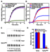
Mutant ypt1-G80D Yeast Cells are Sensitive to Heat-Stress. (A) Effect of heat treatment on cell viability. YPT1 and ypt1-G80D cells (5 × 10⁷ cells/mL) grown in YPD medium were incubated at 27 °C or 45 °C, and samples were withdrawn at the indicated times for measurement of viable counts. Cell survival (percent) at each time point was calculated as 100× the ratio of the viable count at that time to the viable count at time zero. Error bars: means ± SD of at least 3 independent experiments. (B) Trypan blue (TB) exclusion assay of heat-shock-induced cell death. Samples of the YPT1 and ypt1-G80D cells at the 60 min time point in (A) were visualized by fluorescence microscopy after staining with TB. The percentages of TB-negative cells are shown below the images. Data are represented as the mean ± SD of at least three independent experiments. White and black arrowheads show examples of TB-negative and -positive cells, respectively. Scale bar, 50 μm.
Figure 2
Figure 2
Heat-Shock-Induced Cytosolic Protein Aggregation in Yeast Cells. (A) Heat-shock-induced cytosolic protein aggregation in YPT1 and ypt1-G80D cells. The cells (5 × 10⁷ cells/mL) were grown in YPD medium and incubated at 27 °C or 45 °C. Samples were withdrawn at the 60 min time point, and the amounts of insoluble cytosolic protein were determined. The insoluble fractions were subjected to SDS-PAGE followed by silver-staining. Each lane represents the insoluble fraction of 3 × 10⁶ cells. (B) Proportions of insoluble fractions in the total cytosolic proteins extracted in (A). Red line indicates the comparison between YPT1 and ypt1-G80D samples at 45 °C, blue line shows the comparison of two ypt1-G80D samples at 27 °C and 45 °C, whereas black lines represents the comparison of two YPT1 samples at 27 °C and 45 °C or the comparison between YPT1 and ypt1-G80D samples at 27 °C. Error bars: means ± SD of at least 3 independent experiments. * p < 0.05, *** p < 0.001, and NS, no significance, according to a 2-tailed Student’s t test.
Figure 3
Figure 3
Heat-Shock Induces Changes in the Molecular State of Ypt1p but not Ypt1pG80D. (A) Changes in the molecular state of Ypt1p and Ypt1pG80D in vivo. YPT1 and ypt1-G80D cells were grown in YPD medium (1 × 10⁸ cells/mL) and incubated at 27 °C or 45 °C for 45 min. Half of the heat-treated cells were transferred to an equal volume of fresh YPD medium containing 100 μg/mL cycloheximide and allowed to recover for 10 h at 27 °C (45/27). Total protein extracts (10 μg) were analyzed by SDS-PAGE, and proteins were visualized by CBB staining (left image). In addition, total protein extracts (50 μg) were analyzed by immunoblotting with a polyclonal anti-Ypt1p antibody after fractionation by SDS-PAGE (middle image) or native-PAGE (right image). W.B., western blotting. (B,C) Changes in the molecular state of Ypt1p and Ypt1pG80D in vitro. (B) Purified bacterially expressed Ypt1p and Ypt1pG80D (3 μg/μL) were incubated at 25 °C or 45 °C for 30 min, subjected to native-PAGE (upper image) or SDS-PAGE (lower image), and then silver-stained. (C) SEC analysis of the protein solutions described in (B). (D) A bis-ANS binding assay to identify heat-shock-induced exposure of hydrophobic domains in Ypt1p and Ypt1pG80D. Fluorescence spectra of bis-ANS were measured with excitation at 380 nm and emission scanning at 400–600 nm. The samples used were 10 μM bis-ANS (a), 10 μM bis-ANS plus 30 μM Ypt1p incubated at 25 °C (b) or 45 °C (b′), and 10 μM bis-ANS plus 30 μM Ypt1pG80D incubated at 25 °C (c) or 45 °C (c′) for 20 min.
Figure 4
Figure 4
Ypt1pG80D has GTPase Activity but not Molecular Chaperone Activity. (A,B) Chaperone activity assay. Light scattering was monitored at 340 nm over a 15 min incubation period. Shown are representative data out of at least three independent experiments. (A) Solutions of MDH (1.67 μM) alone (-o-) or with 8.35 μM GST (-●-), Ypt1p (-▲-), or Ypt1pG80D (-■-) in 50 mM HEPES (pH 8.0) were incubated in a spectrophotometer cell at 45 °C. (B) Solutions of MDH (1.67 μM) alone (-o-) or with 3.34 μM Ypt1p pretreated at 25 °C (-▲-) or 45 °C (-▲-) or 3.34 μM Ypt1pG80D pretreated at 25 °C (-■-) or 45 °C (-■-) in 50 mM HEPES (pH 8.0) were incubated in a spectrophotometer cell at 45 °C. (C) GTPase activity assay. Recombinant Ypt1p and Ypt1pG80D (2 μg each) were incubated with [α-32P]GTP at 30 °C, and samples of the reaction mixtures were withdrawn at different time points for analysis by TLC. Shown is a representative image out of at least three independent experiments. (D) Relative GTPase (240 min) and chaperone (15 min) activities of Ypt1p and Ypt1pG80D. The activities of Ypt1p were set to 100%. Data are represented as the mean ± SD of at least three independent experiments. **** P < 0.0001, and NS, no significance, according to a Student’s t test.
Figure 5
Figure 5
PBA Increases the Thermo Tolerance of ypt1-G80D Cells. (A) Yeast spot assay. Yeast cells (5 × 7 cells/mL) were grown in YPD medium and incubated at 27 °C or 45 °C for 1 h in the presence (+PBA) or absence (-PBA) of 1 mM PBA. Samples of the YPT1 and ypt1-G80D cells were withdrawn for analysis. Aliquots (6 μL) of 10-fold serial dilutions of these cell suspensions were spotted onto YPD plates, and the plates were photographed after incubation at 27 °C for 3 days. (B) Effect of PBA on heat-shock resistance of the YPT1 and ypt1-G80D cells. Yeast cells (5 × 7 cells/mL) were grown in YPD medium and incubated at 45 °C in the presence (+PBA) or absence (-PBA) of 1 mM PBA. Samples were withdrawn at the indicated times for measurement of viable counts. Cell survival (percent) at each time point was calculated as 100× the ratio of the viable count at that time to the viable count at time zero. Error bars: means ± SD of at least 3 independent experiments. (C) Trypan blue exclusion assay of heat-shock-induced cell death. Samples of the YPT1 and ypt1-G80D cells at the 60 min time point in (B) were visualized by fluorescence microscopy after staining with trypan blue. The percentages of TB-negative cells are shown below the images. Data are represented as the mean ± SD of at least three independent experiments.
Figure 6
Figure 6
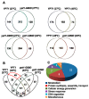
Analyses of LC/MS Data. (A) Venn diagrams showing the numbers of proteins identified by the LC/MS analysis in extracts of YPT1 and ypt1-G80D cells incubated at 27 °C (upper left), extracts of YPT1 cells incubated at 27 °C or 45 °C (upper right), extracts of ypt1-G80D cells incubated at 27 °C or 45 °C (lower left), and extracts of YPT1 and ypt1-G80D cells incubated at 45 °C (lower right). (B) Venn diagram showing the relationships between the proteins identified by the LC/MS analysis in extracts of YPT1 and ypt1-G80D cells incubated at 27 °C or 45 °C. The group of 49 proteins that was induced at 45 °C in YPT1 cells but not ypt1-G80D cells is indicated by the red outline. (C) Pie chart showing distribution of these 49 proteins among different cellular processes.

Similar articles

Cited by

References

    1. Jedd G., Richardson C., Litt R., Segev N. The Ypt1 GTPase is essential for the first two steps of the yeast secretory pathway. J. Cell Biol. 1995;131:583–590. doi: 10.1083/jcb.131.3.583. - DOI - PMC - PubMed
    1. Segev N., Botstein D. The ras-like yeast YPT1 gene is itself essential for growth, sporulation, and starvation response. Mol. Cell Biol. 1987;7:2367–2377. doi: 10.1128/MCB.7.7.2367. - DOI - PMC - PubMed
    1. Nuoffer C., Davidson H.W., Matteson J., Meinkoth J., Balch W.E. A GDP-bound of rab1 inhibits protein export from the endoplasmic reticulum and transport between Golgi compartments. J. Cell Biol. 1994;125:225–237. doi: 10.1083/jcb.125.2.225. - DOI - PMC - PubMed
    1. Richardson C.J., Jones S., Litt R.J., Segev N. GTP hydrolysis is not important for Ypt1 GTPase function in vesicular transport. Mol. Cell Biol. 1998;18:827–838. doi: 10.1128/MCB.18.2.827. - DOI - PMC - PubMed
    1. Du L.L., Novick P. Yeast rab GTPase-activating protein Gyp1p localizes to the Golgi apparatus and is a negative regulator of Ypt1p. Mol. Biol. Cell. 2001;12:1215–1226. doi: 10.1091/mbc.12.5.1215. - DOI - PMC - PubMed

MeSH terms

LinkOut - more resources