. 2019 Jan 2;8(1):bio037622.
doi: 10.1242/bio.037622.

Glucose starvation triggers filamentous septin assemblies in an S. pombe septin-2 deletion mutant

Affiliations

Glucose starvation triggers filamentous septin assemblies in an S. pombe septin-2 deletion mutant

Minghua Liu et al. Biol Open. .

Abstract

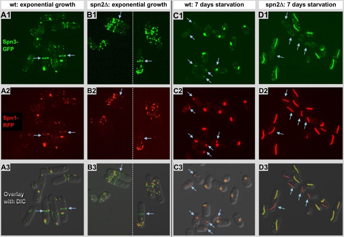
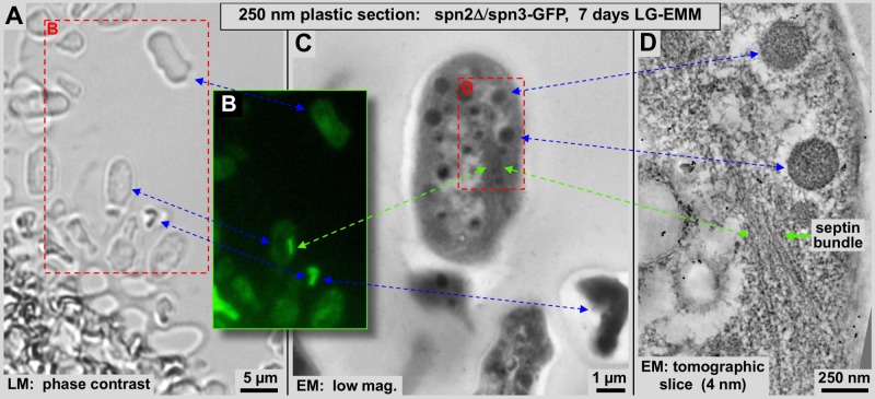
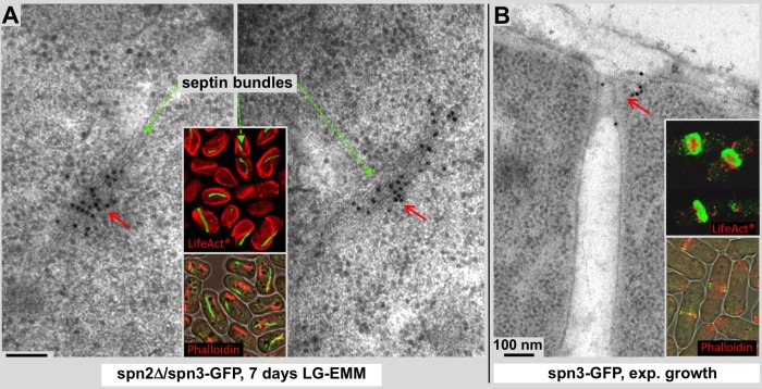
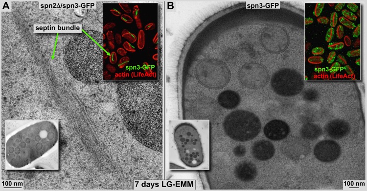
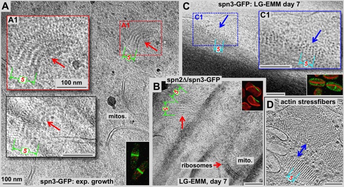
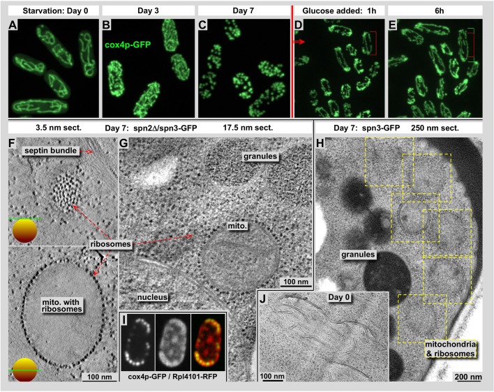
Using correlative light and electron microscopy (CLEM), we studied the intracellular organization by of glucose-starved fission yeast cells (Schizosaccharomyces pombe) with regards to the localization of septin proteins throughout the cytoplasm. Thereby, we found that for cells carrying a deletion of the gene encoding septin-2 (spn2Δ), starvation causes a GFP-tagged version of septin-3 (spn3-GFP) and family members, to assemble into a single, prominent filamentous structure. It was previously shown that during exponential growth, spn2Δ cells form septin-3 polymers. However, the polymers we observed during exponential growth are different from the spn3p-GFP structure we observed in starved cells. Using CLEM, in combination with anti-GFP immunolabeling on plastic-sections, we could assign spn3p-GFP to the filaments we have found in EM pictures. Besides septin-3, these filamentous assemblies most likely also contain septin-1 as an RFP-tagged version of this protein forms a very similar structure in starved spn2Δ cells. Our data correlate phase-contrast and fluorescence microscopy with electron micrographs of plastic-embedded cells, and further on with detailed views of tomographic 3D reconstructions. Cryo-electron microscopy of spn2Δ cells in vitrified sections revealed a very distinct overall morphology of the spn3p-GFP assembly. The fine-structured, regular density pattern suggests the presence of assembled septin-3 filaments that are clearly different from F-actin bundles. Furthermore, we found that starvation causes substantial mitochondria fission, together with massive decoration of their outer membrane by ribosomes.

Keywords: Conventional and cryo-electron microscopy; Correlative light and electron microscopy; Filamentous septin assemblies; Glucose starvation in S. pombe; Septins; Vitrified sectioning.


Conflict of interest statement

Competing interestsThe authors declare no competing or financial interests.

Figures

Fig. 1.
Fig. 1.
Spn1p-RFP and spn3p-GFP expression and localization patterns. (A1–A3) Exponentially growing cells expressing spn3p-GFP (A1, green), spn1p-RFP (A2, red), overlaid with a DIC image in A3. Both proteins can be found together, evenly distributed throughout the entire cytosol forming little clusters and accumulated at the periphery of septa in dividing cells (arrows; also see Fig. 4B). (B1–B3) Exponentially growing spn2Δ cells expressing spn3p-GFP (B1) and spn1p-RFP (B2). The panels are merges of two different images indicated by the dotted line. Both proteins assemble into globular clusters (also see Fig. 5A) or short filamentous assemblies. Spn3p-GFP can be found on septa while spn1p-RFP seems absent, or only present at a very low concentration [also see overlay (B3) and arrows]. (C1–C3) Starved cells expressing spn3p-GFP (C1) and spn1p-RFP (C2), after 7 days of culturing in low glucose medium (LMM). Both proteins aggregate and merge together into one single clump per cell, except for minor traces of protein that remain distributed throughout the cytosol. Evident from the overlay panel; not all clusters contain both proteins (arrows). (D1–D3) Starved spn2Δ cells expressing spn3p-GFP (D1) and spn1p-RFP (D2) after 7 days of culturing in LMM. Both proteins form prominent elongated filamentous structures, typically only one per cell. The large elongated assemblies in each cell all seem to contain spn1p-RFP or both, but interestingly, some cells lack the spn3p-GFP component (arrows; see overlay D3).
Fig. 2.
Fig. 2.
Correlative light and electron microscopy performed on spn2Δ/spn3-GFP cells. Arrows connect identical features that we can identify in the phase contrast image, fluorescence image and tomographic reconstruction of starved cells that have formed filamentous spn3p-GFP assemblies after 7 days of culturing LMM. A 250 nm thick plastic section of high-pressure-frozen, lowicryl-K4M embedded cells were mounted on an EM grid. An identical region on the grid was imaged on the grid by phase contrast (A: LM), by fluorescence light microscopy (B) and, after transfer to a 300 kV Tecnai-F30, with low-magnification as micrograph (C: EM) and a thin (4.0 nm) computational section through an electron tomogram (D). Green arrows connect the sites of the spn3p-GFP assemblies. Blue arrows connect other easily recognizable common features of the different panels such as high-density polymers, granules and entire cells. The red frame in A corresponds to the image area shown in B. The red frame in C corresponds to the image area shown in D.
Fig. 3.
Fig. 3.
Immunogold labeling on thin sections of spn2Δ/spn3-GFP and spn3-GFP cells. Labeling was achieved with an anti-GFP primary antibody and a secondary antibody conjugated to 15 nm gold particles (arrows). (A) Immunolabeling provides another mode of correlation between the fluorescence signals (inset panel) and EM density data and confirms the presence of spn3p-GFP in the filamentous structures observed in starved spn2Δ cells after 7 days of culturing in low glucose medium (red arrows in both panels). Green dashed arrows indicate comparable septin bundles on electron micrograph and fluorescence images. The inset panels in A and B show spn3p-GFP fluorescence images of the corresponding cells in green, and actin staining in red (upper panels, LifeAct®-mCherry; lower panels, Rhodamine-Phalloidin). The lower insets display actin staining with Rhodamine-Phalloidin, overlaid with a phase-contrast image, to test for potential differences with LifeAct®-mCherry (also see Fig. 4). (B) The anti-GFP primary antibody binds to spn3p-GFP at the outer ring formed by the septa and the old cell wall in dividing wild-type cells during exponential growth (red arrow). The inset panels show dividing cells in a projection of a thin confocal slice (lower insets) and an entire confocal 3D image stack that is slightly tilted to better visualize the ring shaped spn3p-GFP and inner actin distribution (upper inset). In septa, actin (red, Phalloidin, or LifeAct®-mCherry labeled) is surrounded by spn3p-GFP (green), confirming that the latter mostly locates closer to the cell periphery, forming a ring-like structure.
Fig. 4.
Fig. 4.
Filamentous septin assemblies in plastic sections. Comparison of 80 nm plastic sections of high-pressure frozen, freeze-substituted and plastic-embedded starved spn2Δ/spn3-GFP cells (A) and spn3-GFP cells (B). Both cells were starved for 7 days in low glucose medium as described. The upper inset panels show the corresponding spn3p-GFP fluorescence (green) as well as LifeAct®-mCherry, which marks F-actin (also see Fig. 3). In starvation, F-actin forms long filamentous structures in both cell types. The lower inset panels show EM overviews of cells at corresponding conditions. (A) In spn2Δ/spn3-GFP cells, the F-actin bundles and filamentous spn3p-GFP assemblies do not overlap (upper inset panel). Green arrows point to comparable septin bundles on electron micrograph and fluorescence images. (B) None of the filamentous spn3p-GFP assemblies were present in starved wild-type cells even though F-actin forms the same type of bundles as in spn2Δ cells (upper inset panel). Otherwise, the cytosol of both cell types look very similar. Note the dense vacuoles and the lighter stained fragmented mitochondria, which are densely decorated with ribosomes.
Fig. 5.
Fig. 5.
Septin and F-actin bundles observed in sections of unstained, frozen-hydrated specimens. (A–C) We have taken advantage of the superb molecular structure preservation of vitrified sections to compare the morphology of F-actin bundles with that of spn3p-GFP aggregates in wild-type (A, exponentially growing; C, starved) and spn2Δ cells, and after 7 days of glucose starvation (B). C shows a starved wild-type cell that shows F-actin bundles, but no septin assemblies (also see Fig. 4B). D shows a vitrified section through the stress fibers of a 3T3 fibroblast, allowing for a direct comparison of shape and dimensions with the F-actin bundles in C. The excellent molecular preservation in frozen-hydrated specimens reveals distinct differences between septin (A, including insets; B) and actin bundles (C) or actin stress fibers (D). Both F-actin bundles and stress fibers are morphologically quite different from spn3p-GFP assemblies, which form tighter curves and show a fine, but well visible, repetitive pattern of globular domains while appearing less ordered at the overall bundle level, especially in the clusters found in exponentially growing state (A). The lateral packing of F-actin bundles is much tighter than that of septin bundles [compare the width of five strands within actin bundles (visible in panels C and D, indicated in blue) and septin bundles (panels A and B, indicated in green)]. Insets in A, B and C show corresponding cells with fluorescence labeling of septin-3 (spn3p-GFP) and actin (LifeAct®-mCherry).
Fig. 6.
Fig. 6.
During prolonged glucose starvation, mitochondria undergo substantial fission and show a massive decoration of their outer membranes by ribosomes. (A–E) Mitochondria in S. pombe at different stages of starvation (A–C) and recovering after adding back glucose (D,E). Mitochondria were visualized with cox4-GFP (Sesaki and Jensen, 1999). A–D show different cells in each panel due to the length of the process. Recovery after adding glucose is much faster than entering starvation, and we could demonstrate how cells start growing and dividing again and elongate their shape gradually (see cell-length markers in D and E, which point to identical cells. The green bar indicates the growth during that time.) EM pictures (F–I) were either tomographic X-Y slices of 250 nm plastic sections (F,G) at 3.5 nm (F) and 17.5 nm thickness (G), a full 250 nm plastic section (H) or a tomographic slice from a cryo-electron tomogram of a vitrified section (J). Panels F–H show cells after 7 days of starvation, while panel J functions as a control of cells during exponential growth. Starved cells show mitochondria outer membranes which are densely packed with ribosomes (F–H), while the membranes of exponentially growing cells are smooth (J). (I) Co-localization of mitochondria, labeled with cox4p-GFP (left), ribosomes labeled with Rpl4101-RFP (center) and the overlay of both (right).
Fig. 7.
Fig. 7.
Western blot with anti-GFP antibodies marking septin-GFP constructs (spn1-GFP to spn7-GFP) and, as control, wild type during the exponential growing phase and after 6 days of starvation. Septins 5–7 are expressed mostly during meiosis, and therefore are relatively sparse, especially in non-dividing, starved cell cultures. Septins 1–4 are strongly expressed under both conditions – exponential growth (EG) and after 6 days of starvation (SD6) – but seem to fragment much more during starvation, most likely caused by a tuned-down expression of fresh protein. Tubulin and actin expressions are significantly reduced after 6 days of starvation.

Similar articles

Cited by

References

    1. Al-Amoudi A., Chang J.-J., Leforestier A., McDowall A., Salamin L. M., Norlén L. P. O., Richter K., Blanc N. S., Studer D. and Dubochet J. (2004). Cryo-electron microscopy of vitreous sections. EMBO J. 23, 3583-3588. 10.1038/sj.emboj.7600366 - DOI - PMC - PubMed
    1. An H., Morrell J. L., Jennings J. L., Link A. J. and Gould K. L. (2004). Requirements of fission yeast septins for complex formation, localization, and function. Mol. Biol. Cell. 15, 5551-5564. 10.1091/mbc.e04-07-0640 - DOI - PMC - PubMed
    1. Bähler J., Wu J.-Q., Longtine M. S., Shah N. G., McKenzie A. III, Steever A. B., Wach A., Philippsen P. and Pringle J. R. (1998). Heterologous modules for efficient and versatile PCR-based gene targeting in Schizosaccharomyces pombe. Yeast 14, 943-951. 10.1002/(SICI)1097-0061(199807)14:10<943::AID-YEA292>3.0.CO;2-Y - DOI - PubMed
    1. Bertin A., McMurray M. A., Thai L., Garcia G. III, Votin V., Grob P., Allyn T., Thorner J. and Nogales E. (2010). Phosphatidylinositol-4,5-bisphosphate promotes budding yeast septin filament assembly and organization. J. Mol. Biol. 404, 711-731. 10.1016/j.jmb.2010.10.002 - DOI - PMC - PubMed
    1. Bouchet-Marquis C. and Hoenger A. (2011). Cryo-electron tomography on vitrified sections: a critical analysis of benefits and limitations for structural cell biology. Micron 42, 152-162. 10.1016/j.micron.2010.07.003 - DOI - PubMed