

IRF5 Is Required for Bacterial Clearance in Human M1-Polarized Macrophages, and IRF5 Immune-Mediated Disease Risk Variants Modulate This Outcome
- PMID: 30593537
- PMCID: PMC6585431
- DOI: 10.4049/jimmunol.1800226
IRF5 Is Required for Bacterial Clearance in Human M1-Polarized Macrophages, and IRF5 Immune-Mediated Disease Risk Variants Modulate This Outcome
Abstract
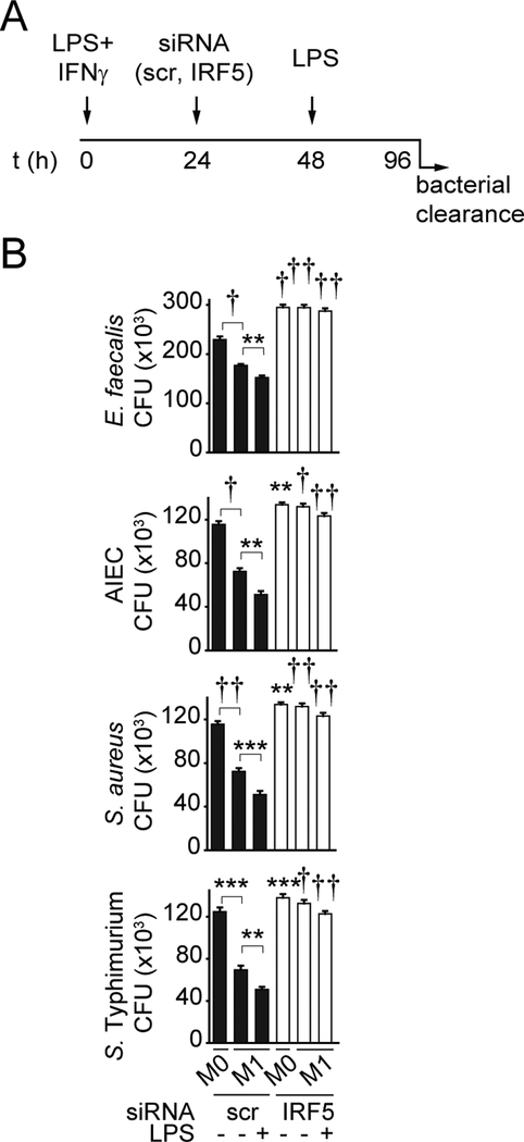
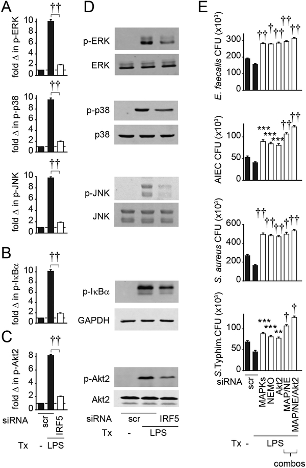
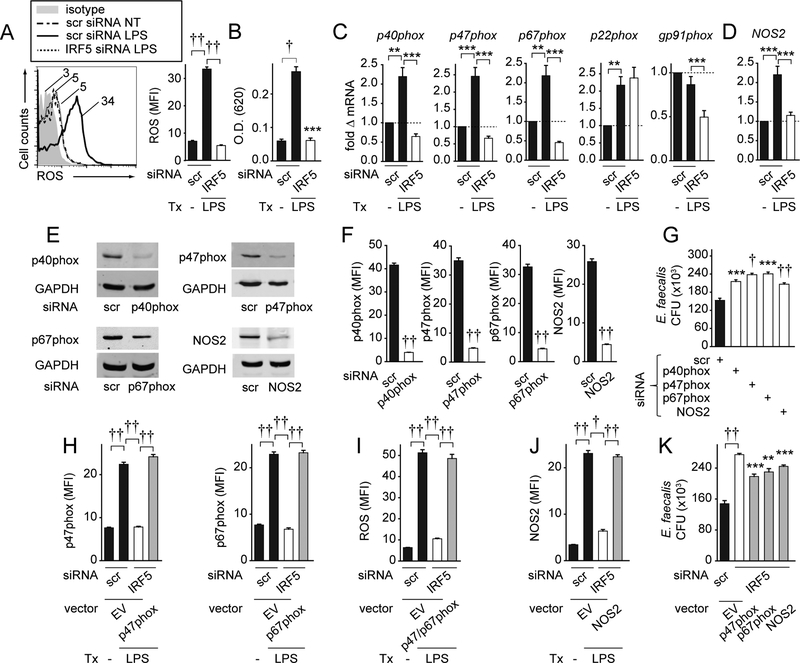
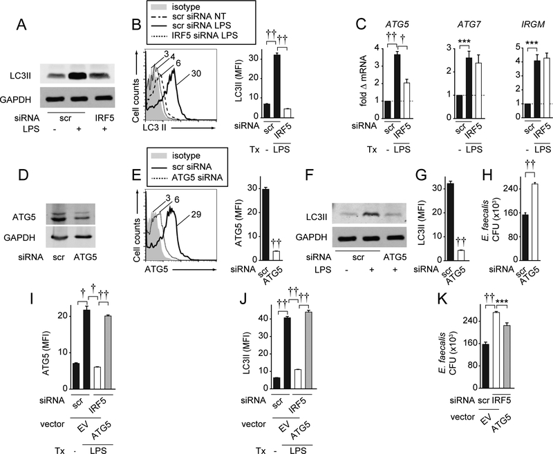
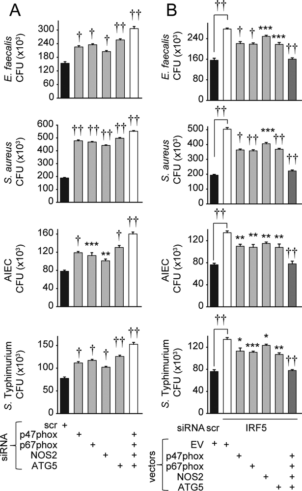
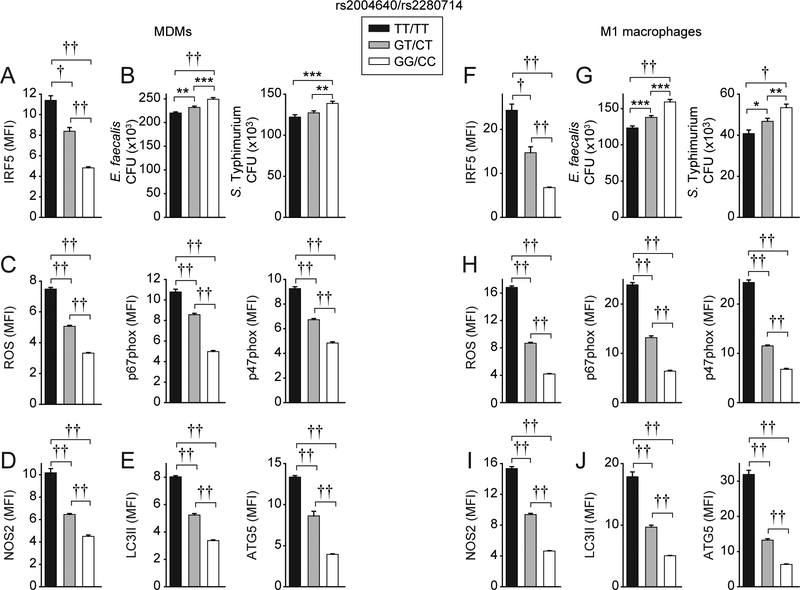
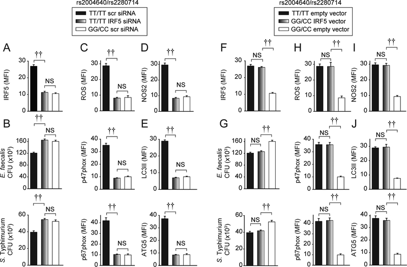
Common IFN regulatory factor 5 (IRF5) variants associated with multiple immune-mediated diseases are a major determinant of interindividual variability in pattern recognition receptor (PRR)-induced cytokines in macrophages. PRR-initiated pathways also contribute to bacterial clearance, and dysregulation of bacterial clearance can contribute to immune-mediated diseases. However, the role of IRF5 in macrophage-mediated bacterial clearance is not well defined. Furthermore, it is unclear if macrophages from individuals who are carriers of low IRF5-expressing genetic variants associated with protection for immune-mediated diseases might be at a disadvantage in bacterial clearance. We found that IRF5 was required for optimal bacterial clearance in PRR-stimulated, M1-differentiated human macrophages. Mechanisms regulated by IRF5 included inducing reactive oxygen species through p40phox, p47phox and p67phox, NOS2, and autophagy through ATG5. Complementing these pathways in IRF5-deficient M1 macrophages restored bacterial clearance. Further, these antimicrobial pathways required the activation of IRF5-dependent MAPK, NF-κB, and Akt2 pathways. Importantly, relative to high IRF5-expressing rs2004640/rs2280714 TT/TT immune-mediated disease risk-carrier human macrophages, M1-differentiated GG/CC carrier macrophages demonstrated less reactive oxygen species, NOS2, and autophagy pathway induction and, consequently, reduced bacterial clearance. Increasing IRF5 expression to the rs2004640/rs2280714 TT/TT levels restored these antimicrobial pathways. We define mechanisms wherein common IRF5 genetic variants modulate bacterial clearance, thereby highlighting that immune-mediated disease risk IRF5 carriers might be relatively protected from microbial-associated diseases.
Copyright © 2019 by The American Association of Immunologists, Inc.
Figures
Similar articles
-
IRF5 and IRF5 Disease-Risk Variants Increase Glycolysis and Human M1 Macrophage Polarization by Regulating Proximal Signaling and Akt2 Activation.Cell Rep. 2016 Aug 30;16(9):2442-55. doi: 10.1016/j.celrep.2016.07.060. Epub 2016 Aug 18. Cell Rep. 2016. PMID: 27545875 Free PMC article.
-
IRF5 risk polymorphisms contribute to interindividual variance in pattern recognition receptor-mediated cytokine secretion in human monocyte-derived cells.J Immunol. 2012 Jun 1;188(11):5348-56. doi: 10.4049/jimmunol.1103319. Epub 2012 Apr 27. J Immunol. 2012. PMID: 22544929 Free PMC article.
-
Myeloid Cell-Intrinsic IRF5 Promotes T Cell Responses through Multiple Distinct Checkpoints In Vivo, and IRF5 Immune-Mediated Disease Risk Variants Modulate These Myeloid Cell Functions.J Immunol. 2020 Aug 15;205(4):1024-1038. doi: 10.4049/jimmunol.1900743. Epub 2020 Jul 20. J Immunol. 2020. PMID: 32690658 Free PMC article.
-
IRF5-mediated signaling and implications for SLE.Clin Immunol. 2014 Aug;153(2):343-52. doi: 10.1016/j.clim.2014.06.001. Epub 2014 Jun 10. Clin Immunol. 2014. PMID: 24928322 Review.
-
Activation and function of interferon regulatory factor 5.J Interferon Cytokine Res. 2015 Feb;35(2):71-8. doi: 10.1089/jir.2014.0023. Epub 2014 Sep 26. J Interferon Cytokine Res. 2015. PMID: 25259415 Review.
Cited by
-
A genome-wide screen identifies IRF2 as a key regulator of caspase-4 in human cells.EMBO Rep. 2019 Sep;20(9):e48235. doi: 10.15252/embr.201948235. Epub 2019 Jul 29. EMBO Rep. 2019. PMID: 31353801 Free PMC article.
-
Physiological and Pathological Inflammation Induced by Antibodies and Pentraxins.Cells. 2021 May 12;10(5):1175. doi: 10.3390/cells10051175. Cells. 2021. PMID: 34065953 Free PMC article. Review.
-
Neutralization of excessive levels of active TGF-β1 reduces MSC recruitment and differentiation to mitigate peritendinous adhesion.Bone Res. 2023 May 8;11(1):24. doi: 10.1038/s41413-023-00252-1. Bone Res. 2023. PMID: 37156778 Free PMC article.
-
T Cell-Intrinsic IRF5 Regulates T Cell Signaling, Migration, and Differentiation and Promotes Intestinal Inflammation.Cell Rep. 2020 Jun 30;31(13):107820. doi: 10.1016/j.celrep.2020.107820. Cell Rep. 2020. PMID: 32610123 Free PMC article.
-
Diamond-Like Carbon Depositing on the Surface of Polylactide Membrane for Prevention of Adhesion Formation During Tendon Repair.Nanomicro Lett. 2024 Apr 30;16(1):186. doi: 10.1007/s40820-024-01392-7. Nanomicro Lett. 2024. PMID: 38687411 Free PMC article.
References
-
- Liu H, Irwanto A, Fu X, Yu G, Yu Y, Sun Y, Wang C, Wang Z, Okada Y, Low H, et al. 2015. Discovery of six new susceptibility loci and analysis of pleiotropic effects in leprosy. Nat Genet 47: 267–271. - PubMed
-
- Wajant H, Pfizenmaier K, and Scheurich P. 2003. Tumor necrosis factor signaling. Cell Death Differ 10: 45–65. - PubMed
-
- Pasparakis M, Alexopoulou L, Episkopou V, and Kollias G. 1996. Immune and inflammatory responses in TNF alpha-deficient mice: a critical requirement for TNF alpha in the formation of primary B cell follicles, follicular dendritic cell networks and germinal centers, and in the maturation of the humoral immune response. J Exp Med 184: 1397–1411. - PMC - PubMed
Publication types
MeSH terms
Substances
Grants and funding
LinkOut - more resources
Full Text Sources
Research Materials
Miscellaneous