. 2019 Feb 15;30(4):427-440.
doi: 10.1091/mbc.E18-10-0609. Epub 2018 Dec 26.

Dissociation of membrane-chromatin contacts is required for proper chromosome segregation in mitosis

Affiliations

Dissociation of membrane-chromatin contacts is required for proper chromosome segregation in mitosis

Lysie Champion et al. Mol Biol Cell. .

Abstract

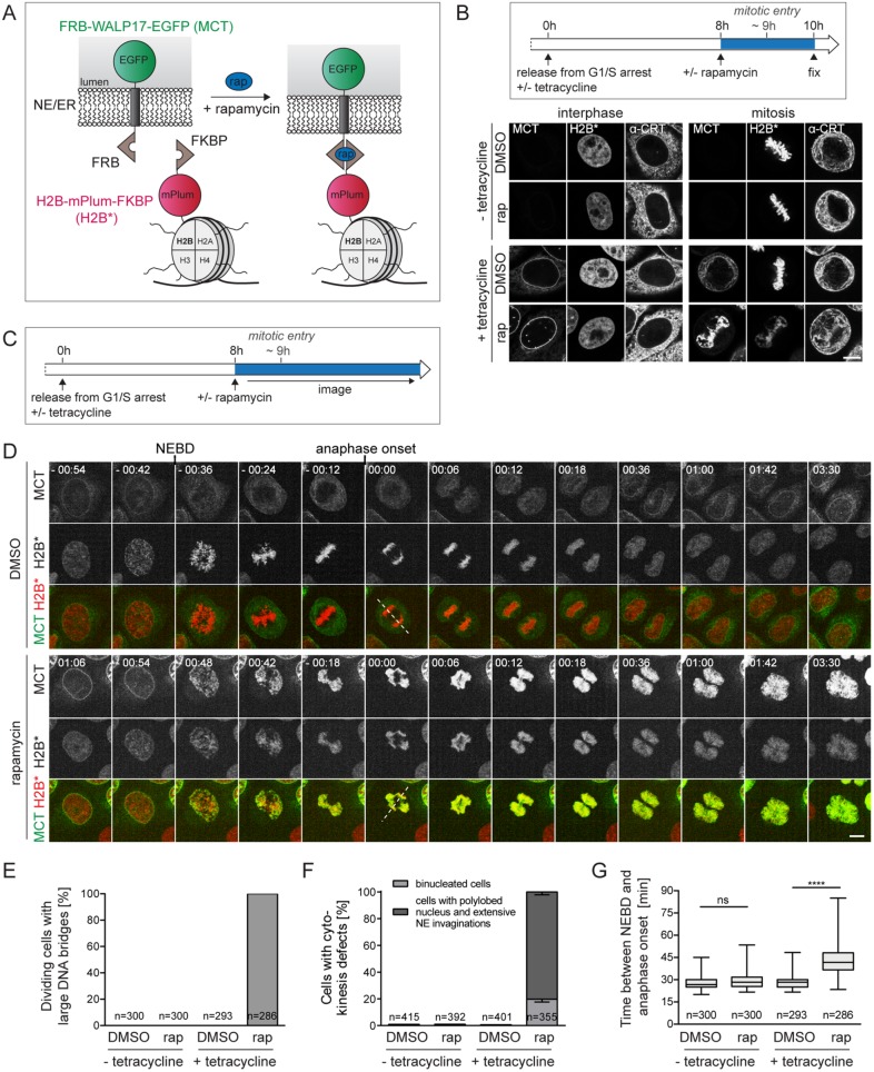
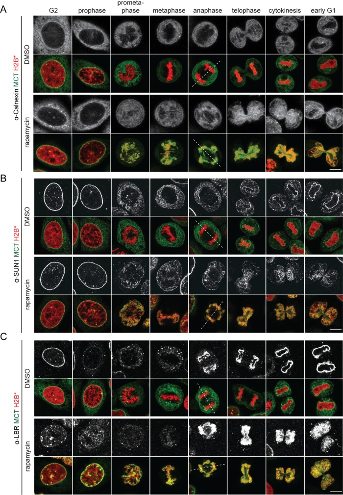
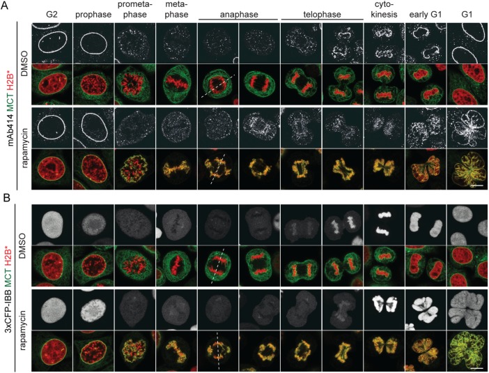
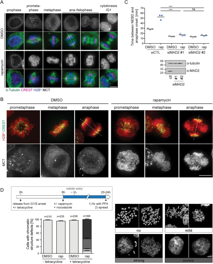
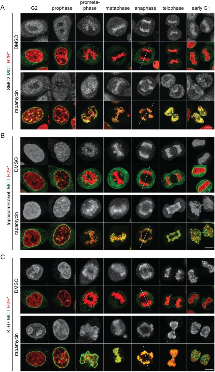
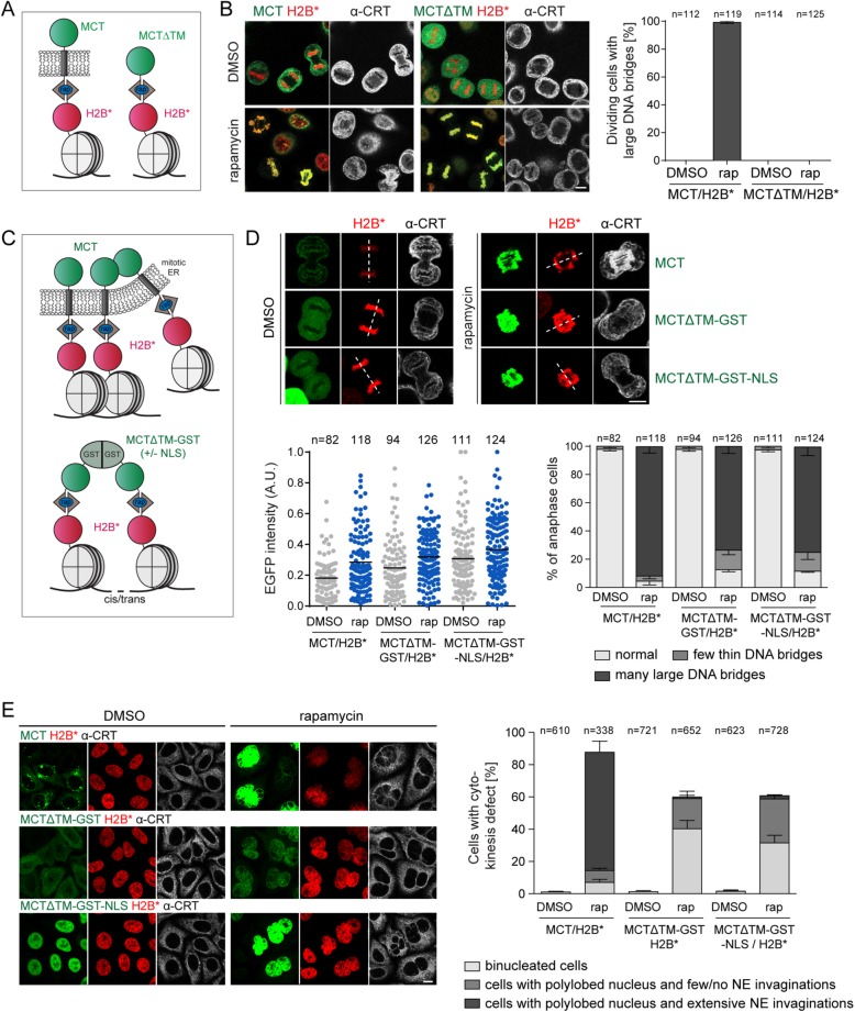
The nuclear envelope (NE) aids in organizing the interphase genome by tethering chromatin to the nuclear periphery. During mitotic entry, NE-chromatin contacts are broken. Here, we report on the consequences of impaired NE removal from chromatin for cell division of human cells. Using a membrane-chromatin tether that cannot be dissociated when cells enter mitosis, we show that a failure in breaking membrane-chromatin interactions impairs mitotic chromatin organization, chromosome segregation and cytokinesis, and induces an aberrant NE morphology in postmitotic cells. In contrast, chromosome segregation and cell division proceed successfully when membrane attachment to chromatin is induced during metaphase, after chromosomes have been singularized and aligned at the metaphase plate. These results indicate that the separation of membranes and chromatin is critical during prometaphase to allow for proper chromosome compaction and segregation. We propose that one cause of these defects is the multivalency of membrane-chromatin interactions.


Figures

FIGURE 1:
FIGURE 1:
Tethering of ER membranes to mitotic chromatin impairs chromosome segregation and cell division. (A) Scheme of the chromatin-ER membrane tethering system. FRB-WALP17-EGFP (membrane–chromatin tether, MCT); H2B-mPlum-FKBP (chromatin anchor, H2B*). (B) Flowchart of cell synchronization and drug treatment. Confocal images of interphase and mitotic MCT/H2B* cells. The ER was stained for calreticulin (α-CRT). (C) Flowchart of the experimental setup for D. (D) Time-lapse confocal microscopy of synchronized MCT/H2B* cells progressing through mitosis in the presence of DMSO or 200 nM rapamycin at the indicated times relative to anaphase onset. Quantification of anaphase DNA bridges (E), cytokinesis defects (F), and the time span between NEBD and anaphase onset (G) from time-lapse wide-field microscopy of synchronized MCT/H2B* cells. Note that only 70–80% of all cells express MCT in the presence of tetracycline. Only GFP-positive cells were analyzed. N = 3; mean ± SEM; n, number of cells. Dashed lines represent spindle axes. ****p < 0.0001; ns, not significant. Bars, 10 μm.
FIGURE 2:
FIGURE 2:
ER and NE membrane proteins are distributed throughout the mitotic ER in the presence of MCT-induced chromatin–membrane contacts. MCT/H2B* cells were treated as outlined in Figure 1B, immunostained using anti-calnexin (A), anti-SUN1 (B), and anti-LBR (C) antibodies, and analyzed by confocal microscopy. Chromatin configuration was used to assign cells to the indicated cell cycle stages. Dashed lines represent spindle axes. Bars, 10 μm.
FIGURE 3:
FIGURE 3:
Persistent, MCT-induced chromatin–membrane contacts induce an aberrant postmitotic nuclear morphology but do not affect postmitotic NPC assembly. MCT/H2B* cells were treated as in Figure 1B. (A) Immunostaining of FG repeat Nups using the mAb414 antibody. (B) Analysis of MCT/H2B*-3xCFP-IBB expressing cells. Dashed lines represent spindle axes. Bars, 10 μm.
FIGURE 4:
FIGURE 4:
Induced membrane-chromatin-contacts perturb the organization of mitotic chromatin. (A) Wide-field fluorescence microscopy of MCT/H2B* cells treated as in Figure 1B. Cells were stained for α-tubulin and kinetochores (CREST). Dashed lines represent spindle axes. (B) Representative wide-field fuorescent images of chromatin organization of DMSO- or rapamycin-treated MCT/H2B* cells progressing through mitosis. Kinetochores were immunostained using the CREST antibody. (C) Quantification of the time span between NEBD and anaphase onset of MCT/H2B* cells (as in Figure 1G) treated with either control or MAD2 siRNAs for 48 h. N = 3; mean ± SEM; n = 150 per condition. Bottom, immunoblot analysis of MAD2 depletion. (D) Analysis of MCT/H2B* cells with respect to mitotic chromatin structure. Flowchart of the cell synchronization protocol combined with drug treatment used for the generation of mitotic chromatin spreads (top). Representative confocal images of MeOH-fixed metaphase spreads from nocodazole-arrested MCT/H2B* cells. Spreads were counterstained with Hoechst. The chromosome structure defects were classified into four categories, ranging from a classical thread-like shape (“no”) to a disorganized amorphous cloud (“severe”). The quantification of each phenotype observed after the indicated treatment is shown in the bottom panel (N = 4; mean ± SEM; n, number of analyzed cells). Bars, 10 μm.
FIGURE 5:
FIGURE 5:
Chromatin condensation factors can be efficiently loaded on mitotic chromatin in the presence of the MCT. Synchronized MCT/H2B* cells were released into mitosis as outlined in Figure 1B. Cells were treated with DMSO or rapamycin 8 h after release, fixed 2 h after the treatment, and immunostained using anti-SMC2 (A), anti-topoisomeraseII (B) and anti–Ki-67 (C) antibodies and analyzed by confocal microscopy. Chromatin configuration was used to assign cells to the indicated cell cycle stages. Dashed lines represent spindle axes. Bars, 10 μm.
FIGURE 6:
FIGURE 6:
Chromosome segregation and cell division are not affected when membrane–chromatin tethering is induced in metaphase. (A) Flowchart of experimental approach in B. Cells were fixed at the indicated time points (see arrowheads). (B) Confocal images of synchronized MCT/H2B* cells fixed at the indicated time points. (C) Quantification of large DNA bridges in cells 1 h after release into anaphase. Note that all dividing cells regardless of MCT expression level were analyzed (without tetracycline: N = 2; mean ± SD; n, number of cells; with tetracycline: N = 4; mean ± SEM). Bars, 10 μm.
FIGURE 7:
FIGURE 7:
Membrane-induced chromatin segregation defects can be mimicked by soluble, bivalent chromatin-binding proteins. (A) Schematic comparison of the MCT and a soluble derivative lacking the transmembrane domain (MCT∆TM). (B) Synchronized MCT/H2B* and MCT∆TM/H2B* cells were released into mitosis as in Figure 1B, fixed, stained for calreticulin, and analyzed by confocal microscopy. The number of dividing cells with large DNA bridges was quantified (N = 3; mean ± SEM; n, number of analyzed GFP-positive cells). (C) Depiction of chromatin cross-linking by the MCT and MCT∆TM-GST(-NLS). (D, E) MCT/H2B*, MCTΔTM-GST/H2B*, and MCTΔTM-GST-NLS/H2B* cells were synchronized, treated with DMSO or 200 nM rapamycin before mitotic entry, and fixed in anaphase or after mitosis. (D) Confocal images of MCT/H2B*, MCTΔTM-GST/H2B* or MCTΔTM-GST-NLS/H2B* anaphase cells. Relative GFP intensities of all analyzed cells (N = 3; mean ± SEM; n, total number of cells) and quantification of chromosome segregation defects. (E) Confocal images of MCT/H2B*, MCTΔTM-GST/H2B*, or MCTΔTM-GST-NLS/H2B* after mitosis. The indicated postmitotic aberrations were quantified (N = 3; mean ± SEM; n, number of cells). CRT: calreticulin; rap: rapamycin. Dashed lines represent spindle axes. Bars, 10 μm.

Similar articles

Cited by

References

    1. Baxter J, Aragon L. (2012). A model for chromosome condensation based on the interplay between condensin and topoisomerase II. Trends Genet , 110–117. - PubMed
    1. Baxter J, Sen N, Martinez VL, De Carandini ME, Schvartzman JB, Diffley JF, Aragon L. (2011). Positive supercoiling of mitotic DNA drives decatenation by topoisomerase II in eukaryotes. Science , 1328–1332. - PubMed
    1. Beaudouin J, Gerlich D, Daigle N, Eils R, Ellenberg J. (2002). Nuclear envelope breakdown proceeds by microtubule-induced tearing of the lamina. Cell , 83–96. - PubMed
    1. Bolhy S, Bouhlel I, Dultz E, Nayak T, Zuccolo M, Gatti X, Vallee R, Ellenberg J, Doye V. (2011). A Nup133-dependent NPC-anchored network tethers centrosomes to the nuclear envelope in prophase. J Cell Biol , 855–871. - PMC - PubMed
    1. Booth DG, Beckett AJ, Molina O, Samejima I, Masumoto H, Kouprina N, Larionov V, Prior IA, Earnshaw WC. (2016). 3D-CLEM reveals that a major portion of mitotic chromosomes is not chromatin. Mol Cell , 790–802. - PMC - PubMed

Publication types

LinkOut - more resources