. 2019 Mar 5;93(6):e01459-18.
doi: 10.1128/JVI.01459-18. Print 2019 Mar 15.

Functional and Physical Interaction between the Arf Activator GBF1 and Hepatitis C Virus NS3 Protein

Affiliations

Functional and Physical Interaction between the Arf Activator GBF1 and Hepatitis C Virus NS3 Protein

Nadjet Lebsir et al. J Virol. .

Abstract

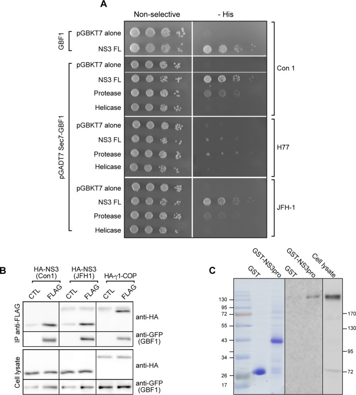
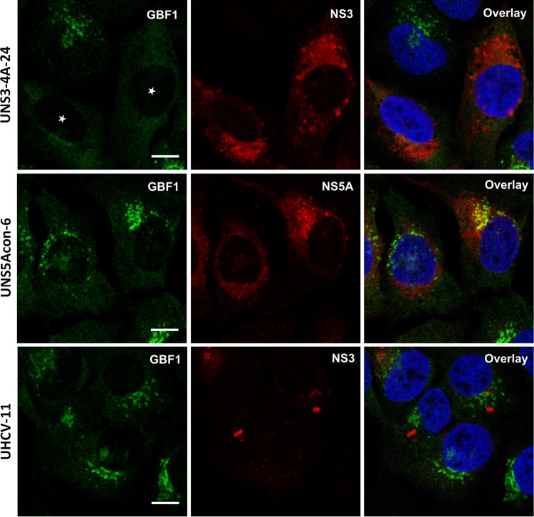
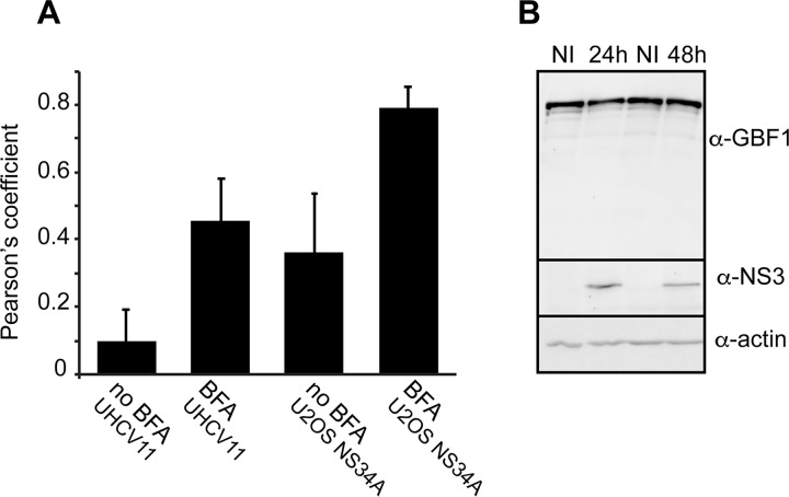
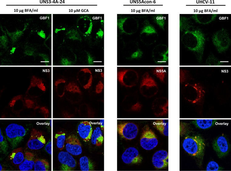
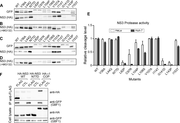
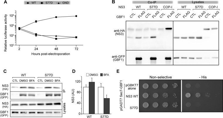
GBF1 has emerged as a host factor required for the genome replication of RNA viruses of different families. During the hepatitis C virus (HCV) life cycle, GBF1 performs a critical function at the onset of genome replication but is dispensable when the replication is established. To better understand how GBF1 regulates HCV infection, we have looked for interactions between GBF1 and HCV proteins. NS3 was found to interact with GBF1 in yeast two-hybrid, coimmunoprecipitation, and proximity ligation assays and to interfere with GBF1 function and alter GBF1 intracellular localization in cells expressing NS3. The interaction was mapped to the Sec7 domain of GBF1 and the protease domain of NS3. A reverse yeast two-hybrid screen to identify mutations altering NS3-GBF1 interaction yielded an NS3 mutant (N77D, Con1 strain) that is nonreplicative despite conserved protease activity and does not interact with GBF1. The mutated residue is exposed at the surface of NS3, suggesting it is part of the domain of NS3 that interacts with GBF1. The corresponding mutation in strain JFH-1 (S77D) produces a similar phenotype. Our results provide evidence for an interaction between NS3 and GBF1 and suggest that an alteration of this interaction is detrimental to HCV genome replication.IMPORTANCE Single-stranded, positive-sense RNA viruses rely to a significant extent on host factors to achieve the replication of their genome. GBF1 is such a cellular protein that is required for the replication of several RNA viruses, but its mechanism of action during viral infections is not yet defined. In this study, we investigated potential interactions that GBF1 might engage in with proteins of HCV, a GBF1-dependent virus. We found that GBF1 interacts with NS3, a nonstructural protein involved in HCV genome replication, and our results suggest that this interaction is important for GBF1 function during HCV replication. Interestingly, GBF1 interaction with HCV appears different from its interaction with enteroviruses, another group of GBF1-dependent RNA viruses, in keeping with the fact that HCV and enteroviruses use different functions of GBF1.

Keywords: ADP ribosylation factor; GBF1; NS3; hepatitis C virus; viral replication.


Figures

FIG 1
FIG 1
NS3 interacts with GBF1. (A) pGBKT7 plasmids carrying full-length NS3 (Con1 strain), the indicated fragments from the Con1, H77, and JFH1 strains, or pGBKT7 alone were cotransformed into yeast strain AH109 with the pGADT7 plasmids carrying full-length GBF1 (GBF1) or the catalytic Sec7 domain as indicated. Transformants were plated onto nonselective medium (left) or onto plates lacking histidine (−His) to monitor expression of the reporter His3 (right). Tenfold serial dilutions of each culture were spotted from left to right for each transformed strain. (B) HA-tagged NS3-4A of Con1 or JFH-1 or HA-tagged γ1-COP was coexpressed with YFP-GBF1 (CTL) or YFP-GBF1-FLAG (FLAG) in HeLa cells. Cells were lysed and lysates precipitated with anti-FLAG beads. Immunoprecipitated material and 5% of lysates were analyzed by immunoblotting with anti-HA and anti-GFP antibodies. (C) GST fused to the NS3 protease domain (GST-pro) or GST (left) were coupled to glutathione-Sepharose beads and incubated with 400 μl of HeLa cell cytoplasmic lysate. (Right) The lysate (10 μl) and the material bound to beads were analyzed by immunoblotting with an anti-GBF1 antibody.
FIG 2
FIG 2
GBF1 localization is altered in cells expressing HCV NS3-4A. UNS3-4A-24, UNS5Acon-6, and UHCV-11 cells were induced for 24 h and processed for immunofluorescence detection of GBF1 (green) and NS3 or NS5A (red), as indicated. White stars indicate cells expressing NS3. Bars, 10 μm.
FIG 3
FIG 3
Quantification of GBF1-NS3 colocalization and GBF1 expression in cells expressing NS3. (A) The colocalization of NS3 and GBF1 in UNS3-4A-24 and UHCV-11 cells induced for 24 h and treated with 10 μg/ml BFA for 30 min or left untreated was quantified by calculating the Pearson's correlation coefficient of at least 10 cells. Error bars represent the standard deviations (SD). (B) The expression of NS3 was induced in UNS3-4A-24 cells for 24 or 48 h. The cells were lysed and GBF1, NS3, and actin expression was analyzed by immunoblotting. NI, not induced.
FIG 4
FIG 4
GBF1 colocalizes with HCV NS3 in cells treated with BFA or GCA. UNS3-4A-24, UNS5Acon-6, and UHCV-11 cells were induced for 24 h, treated with BFA or GCA for 30 min, and processed for immunofluorescence detection of GBF1 (green) and NS3 or NS5A (red), as indicated. Bars, 10 μm.
FIG 5
FIG 5
Functional impact of GBF1-NS3 interaction on secretion and NS3 protease activity. (A) HeLa cells were cotransfected with pCMV-GLuc and increasing amounts of pcDNA3.1-HA-NS3-4A as indicated. The total amount of DNA was kept constant by adding empty pcDNA3.1. At 16 h posttransfection, the culture medium was changed and the cells were further incubated for 4 h. As a control, cells were treated with 1 μg/ml BFA during this 4-h secretion period. Luciferase activity was measured in supernatants and cell lysates. (B and C) HeLa cells were cotransfected with pEGFP-IPS, pcDNA3.1 HA-NS3-4A, and increasing concentrations of pEYFP-GBF1 as indicated. Empty pcDNA3.1 plasmid was used to keep the same final concentration of total transfected DNA constant. At 24 h posttransfection, EGFP-IPS cleavage was monitored by immunoblotting using anti-GFP antibody (B), and the intensities of bands corresponding to cleaved and uncleaved GPF-IPS were measured and the percentage of cleavage was calculated (C). Error bars represent the standard deviations for 3 independent experiments. *, **, and *** correspond to P values below 0.05, 0.01, and 0.001, respectively.
FIG 6
FIG 6
GBF1 interacts with NS3 in cells replicating HCV. (A and B) Huh-7 cells containing a subgenomic replicon of strain JFH-1 were processed for immunofluorescence detection of GBF1 (green) and NS3 (red), as indicated. Note the difference of GBF1 patterns in cells expressing higher levels of NS3 (indicated with a white star in panel A). A higher magnification of the area marked with a square in panel B is shown on the right side. Bars, 20 μm. (C and D) Huh-7 cells containing a subgenomic replicon of JFH-1 strain and naive Huh-7 cells were processed for proximity ligation assay using antibodies to NS3 and GBF1. Stacks of images corresponding to the total volume of the cells were acquired, and maximum intensity projections of the stacks were generated. (C) Representative images. PLA signal (white dots) and nuclei (blue). (D) Quantification of dots of 12 images from 2 independent experiments. ***, P < 0.001.
FIG 7
FIG 7
Interaction of Sec7‐GBF1 with HCV NS3 protease mutants. (A) Protocol for selection of NS3 mutants that do not interact with GBF1 in the yeast two-hybrid assay. (B) Yeast strain AH109 cotransformed with pGADT7-Sec7-GBF1 and pGBKT7, carrying mutants of the NS3 protease domain, were grown on nonselective and selective (−His) media to monitor reporter expression. (B) Localization of mutated residues in the structure of the NS3 protease domain. Mutated residues are shown in red, NS3 active site is shown in pink, NS3 membrane-associated α-helix is in green, NS4A is in yellow, and other NS3 residues are in blue. Three residues mutated in the yeast reverse two-hybrid assay, which are exposed at the surface of the protein, are indicated.
FIG 8
FIG 8
Replication and BFA sensitivity of NS3 protease domain mutants. (A) In vitro-transcribed RNA of Con1 replicon pFK-rep-PI-luc/ET containing NS3 protease mutants was electroporated into Huh-7.5 cells. Cells were lysed at the indicated times postelectroporation for luciferase assay. Data represent means from 4 independent experiments performed in triplicate. (B) Huh-7.5 cells electroporated with the indicated replicons were cultured for 8 h in the presence of the indicated concentrations of BFA and lysed at 48 h postelectroporation for luciferase assay. Luciferase activities were expressed relative to the luciferase activity measured at 2 h postelectroporation. Data are means from 3 independent experiments performed in triplicate. (C) WT HA-tagged NS3-4A or the indicated mutants were coexpressed with YFP-GBF1 (CTL) or YFP-GBF1-FLAG (FLAG) in HeLa cells. Cells were lysed and lysates precipitated with anti-FLAG beads. Immunoprecipitated material and 5% of lysates were analyzed by immunoblotting with anti-HA and anti-GFP antibodies.
FIG 9
FIG 9
Protease activity of NS3 protease domain mutants. (A) GFP-IPS was expressed in Huh-7 cells with the indicated HA-tagged NS3-4A mutants. Cell lysates were analyzed by immunoblotting with anti-GFP and anti-HA antibodies. (B) Immunoblot analysis of Huh-7 cells expressing the indicated NS3-4A mutants and treated for 8 h with MG132. (C) Immunoblot analysis of GFP-IPS cleavage in HeLa cells. (D) Immunoblot analysis of NS3 in HeLa cells treated with MG132. (E) Quantification of GFP-IPS cleavage by NS3-4A mutants in Huh-7 and HeLa cells. Data are means and standard deviations from 3 independent experiments. (F) WT or S77D HA-tagged NS3-4A was coexpressed with YFP-GBF1 (CTL) or YFP-GBF1-FLAG (FLAG) in HeLa cells. Cells were lysed and lysates precipitated with anti-FLAG beads. Immunoprecipitated material and 5% of lysates were analyzed by immunoblotting with anti-HA and anti-GFP antibodies.
FIG 10
FIG 10
Phenotype of the S77D mutation in NS3 of strain JFH-1. In vitro-transcribed RNA of ΔE1E2 JFH-1 containing the indicated mutations was electroporated in Huh-7.5 cells. Cells were lysed at the indicated times postelectroporation and luciferase activity was measured. Data represent means from 3 independent experiments performed in triplicate. (B and C) HeLa cells were cotransfected with expression plasmids for HA-tagged WT or S77D NS3-4A or HA-tagged γ1-COP with YFP-GBF1 (CTL) or YFP-GBF1-FLAG (FLAG). Cells were left untreated (B) or were treated with 10 μg/ml BFA or 0.1% DMSO (C). Cells were lysed and lysates incubated with anti-FLAG beads. Immunoprecipitated material and 5% of lysates were analyzed by immunoblotting with anti-HA and anti-GFP antibodies. (D) NS3 signals from 4 independent experiments were quantified and normalized to values of DMSO-treated cells, which were expressed as 100. Errors bars represent SD. (E) Yeast strain AH109 cotransformed with pGADT7-Sec7-GBF1 and pGBKT7 carrying the WT or the N77D mutant of the NS3 protease domain were grown on nonselective and selective (−His) media to monitor reporter expression.

Similar articles

Cited by

References

    1. Gosert R, Egger D, Lohmann V, Bartenschlager R, Blum HE, Bienz K, Moradpour D. 2003. Identification of the hepatitis C virus RNA replication complex in Huh-7 cells harboring subgenomic replicons. J Virol 77:5487–5492. doi:10.1128/JVI.77.9.5487-5492.2003. - DOI - PMC - PubMed
    1. Ferraris P, Blanchard E, Roingeard P. 2010. Ultrastructural and biochemical analyses of hepatitis C virus-associated host cell membranes. J Gen Virol 91:2230–2237. doi:10.1099/vir.0.022186-0. - DOI - PubMed
    1. Romero-Brey I, Merz A, Chiramel A, Lee JY, Chlanda P, Haselman U, Santarella-Mellwig R, Habermann A, Hoppe S, Kallis S, Walther P, Antony C, Krijnse-Locker J, Bartenschlager R. 2012. Three-dimensional architecture and biogenesis of membrane structures associated with hepatitis C virus replication. PLoS Pathog 8:e1003056. doi:10.1371/journal.ppat.1003056. - DOI - PMC - PubMed
    1. Egger D, Wölk B, Gosert R, Bianchi L, Blum HE, Moradpour D, Bienz K. 2002. Expression of hepatitis C virus proteins induces distinct membrane alterations including a candidate viral replication complex. J Virol 76:5974–5984. doi:10.1128/JVI.76.12.5974-5984.2002. - DOI - PMC - PubMed
    1. Gao L, Aizaki H, He J-W, Lai MMC. 2004. Interactions between viral nonstructural proteins and host protein hVAP-33 mediate the formation of hepatitis C virus RNA replication complex on lipid raft. J Virol 78:3480–3488. doi:10.1128/JVI.78.7.3480-3488.2004. - DOI - PMC - PubMed

Publication types

Substances

LinkOut - more resources