. 2019 Jan 7;216(1):176-194.
doi: 10.1084/jem.20181170. Epub 2018 Dec 19.

Neutrophils facilitate ovarian cancer premetastatic niche formation in the omentum

Affiliations

Neutrophils facilitate ovarian cancer premetastatic niche formation in the omentum

WonJae Lee et al. J Exp Med. .

Abstract

Ovarian cancer preferentially metastasizes to the omentum, a fatty tissue characterized by immune structures called milky spots, but the cellular dynamics that direct this tropism are unknown. Here, we identified that neutrophil influx into the omentum is a prerequisite premetastatic step in orthotopic ovarian cancer models. Ovarian tumor-derived inflammatory factors stimulated neutrophils to mobilize and extrude chromatin webs called neutrophil extracellular traps (NETs). NETs were detected in the omentum of ovarian tumor-bearing mice before metastasis and of women with early-stage ovarian cancer. NETs, in turn, bound ovarian cancer cells and promoted metastasis. Omental metastasis was decreased in mice with neutrophil-specific deficiency of peptidylarginine deiminase 4 (PAD4), an enzyme that is essential for NET formation. Blockade of NET formation using a PAD4 pharmacologic inhibitor also decreased omental colonization. Our findings implicate NET formation in rendering the premetastatic omental niche conducive for implantation of ovarian cancer cells and raise the possibility that blockade of NET formation prevents omental metastasis.


Figures

None
Graphical abstract
Figure 1.
Figure 1.
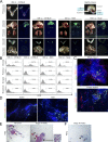
Ovarian cancer cells preferentially implant on the omentum, and this tropism is not affected by lymphoid deficiency. (A and B) GFP-expressing ID8 cells were injected i.b., between the left ovary and oviduct and contralateral to the omentum, into immunocompetent C57BL/6 mice, and tumors were evaluated at 6 wk thereafter. ID8 cells were injected i.p. into female immunocompetent C57BL/6 and NSG mice, and tumors were evaluated at 10 wk thereafter. GFP-expressing ES2 cells were injected i.p. into female nude mice, and tumors were evaluated at 9 d thereafter. Five mice per group were evaluated. (A) Representative images of tissues in each group viewed under light and fluorescence microscopy. Location of the omentum in a healthy mouse is also shown. Bars, 10 mm. (B) Representative flow cytometric analyses of the abundance of GFP+ tumor cells in each visceral fat tissue (omental, perirenal, gonadal, and mesenteric), expressed as a percentage of the nonfat cellular content in each tissue. (C) Visualization of total vasculature (CD31+; red) and HEVs in milky spots (PNAd+; green) in the naive mouse omentum by immunofluorescence staining of whole mount (top) and frozen (bottom) sections. Bars, 100 µm. Shown are representative results of three independent experiments. (D) Omental tissues were collected from nude mice at 5 d following i.p. injection of ES2 cells or saline (n = 5 mice per group). Frozen sections were stained to detect cancer cells (red) and PNAd+ vessels in milky spots (green). Bar, 100 µm. Shown are representative examples of staining. (E and F) Omental tissues of women without cancer and with stage III HGSC were stained with (E) H&E and with (F) Ab to PNAd (n = 3 cases per group). Bar, 200 µm. Indicated are vessels within or adjacent to milky spots (arrowheads) and tumor foci (arrows). Inset shows foci at higher magnification. Bar, 20 µm. Shown are representative examples of staining.
Figure 2.
Figure 2.
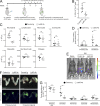
Early-stage ovarian tumors trigger an increase in neutrophil abundance in the premetastatic omental niche. (A and B) Immunocompetent C57BL/6 mice were injected i.b. with ID8 cells that express GFP and luciferase. Control siblings were injected i.b. with saline. Mice were evaluated at 1, 2, and 3 wk thereafter (n = 6 mice per group at each time point). Absence of palpable metastasis was confirmed in the ID8 groups by bioluminescence imaging and by flow cytometric analysis of tissues ex vivo. Visceral fat tissues of each mouse in the ID8 and control groups were stained with Abs to CD11b, Ly6G, and F4/80, and staining was analyzed by flow cytometry. Shown are the abundance of CD11b+Ly6G+ cells (neutrophils; A) and CD11b+F4/80+ cells (macrophages; B), expressed as percentages of the nonfat cellular content in each tissue. **, P < 0.01 (unpaired t test). (C) Staining of Ly6G+ cells (red) and PNAd+ vessels (green) in omental tissues collected from C57BL/6 mice at 3 wk following i.b. injection of nonfluorescent ID8 cells or saline (n = 5 mice per group). Bar, 100 µm. Shown are representative examples of staining. (D) Circulating neutrophil counts in C57BL/6 mice were determined by CBC test at 1, 2, and 3 wk following i.b. injection of ID8 cells or saline (n = 6 mice per group at each time point; unpaired t test). (E–G) Analysis of omental tissues of women with stage I/II HGSC and without cancer (n = 10 cases per group). (E) Average number of neutrophil elastase+ cells for each case in each group. For each case, the average number of neutrophil elastase+ cells was calculated by scoring four random ×100 microscopic fields in stained sections. **, P < 0.01 (Mann–Whitney U test). (F) Representative examples of neutrophil elastase staining. Bar, 100 µm. (G) H&E-stained sections. Cells with segmented morphology characteristic of neutrophils (bracket) are seen surrounding a vessel in omental tissue of a woman with HGSC. Bar, 20 µm. Error bars in A, B, D, and E represent SD.
Figure 3.
Figure 3.
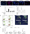
Neutrophil depletion decreases omental metastasis. Immunocompetent C57BL/6 mice were injected i.b. with ID8 cells that express GFP and luciferase. At 3 wk thereafter, formation of primary tumors was confirmed by bioluminescence imaging. Mice were then randomized into groups that were administered either Ly6G Ab or control Ig twice a week for 3 wk (n = 6 mice per group). Following Ab treatment, peripheral blood and tumors in each mouse of each group were analyzed. (A) Experimental scheme. (B) Abundance of neutrophils in peripheral blood, quantified by flow cytometric analysis. **, P < 0.01 (unpaired t test). (C) Analysis of peripheral blood by CBC test. ****, P < 0.0001 (unpaired t test). (D) Sizes of primary tumors and omental implants detected by bioluminescence imaging and calculated from emitted signals. *, P < 0.05 (unpaired t test). (E) Representative images of mice with primary tumors (arrowheads) and omental implants (arrows) detected by bioluminescence imaging. (F) Representative images of tissues viewed under light and fluorescence microscopy. Bars, 10 mm. (G) Abundance of tumor in tissues of the primary site (left ovary and oviduct) and in visceral fat tissues, quantified by flow cytometric analysis of GFP+ cells. *, P < 0.05; **, P < 0.01 (unpaired t test). Error bars in B–D and G represent SD.
Figure 4.
Figure 4.
Ovarian cancer cell–derived factors induce neutrophils to form NETs. Normal neutrophils were stimulated for 4 h with media conditioned by ovarian cancer cells or with nonconditioned media, without or with the addition of PMA. Thereafter, neutrophils were stained with Sytox Green dye to detect extruded DNA (green) and with cit-H3 Ab (red). Nuclei were visualized by staining with DAPI (blue). Bars, 100 µm (top) and 50 µm (bottom). (A) Neutrophils were isolated from peripheral blood of healthy adult donors. Media conditioned by ES2 cells were used. (B) Neutrophils were isolated from bone marrow of healthy adult C57BL/6 mice. Media conditioned by ID8 cells were used. In A and B, three independent experiments were performed, with neutrophils from a different donor used in each experiment. Shown are representative images.
Figure 5.
Figure 5.
Early-stage ovarian tumors induce NET formation in the premetastatic omental niche. (A and B) Analysis of mouse omental tissues. (A) C57BL/6 mice were injected i.b. with ID8 cells or with saline (n = 6 mice per group) and euthanized at 3 wk thereafter. Fresh omental tissues were stained with Sytox Red dye to detect extruded DNA and with Ly6G Ab, and staining was analyzed by flow cytometry. Shown are proportions of omental Ly6G+ cells that were Sytox Red+ in each mouse of each group. **, P < 0.01 (unpaired t test). (B) Frozen sections of omental tissues were stained to detect Ly6G (green) and cit-H3 (red). Bar, 100 µm. Nonfluorescent ID8 cells were used to inject mice. Shown are representative examples of staining. (C and D) Analysis of human omental tissues. Immunofluorescence staining of MPO (green) and cit-H3 (red) was performed on FFPE sections of omental tissues of women with stage I/II HGSC, with stage I/II SLMP, or without cancer history. (C) Average numbers of MPO+cit-H3+ cells are shown for each case in each group (n = 10 cases per group). For each case, the average number of MPO+cit-H3+ cells was calculated by counting double-positive cells in five random ×200 microscopic fields. **, P < 0.01; ***, P < 0.001 (Mann-Whitney U test). (D) Representative examples of staining. Bar, 100 µm. Error bars in A and C represent SD.
Figure 6.
Figure 6.
Ovarian cancer cells attach to NETs. (A and B) Bone marrow neutrophils of immunocompetent C57BL/6 mice were stimulated for 4 h with ID8-conditioned media or with nonconditioned media without or with addition of PMA. Fluorescently labeled ID8 cells were then seeded onto neutrophils. Attached ID8 cells were counted at 1 h thereafter. (A) Shown are means ± SD of three independent experiments, where attached cancer cells were counted in four random ×200 microscopic fields in each experiment. Each experiment used neutrophils from a different donor. **, P < 0.01 (unpaired t test). (B) Representative image of ID8 cells (red) bound to DNA extruded by neutrophils and stained with Sytox Green dye (green). Bar, 100 µm. (C) Human peripheral blood neutrophils were stimulated for 4 h with ES2-conditioned media or with nonconditioned media without or with addition of PMA. ES2 cells were then seeded onto neutrophils and assayed for attachment, as in A. Shown are means ± SD of three independent experiments. Each experiment used neutrophils from a different healthy donor. **, P < 0.01 (unpaired t test). (D) Omental tissues were collected from nude mice at 6 d following i.p. injection of ES2 cells or saline (n = 3 mice per group). Frozen sections were stained to detect cit-H3 (red) and tumor cells (green). Bar, 100 µm. Shown are representative examples of staining. (E and F) Bone marrow neutrophils of neutrophil-specific Padi4−/− mice and control siblings were stimulated for 4 h with ID8-conditioned media or with nonconditioned media. (E) Neutrophils were stained with Sytox Green dye (green) and with cit-H3 Ab (red). Bars, 100 µm (top) and 20 µm (bottom). (F) ID8 cells were seeded onto stimulated neutrophils and assayed for attachment, as in A. Shown are representative images and means ± SD of three independent experiments. **, P < 0.01 (unpaired t test).
Figure 7.
Figure 7.
Omental metastasis is decreased in NET-defective, neutrophil-specific, Padi4-deficient mice. Female neutrophil-specific Padi4−/− mice and control siblings were injected i.b. with ID8 cells that express RFP and luciferase and thereafter were evaluated for NET formation and tumor progression. (A) At 3 wk, mice in both groups were euthanized (n = 6 mice per group). Fresh omental tissues were stained with Ly6G Ab and Sytox Red dye, and staining was analyzed by flow cytometry. Shown is the proportion of omental Ly6G+ cells that were Sytox Red+ in each mouse of each group. ***, P < 0.001 (unpaired t test). (B and C) Representative examples of primary tumors detected at 3 wk by bioluminescence imaging (arrowheads; B) and by light microscopy (circled; C). Bar, 10 mm. (D–G) Analysis of tumor progression at 6 wk (n = 6 mice per group). (D) Sizes of primary tumors and omental implants detected in each mouse by bioluminescence imaging. **, P < 0.01 (unpaired t test). (E) Representative images of mice with primary tumors (arrowheads) and omental implants (arrows). (F) Representative images of tumors viewed under light microscopy (circled). Bars, 10 mm. (G) Abundance of RFP+ tumor cells in tissues of the primary site, visceral fat tissues, and peritoneal fluid in each mouse, quantified by flow cytometric analysis. *, P < 0.05; ***, P < 0.001 (unpaired t test). Error bars in A, D, and G represent SD.
Figure 8.
Figure 8.
NET-inhibiting agents decrease omental metastasis. (A) Normal bone marrow neutrophils of C57BL/6 mice were stimulated for 4 h with nonconditioned media or with ID8-conditioned media without or with addition of CI-amidine (100 µM), GSK484 (10 µM), or DNase (1 µg/ml) and then stained with cit-H3 Ab (red). Bar, 50 µm. Three independent experiments were performed. Shown are representative examples of staining. (B) ID8 cells were assayed for attachment to neutrophils that had been prestimulated, as described in A. Shown are means ± SD of three independent experiments, where attached cancer cells were counted in four random ×200 microscopic fields in each experiment. Each experiment used neutrophils from a different donor. **, P < 0.01 (unpaired t test). (C–F) Female nude mice were injected i.p. with GFP-expressing ES2 cells. At 1 d thereafter, mice were randomized into groups and then administered saline, GSK484 (20 mg/kg), or DNase (5 mg/kg) i.p. daily for 9 d. A nontumor control group of mice was administered saline i.p. daily for 9 d. Thereafter, mice in all groups were euthanized (n = 6 mice per group). (C) Omental tissues were stained with Ly6G Ab and Sytox Red dye, and staining was analyzed by flow cytometry. Shown are the proportion of omental Ly6G+ cells that were Sytox Red+ in each mouse of each group. **, P < 0.01 (unpaired t test). (D) Representative examples of Ly6G and Sytox Red staining analysis. (E) Representative images of GFP+ tumors on the omentum. Bar, 10 mm. (F) The abundance of GFP+ tumor cells in fat tissues and in peritoneal fluid of each mouse in the ES2 groups was quantified by flow cytometric analysis. *, P < 0.05; **, P < 0.01; ***, P < 0.001 (unpaired t test). Error bars in C and F represent SD.
Figure 9.
Figure 9.
Proposed mechanism for the metastatic tropism of ovarian cancer for the omentum. Early-stage ovarian tumors secrete factors that stimulate influx of neutrophils into the premetastatic omental niche and induce these neutrophils to form NETs. Cancer cells that are shed by tumors into the circulating peritoneal fluid become trapped by NETs and then form implants on the omentum.

Comment in

  • Neutrophil: A mobile fertilizer.
    Ng LG. Ng LG. J Exp Med. 2019 Jan 7;216(1):4-6. doi: 10.1084/jem.20182059. Epub 2018 Dec 19. J Exp Med. 2019. PMID: 30567720 Free PMC article.

Similar articles

Cited by

References

    1. Abram C.L., Roberge G.L., Hu Y., and Lowell C.A.. 2014. Comparative analysis of the efficiency and specificity of myeloid-Cre deleting strains using ROSA-EYFP reporter mice. J. Immunol. Methods. 408:89–100. 10.1016/j.jim.2014.05.009 - DOI - PMC - PubMed
    1. Albrengues J., Shields M.A., Ng D., Park C.G., Ambrico A., Poindexter M.E., Upadhyay P., Uyeminami D.L., Pommier A., Küttner V., et al. . 2018. Neutrophil extracellular traps produced during inflammation awaken dormant cancer cells in mice. Science. 361:eaao4227 10.1126/science.aao4227 - DOI - PMC - PubMed
    1. Aune G., Stunes A.K., Lian A.M., Reseland J.E., Tingulstad S., Torp S.H., and Syversen U.. 2012. Circulating interleukin-8 and plasminogen activator inhibitor-1 are increased in women with ovarian carcinoma. Results Immunol. 2:190–195. 10.1016/j.rinim.2012.10.003 - DOI - PMC - PubMed
    1. Brinkmann V., Reichard U., Goosmann C., Fauler B., Uhlemann Y., Weiss D.S., Weinrauch Y., and Zychlinsky A.. 2004. Neutrophil extracellular traps kill bacteria. Science. 303:1532–1535. 10.1126/science.1092385 - DOI - PubMed
    1. Buscher K., Wang H., Zhang X., Striewski P., Wirth B., Saggu G., Lütke-Enking S., Mayadas T.N., Ley K., Sorokin L., and Song J.. 2016. Protection from septic peritonitis by rapid neutrophil recruitment through omental high endothelial venules. Nat. Commun. 7:10828 10.1038/ncomms10828 - DOI - PMC - PubMed

Publication types

MeSH terms

Substances