. 2018 Dec 17;14(12):e1007502.
doi: 10.1371/journal.ppat.1007502. eCollection 2018 Dec.

Gluconeogenesis is essential for trypanosome development in the tsetse fly vector

Affiliations

Gluconeogenesis is essential for trypanosome development in the tsetse fly vector

Marion Wargnies et al. PLoS Pathog. .

Abstract

In the glucose-free environment that is the midgut of the tsetse fly vector, the procyclic form of Trypanosoma brucei primarily uses proline to feed its central carbon and energy metabolism. In these conditions, the parasite needs to produce glucose 6-phosphate (G6P) through gluconeogenesis from metabolism of non-glycolytic carbon source(s). We showed here that two phosphoenolpyruvate-producing enzymes, PEP carboxykinase (PEPCK) and pyruvate phosphate dikinase (PPDK) have a redundant function for the essential gluconeogenesis from proline. Indeed, incorporation of 13C-enriched proline into G6P was abolished in the PEPCK/PPDK null double mutant (Δppdk/Δpepck), but not in the single Δppdk and Δpepck mutant cell lines. The procyclic trypanosome also uses the glycerol conversion pathway to feed gluconeogenesis, since the death of the Δppdk/Δpepck double null mutant in glucose-free conditions is only observed after RNAi-mediated down-regulation of the expression of the glycerol kinase, the first enzyme of the glycerol conversion pathways. Deletion of the gene encoding fructose-1,6-bisphosphatase (Δfbpase), a key gluconeogenic enzyme irreversibly producing fructose 6-phosphate from fructose 1,6-bisphosphate, considerably reduced, but not abolished, incorporation of 13C-enriched proline into G6P. In addition, the Δfbpase cell line is viable in glucose-free conditions, suggesting that an alternative pathway can be used for G6P production in vitro. However, FBPase is essential in vivo, as shown by the incapacity of the Δfbpase null mutant to colonise the fly vector salivary glands, while the parental phenotype is restored in the Δfbpase rescued cell line re-expressing FBPase. The essential role of FBPase for the development of T. brucei in the tsetse was confirmed by taking advantage of an in vitro differentiation assay based on the RNA-binding protein 6 over-expression, in which the procyclic forms differentiate into epimastigote forms but not into mammalian-infective metacyclic parasites. In total, morphology, immunofluorescence and cytometry analyses showed that the differentiation of the epimastigote stages into the metacyclic forms is abolished in the Δfbpase mutant.


Conflict of interest statement

The authors have declared that no competing interests exist.

Figures

Fig 1
Fig 1. Proline, glucose and glycerol metabolism of the PCF trypanosomes.
Panel A shows a schematic representation of the proline metabolism (blue) in PCF grown in glucose-depleted medium, with glycerol contribution possibly present in the medium (black for the glycerol-specific steps). Panel B corresponds to cells incubated in glucose-rich medium, with contribution of glucose to central carbon metabolism indicated in red. End products excreted from catabolism of proline and glucose are shown in a rectangle, the number corresponding to enzymes under investigation are circled and metabolites analysed by IC-MS/MS are underlined and in italic. For the sake of clarity, the reversible non-oxidative branch of the pentose phosphate pathway (PPP) is represented by double lines and neither the glycerol 3-phosphate (Gly3P)/dihydroxyacetone phosphate (DHAP) shuttle nor the cofactors and nucleotides are shown. Abbreviations: DHAP, dihydroxyacetone phosphate; F1,6BP, fructose 1,6-bisphosphate; 1,3BPG, 1,3-bisphosphoglycerate; F6P, fructose 6-phosphate; G3P, glyceraldehyde 3-phosphate; G6P, glucose 6-phosphate; Gly3P, glycerol 3-phosphate; 6PG, 6-phosphogluconolactone; M6P, mannose 6-phosphate; OAA, oxaloacetate; PEP, phosphoenolpyruvate; 2PG, 2-phosphoglycerate; 3PG, 3-phosphoglycerate; Pen5P, pentose 5-phosphate (ribulose 5-phosphate, ribose 5-phosphate and xylose 5-phosphate). Indicated enzymes are: 1, proline dehydrogenase; 2, pyrroline-5 carboxylate dehydrogenase; 3, L-alanine aminotransferase; 4, α-ketoglutarate dehydrogenase complex; 5, succinyl-CoA synthetase; 6a, succinate dehydrogenase (complex II of the respiratory chain); 6b, mitochondrial NADH-dependent fumarate reductase; 7, mitochondrial fumarase; 8, mitochondrial malic enzyme; 9, pyruvate dehydrogenase complex; 10, acetate:succinate CoA-transferase (ASCT); 11, acetyl-CoA thioesterase; 12, cytosolic malic enzyme; 13, cytosolic fumarase; 14, glycosomal NADH-dependent fumarate reductase; 15, glycosomal malate dehydrogenase; 16, phosphoenolpyruvate carboxykinase (PEPCK); 17, pyruvate phosphate dikinase (PPDK); 18, pyruvate kinase; 19, enolase (ENO); 20, phosphoglycerate mutase; 21, cytosolic phosphoglycerate kinase; 22, glyceraldehyde-3-phosphate dehydrogenase; 23, triose-phosphate isomerase (TIM); 24, aldolase; 25, fructose-1,6-bisphosphatase (FBPase); 26, phosphofructokinase; 27, glucose-6-phosphate isomerase; 28, glucose-6-phosphate dehydrogenase (G6PDH); 29, 6-phosphogluconolactonase, 6-phosphogluconate dehydrogenase, ribose-5-phosphate isomerase and ribulose-5-phosphate epimerase; 30, phosphomannose isomerase; 31, hexokinase; 32, glycerol kinase (GK); 33, NADH-dependent glycerol-3-phosphate dehydrogenase (GPDH).
Fig 2
Fig 2. IC-MS/MS analysis of intracellular metabolites after isotopic labelling with [U-13C]-proline.
The EATRO1125.T7T parental (WT), Δpepck, Δppdk and Δppdkpepck cell lines were incubated for 2 h in PBS containing 2 mM [U-13C]-proline with 2 mM glucose (+G, only the parental cells) or without glucose (-G) prior to metabolite extraction. The figure shows enrichment of key glycolytic intermediates at 0 to 6 carbon positions (m0 to m6) with 13C expressed as percentage of all corresponding molecules (MID; Mass Isotopomer Distribution). For abbreviations, see Fig 1: S7P, sedoheptulose 7-phosphate; 2/3PG means that 2-phosphoglycerate (2PG) and 3-phosphoglycerate (3PG) are undistinguished by IC-MS/MS. The asterisks (*) in the F6P column for the Δpepck mutant highlights that the absolute amount of this metabolite being particularly low in this incubation conditions, solely the m0 and m3 isotopomer were detected.
Fig 3
Fig 3. Functional analysis of Δppdkpepck cell lines.
This figure represents growth curves of the Δppdkpepck cell line (panel A) and tetracycline-induced Δppdkpepck/RNAiGK.i, Δppdkpepck/RNAiGPDH.i and Δppdkpepck/RNAiTIM.i triple mutants (panels B-D) grown in SDM79-GlcFree medium containing 10 mM glucose (+G) or not (-G). In glucose-free conditions, the SDM79-GlcFree medium was supplemented with 50 mM N-acetyl-D-glucosamine that inhibits uptake of residual glucose. Cells were maintained in the exponential growth phase (between 106 and 107 cells ml-1). The insets show western blot analyses of the parental (WT), Δppdkpepck (ΔΔ) and tetracycline-induced (.i) and non-induced (.ni) triple mutants with the immune sera indicated in the right margin (ASCT, acetate:succinate CoA-transferase; ENO, enolase). The cross in panel B means that the mutant ultimately dies.
Fig 4
Fig 4. Proton (1H) NMR analysis of end products excreted from glucose and [U-13C]-glycerol metabolism.
The Δppdkpepck and tetracycline-induced (.i) Δppdkpepck/RNAiGK cell lines were incubated for 6 h in PBS/NaHCO3 buffer containing 4 mM glucose or 4 mM [U-13C]-glycerol. Panel A shows the quantitative data of excreted end products from glucose or [U-13C]-glycerol metabolism expressed as nmol of acetate, pyruvate, alanine and lactate excreted per h and per mg of protein. Panel B shows one representative NMR spectrum (out of 3 spectra and ranging from 1.2 to 4 ppm) of end products excreted from [U-13C]-glycerol metabolism of the Δppdkpepck and Δppdkpepck/RNAiGK.i cell lines. The resonance massif corresponding to unconsumed [U-13C]-glycerol as indicated (from 3.3 to 3.8 ppm) and resonances indicated by asterisks correspond to [13C]-enriched pyruvate and acetate. The resonances labelled 1 and 2 correspond to non-[13C]-enriched succinate and acetate, respectively, produced from the catabolism of an unknown internal carbon source [6, 23].
Fig 5
Fig 5. Analysis of FBPase mutants cell lines.
Panel A shows a PCR analysis of genomic DNA isolated from the parental (WT) and Δfbpase cell lines. The 1 to 10 lanes of the gel picture correspond to different PCR products described in the lower panel with circled numbers. As expected, PCR amplification of the FBPase gene (lane 10) was only observed in the parental cell line, while BSD and PAC PCR-products were observed only in the Δfbpase mutant (lanes 1–2 and 7–9). As control, both the 5'- and 3'-untranslated regions (UTRs) are PCR amplified from the parental and Δfbpase samples (lanes 3–6). Panels B-D show the growth curves of the Δfbpase (B), RNAiFPBase.i (C) and Δfbpase/FPBase.i (D) mutant cell lines, together with the parental (WT) cell line, incubated in SDM79-GlcFree medium containing 10 mM glucose (+G) or not (-G). Western blot analyses with the immune sera indicated in the right margin of the parental (WT), Δfbpase, the tetracycline-induced (.i) and non-induced (.ni) RNAiFPBase and/or Δfbpase/FPBase.i mutant are shown in the insets.
Fig 6
Fig 6. IC-MS/MS analysis of intracellular metabolites of the Δfbpase mutant incubated with [U-13C]-proline.
The parental (EATRO1125.T7T), Δfbpase and Δfbpase/FBPase.i cell lines were incubated for 2 h in PBS containing 2 mM [U-13C]-proline. Enrichment of key glycolytic intermediates at carbon positions 0 to 6 (m0 to m6) is expressed as percentage of all the corresponding molecules. For abbreviations, see Figs 1 and 2.
Fig 7
Fig 7. The glycosomal FBPase activity is not abolished in the Δfbpase mutant.
Panel A shows the FBPase activity (top panel) and western blotting analysis (lower panel) of glycosomal fractions of the parental (WT), Δfbpase and Δfbpase/FBPase.i (rescue) cell lines. The FBPase activities were normalised with the glycerol kinase activities. In panel B the glycosomal localisation of FBPase and SBPase was confirmed by western blotting analyses on glycosomal and cytosolic fractions, using control immune sera against glycosomal (PPDK) and cytosolic (enolase) markers.
Fig 8
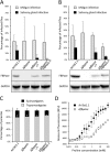
Fig 8. The deletion of the FBPase gene prevents parasites to colonise the tsetse salivary glands.
In this experiment, batches of 50 Glossina morsitans morsitans teneral males were artificially fed with either the parental AnTat1.1E wild-type, the Δfbpase-/+ (one allele deleted), the Δfbpase (null mutant) or the Δfbpase/FBPase-1 and -2 (rescue cell lines) PCF cell lines in culture medium as previously described [28]. Four weeks after the infective meal, a total of 790 flies were dissected to assess the presence of parasites in their midgut and salivary glands by microscopic examination. Midgut and salivary gland infection rates (in % ±SD) are presented for each cell line as the mean of 4 independent biological replicates done with the rescue cell line expressing the FBPase through the pLew100 vector (panel A), and of 6 independent biological replicates done with the rescue cell line expressing the FBPase from an ectopic FBPase copy in the FBPase locus (panel B). The proportion of flies developing a salivary gland infection are: 12/96 for the parental AnTat1.1E, 0/60 for Δfbpase, 3/104 for Δfbpase-/+ and 1/84 for Δfbpase/FBPase-1 in panel A and 33/163 for the parental AnTat1.1E, 0/91 for Δfbpase, 8/65 for Δfbpase-/+ and 6/125 for Δfbpase/FBPase-2 in panel B. Below the graphs are shown western blot analyses of the FBPase expression using the glucose-6-phosphate dehydrogenase (G6PDH) as loading control. (C) The proportions of cells in the different morphotypes found in the proventriculus and midgut were compared after dissection of flies infected with either one the four different cell lines from at least two independent biological replicates. The proportion of trypomastigotes and epimastigotes is indicated in grey and black respectively. 692, 373, 432, and 280, cells isolated from 6, 2, 3 and 2 flies have been analysed for the parental AnTat1.1E, Δfbpase, Δfbpase-/+ and Δfbpase/FBPase-2 cell lines, respectively. Panel D shows growth of the AnTat1.1E and Δfbpase cell lines in the SDM79-GlcFree medium containing from less than 20 μM to 100 mM proline, using the Alamar Blue assay. The proline concentration values on the x axis correspond to the amounts of proline added to the proline-depleted SDM79-GlcFree medium, which contains less than 20 μM proline.
Fig 9
Fig 9. FBPase is expressed in all parasite cycle stages yet at variable levels.
(A) Wild-type AnTat1.1E cultured bloodstream (cBSF) and procyclic (cPCF) forms, as well as parasites isolated from infected tsetse flies were fixed in methanol and stained with the anti-FBPase antibody (white) and DAPI (cyan). PC: procyclic trypomastigote, MS: mesocyclic trypomastigote, DE: dividing epimastigote, LE: long epimastigote, SE: short epimastigote, AE: attached epimastigote, and MT: metacyclic trypomastigote. (B-C) A region of interest was drawn on the phase picture of each individual cell and used for the quantification of the fluorescence provided by the anti-FBPase labelling. After background removal, the fluorescent signals were normalised (in %) to that obtained in PCF treated in the same experiment. The maximum fluorescence intensity (B) and total amount of fluorescence (C) corresponding to the FBPase expression levels were plotted according to the parasite stage. The total number of cells analysed from 3 independent experimental infections is indicated below the histogram bars for each stage. The different shades of grey represent groups of stages statistically identical between each other and different from those in the other groups according to a two-tailed ANOVA test with Tukey ad-hoc post-tests at 95% confidence for inter-group comparisons. * indicates the stage statistically different from all others with p<0.005.
Fig 10
Fig 10. FBPase is essential for metacyclogenesis in culture.
RBP6-induced differentiation kinetics is shown for the parental EATRO1125.T7T RBP6.i cell line (A) and the Δfbpase RBP6.i mutant (B). RBP6 over-expression was induced at time 0 by addition of 1 μM tetracycline. Developmental stages were morphologically scored by cell size, shape and the relative position of the kinetoplast to the nucleus within the cell (n >100 cells per time point, biological triplicate, SEM). Expression of the stage-specific proteins EP and calflagin was quantified at the single cell level by flow cytometry and gated into two populations with low or high fluorescence. The left panels show histograms of morphotypes with PCF in yellow, EMF in blue and MF forms in red. The middle and right panels present morphotype fractions from the histogram (solid lines, data points colour matched to histogram) together with the fractions of stage marker expression levels (cells with high fluorescence/total number of cells, dashed lines) for EP and calflagin, respectively. (C) Western blot showing the RBP6 over-expression levels upon tetracycline induction of the indicated cell lines. PFR was used as loading control and the Δfbpase knock out was verified by anti-FBPase. (D) Growth curves of the RBP6.i and Δfbpase RBP6.i populations upon induction of RBP6 over-expression. C and D are from a representative replicate of the three experiments shown in A and B.

Similar articles

Cited by

References

    1. Buscher P, Cecchi G, Jamonneau V, Priotto G. Human African trypanosomiasis. Lancet. 2017;390:2397–2409. 10.1016/S0140-6736(17)31510-6 . - DOI - PubMed
    1. Bringaud F, Barrett MP, Zilberstein D. Multiple roles of proline transport and metabolism in trypanosomatids. Frontiers in Bioscience 2012;17:349–374. - PubMed
    1. Mantilla BS, Marchese L, Casas-Sanchez A, Dyer NA, Ejeh N, Biran M, et al. Proline metabolism is essential for Trypanosoma brucei brucei survival in the tsetse vector. PLoS Pathog. 2017;13:e1006158 10.1371/journal.ppat.1006158 - DOI - PMC - PubMed
    1. Coustou V, Biran M, Breton M, Guegan F, Riviere L, Plazolles N, et al. Glucose-induced remodeling of intermediary and energy metabolism in procyclic Trypanosoma brucei. J Biol Chem. 2008;283:16342–16354. 10.1074/jbc.M709592200 - DOI - PubMed
    1. Bringaud F, Riviere L, Coustou V. Energy metabolism of trypanosomatids: adaptation to available carbon sources. Mol Biochem Parasitol. 2006;149:1–9. 10.1016/j.molbiopara.2006.03.017 - DOI - PubMed

Publication types

Grants and funding

The funders had no role in study design, data collection and analysis, decision to publish, or preparation of the manuscript. FB's and BR's group were funded by the Agence Nationale de la Recherche (ANR) through GLYCONOV (grant number ANR-15-CE15-0025-01) of the ANR-BLANC-2015 call. FB's group was supported by the Centre National de la Recherche Scientifique (CNRS), the Université de Bordeaux, the ANR through the grants ACETOTRYP (grant number ANR-2010-BLAN-1319-02) of the ANR-BLANC-2010 call, the Laboratoire d’Excellence (LabEx) ParaFrap ANR-11-LABX-0024 and the ParaMet PhD programme of Marie Curie Initial Training Network. BR’s group was supported by the Institut Pasteur, the Institut National de la Santé et de la Recherche Médicale (INSERM). EB is funded by a doctoral fellowship from French National Ministry for Research and Technology (Doctoral School CDV515). MB was funded by the University of Munich and MB and FB were supported by a research cooperation grant of the Franco-Bavarian University Cooperation Center (BFHZ/CCUFB).