

Significance of the epidermal growth factor receptor mutation status and differences among molecular subgroups in surgically resected lung microinvasive adenocarcinoma
- PMID: 30546439
- PMCID: PMC6256736
- DOI: 10.3892/ol.2018.9539
Significance of the epidermal growth factor receptor mutation status and differences among molecular subgroups in surgically resected lung microinvasive adenocarcinoma
Abstract
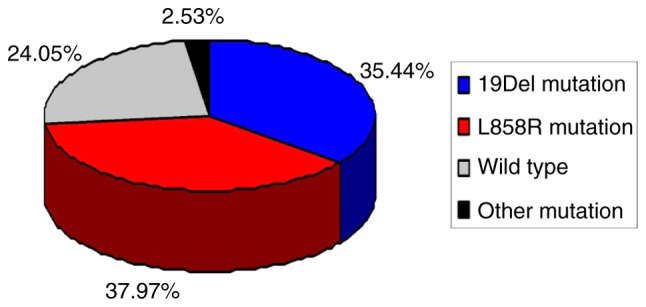
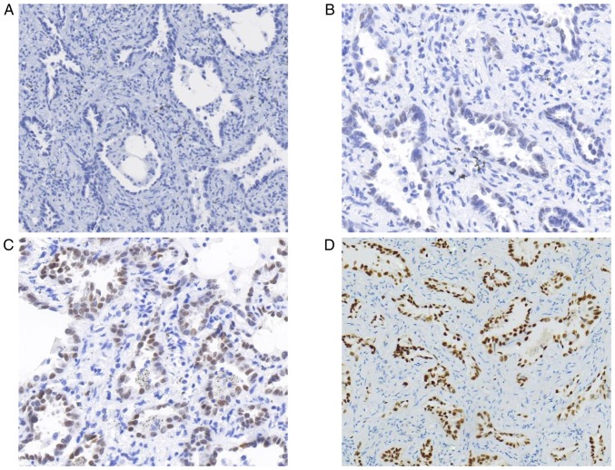
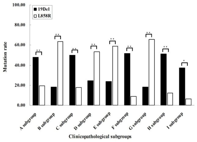
Lung microinvasive adenocarcinoma (MIA) is a newly-defined subtype of early stage non-small cell lung cancer (NSCLC). However, its epidermal growth factor receptor (EGFR) mutation status and clinical significance remain unclear. The present study aimed to determine EGFR mutation characteristics and identify their significance in patients with resected lung MIA. The present study also analyzed clinicopathological differences between EGFR molecular subgroups defined as 19Del and L858R. The present study examined EGFR mutations in 79 consecutive lung MIA resection specimens and compared the differences in clinicopathological features between the EGFR wild-type and mutation groups, as well as between the 19Del and L858R subgroups. EGFR mutations were detected in 60 (75.95%) tumors. The most common mutations were 19Del (28 cases; 35.44%) and L858R (30 cases; 37.97%). Two patients harbored rare mutations and one of them had a concomitant double mutation. EGFR mutations were significantly associated with microinvasion component, thyroid transcription factor 1 (TTF-1) expression, intratumoral fibrosis and inflammatory cell infiltration. Subgroup evaluation indicated that there was a significant association between 19Del and tumor size, maximum diameter of microinvasion, presence of intratumoral fibrosis and inflammatory cell infiltration. Similar associations were observed for the L858R subgroup, and L858R was associated with TTF-1 expression. In particular, 19Del occurred more frequently in MIA with a smaller size, with a smaller microinvasive area, without TTF-1 expression, and lacking intratumoral fibrosis and inflammatory cell infiltration. By contrast, L858R was detected more frequently in MIA with entirely different tumor features. In conclusion, the results of the present study indicated that surgically resected MIA cases harboring different EGFR gene statuses exhibit distinct clinicopathological features. Significant differences in pathological features associated with the tumor microenvironment were identified in MIA with 19Del or L858R mutations. Therefore, the present study proposed that MIA should be classified into molecular subgroups based on EGFR mutation subtypes. The molecular sub-classification should be taken into account for prognostic evaluation and clinical management of MIA.
Keywords: epidermal growth factor receptor; microinvasive adenocarcinoma; molecular subgroup; mutation; non-small cell lung cancer.
Figures
Similar articles
-
EEF1A2 and ERN2 could potentially discriminate metastatic status of mediastinal lymph node in lung adenocarcinomas harboring EGFR 19Del/L858R mutations.Thorac Cancer. 2020 Oct;11(10):2755-2766. doi: 10.1111/1759-7714.13554. Epub 2020 Sep 3. Thorac Cancer. 2020. PMID: 32881299 Free PMC article.
-
Concomitant Mutations in EGFR 19Del/L858R Mutation and Their Association with Response to EGFR-TKIs in NSCLC Patients.Cancer Manag Res. 2020 Sep 18;12:8653-8662. doi: 10.2147/CMAR.S255967. eCollection 2020. Cancer Manag Res. 2020. PMID: 32982456 Free PMC article.
-
Prognostic implication of EGFR mutation status and subtype in resected lung adenocarcinoma patients irrespective of therapy.Clin Transl Oncol. 2019 Mar;21(3):298-303. doi: 10.1007/s12094-018-1922-4. Epub 2018 Jul 18. Clin Transl Oncol. 2019. PMID: 30022385
-
Clinical efficacy of icotinib in lung cancer patients with different EGFR mutation status: a meta-analysis.Oncotarget. 2017 May 16;8(20):33961-33971. doi: 10.18632/oncotarget.15475. Oncotarget. 2017. PMID: 28430623 Free PMC article. Review.
-
Tyrosine kinase inhibitors for epidermal growth factor receptor gene mutation-positive non-small cell lung cancers: an update for recent advances in therapeutics.J Oncol Pharm Pract. 2016 Jun;22(3):461-76. doi: 10.1177/1078155215577810. Epub 2015 Apr 8. J Oncol Pharm Pract. 2016. PMID: 25855240 Review.
Cited by
-
Distinguishing EGFR mutant subtypes in stage IA non-small cell lung cancer using the presence status of ground glass opacity and final histologic classification: a systematic review and meta-analysis.Front Med (Lausanne). 2023 Dec 6;10:1268846. doi: 10.3389/fmed.2023.1268846. eCollection 2023. Front Med (Lausanne). 2023. PMID: 38126071 Free PMC article.
References
-
- Travis WD, Brambilla E, Nicholson AG, Yatabe Y, Austin JHM, Beasley MB, Chirieac LR, Dacic S, Duhig E, Flieder DB, et al. The 2015 world health organization classification of lung tumors: Impact of genetic, clinical and radiologic advances since the 2004 classification. J Thorac Oncol. 2015;10:1243–1260. doi: 10.1097/JTO.0000000000000630. - DOI - PubMed
LinkOut - more resources
Full Text Sources
Research Materials
Miscellaneous