. 2019 Feb 5;93(4):e01984-18.
doi: 10.1128/JVI.01984-18. Print 2019 Feb 15.

Autophagy Promotes Replication of Influenza A Virus In Vitro

Affiliations

Autophagy Promotes Replication of Influenza A Virus In Vitro

Ruifang Wang et al. J Virol. .

Abstract

Influenza A virus (IAV) infection could induce autophagosome accumulation. However, the impact of the autophagy machinery on IAV infection remains controversial. Here, we showed that induction of cellular autophagy by starvation or rapamycin treatment increases progeny virus production, while disruption of autophagy using a small interfering RNA (siRNA) and pharmacological inhibitor reduces progeny virus production. Further studies revealed that alteration of autophagy significantly affects the early stages of the virus life cycle or viral RNA synthesis. Importantly, we demonstrated that overexpression of both the IAV M2 and NP proteins alone leads to the lipidation of LC3 to LC3-II and a redistribution of LC3 from the cytosol to punctate vesicles indicative of authentic autophagosomes. Intriguingly, both M2 and NP colocalize and interact with LC3 puncta during M2 or NP transfection alone and IAV infection, leading to an increase in viral ribonucleoprotein (vRNP) export and infectious viral particle formation, which indicates that the IAV-host autophagy interaction plays a critical role in regulating IAV replication. We showed that NP and M2 induce the AKT-mTOR-dependent autophagy pathway and an increase in HSP90AA1 expression. Finally, our studies provided evidence that IAV replication needs an autophagy pathway to enhance viral RNA synthesis via the interaction of PB2 and HSP90AA1 by modulating HSP90AA1 expression and the AKT-mTOR signaling pathway in host cells. Collectively, our studies uncover a new mechanism that NP- and M2-mediated autophagy functions in different stages of virus replication in the pathogenicity of influenza A virus.IMPORTANCE Autophagy impacts the replication cycle of many viruses. However, the role of the autophagy machinery in IAV replication remains unclear. Therefore, we explored the detailed mechanisms utilized by IAV to promote its replication. We demonstrated that IAV NP- and M2-mediated autophagy promotes IAV replication by regulating the AKT-mTOR signaling pathway and HSP90AA1 expression. The interaction of PB2 and HSP90AA1 results in the increase of viral RNA synthesis first; subsequently the binding of NP to LC3 favors vRNP export, and later the interaction of M2 and LC3 leads to an increase in the production of infectious viral particles, thus accelerating viral progeny production. These findings improve our understanding of IAV pathogenicity in host cells.

Keywords: HSP90AA1; IAV; LC3; M2; NP; autophagy; replication.


Figures

FIG 1
FIG 1
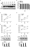
Inhibition of autophagy suppresses influenza HM/06 replication. (A) A549 cells were infected with the HM/06 virus at an MOI of 0.1. Cell lysates were collected at 3, 6, 9, 12, 24, and 36 hpi and were subjected to Western blot analysis. (B) A549 cells plated on the 96-well plates were prepared as described for panel A, and cell viability was detected by CCK-8 assay at the indicated time points postinfection. (C and D) A549 cells were pretreated with a control (DMSO) or 10 μM LY294002 (C) or 5 mM 3-MA (D) for 6 h and then infected with the HM/06 virus at an MOI of 0.1. Cell culture supernatants were collected at 12, 24, and 36 hpi. Virus titers were determined by TCID50 assay on MDCK cells. (E and F) A549 cells were transfected with siATG5 or siATG7 for 36 h and then mock infected or infected with HM/06 at an MOI of 0.1. Cell culture supernatants were collected at 12, 24, and 36 hpi. Virus titers were determined as described for panels C and D. Expression of ATG5 or ATG7 protein was analyzed by Western blotting. siNC, nontargeting siRNA, negative control. Means and SD (error bars) of three independent experiments are indicated (*, P < 0.05; **, P < 0.01; ***, P < 0.001).
FIG 2
FIG 2
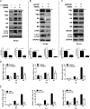
Inhibition of autophagy suppresses the influenza virus protein expression. (A to C) A549 cells treated with the LY294002, siATG5, or siBECN1 were mock infected or infected with the HM/06 virus. Cell lysates were harvested and analyzed by Western blotting (an asterisk next to the blot indicates the protein). (D and E) A549 cells were pretreated with LY294002 or siATG5 and mock infected or infected with the HM/06 virus. Cell lysates were collected at 12, 24, and 36 hpi. Then, total RNA was extracted, and the levels of NP gene vRNA, cRNA, and mRNA were measured by quantitative RT-PCR. GAPDH was used as a control for the normalization of cellular mRNA and intracellular viral RNA. Means and SD (error bars) were determined for triplicates of three independent experiments, and values were standardized to the levels of control-treated or siNC-infected cells at 12 hpi (*, P < 0.05; **, P < 0.01; ***, P < 0.001).
FIG 3
FIG 3
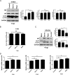
Induction of autophagy with starvation or rapamycin facilitates viral replication. (A) The culture medium of A549 cells was exchanged with Ham’s F-12 medium without FBS for 6 h or pretreated with a control (DMSO) or 500 nM rapamycin for 12 h and then were mock infected or infected with HM/06 at an MOI of 0.1. At 24 hpi, cell lysates were collected and subjected to the Western blot analysis. (B) A549 cells were treated as described for panel A. Cell culture supernatants were collected, and virus titers were determined by TCID50 assay. Means and SD (error bars) of three independent experiments are indicated (*, P < 0.05; **, P < 0.01).
FIG 4
FIG 4
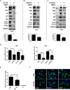
Induction of autophagy promotes the early stages of the viral life cycle or viral gene expression. (A and B) The culture medium of A549 cells was exchanged with Ham’s F-12 medium without FBS for 6 h or pretreated with rapamycin for 12 h and then incubated with the HM/06 virus at an MOI of 10 for 30 min (at 37°C). Cells were washed with PBS-HCl (pH 1.3) to remove all attached virions from the surface of A549 cells. Cell lysates were collected for Western blot analysis (A), and cell culture supernatants (noninfectious virus particles) were collected and determined by TCID50 assay (B). (C) A549 cells were treated as described for panels A and B and then were mock infected or infected with the HM/06 virus for 4 h. Lysates were harvested and analyzed by Western blotting. (D) A549 cells were treated as described for panels A and B and infected with the HM/06 virus for 4 h. The levels of M mRNA, vRNA, and cRNA were determined by RT-PCR. GAPDH was used as a control for the normalization of cellular mRNA and intracellular viral RNA. Means and SD (error bars) were determined for triplicates of three independent experiments and were standardized to the levels of control-infected cells at 4 hpi (*, P < 0.05; ns, nonsignificant).
FIG 5
FIG 5
Inhibition of autophagy disrupts the early stages of the viral life cycle or viral RNA synthesis. (A and B) A549 cells were transfected with siATG5 or siBECN1, as indicated, for 36 h and then were infected with the HM/06 virus. After 4 hpi, cell lysates were collected for Western blot analysis. An asterisk next to the blot indicates the protein. (C) A549 cells were pretreated with LY294002 for 6 h and then were mock-infected or infected with the HM/06 virus for 4 h. Cells were harvested and analyzed by Western blot. An asterisk next to the blot indicates the protein. (D) siATG5- or LY294002-pretreated A549 cells were mock infected or infected with the HM/06 virus for 4 hpi. M mRNA and vRNA levels were determined by RT-PCR. GAPDH was used as a control for the normalization of cellular mRNA and intracellular viral RNA. Means and SD were determined for triplicates of three independent experiments and were standardized to the levels of control-infected cells at 4 hpi. (E) HEK 293T cells were transfected with siATG5. Twelve hours later, cells were further transfected with plasmids encoding the polymerase complex components and NP derived from influenza HM/06 virus, along with a reporter plasmid containing the noncoding sequence from the NP segment as well as the luciferase gene driven by the human polymerase I promoter. Luciferase activity was determined at 36 h posttransfection, and relative activities were compared. (F) A549 cells treated with the indicated siRNAs were infected with the HM/06 virus at an MOI of 10. Cells were fixed at 4, 6, and 8 hpi for immunostaining of NP as a marker for vRNP complex export, and nuclei were stained using 4′,6′-diamidino-2-phenylindole, and then visualized by confocal microscopy. Means and SD (error bars) of three independent experiments are shown (*, P < 0.05; **, P < 0.01; ***, P < 0.001).
FIG 6
FIG 6
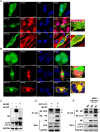
IAV NP and M2 proteins induce autophagy and interact with LC3. (A) A549 cells were transfected with GFP-LC3 for 24 h and then were mock-infected or infected with the HM/06 virus. Twenty-four hours later, cells were fixed and stained with anti-NP or -M2 antibody and then visualized by confocal microscopy. In the zoomed images, yellow spots indicate the merging of M2 or NP with LC3 puncta. The image is representative of 20 cells. Scale bar, 10 μm. (B) A549 cells were cotransfected with GFP-LC3 and HA-M2 or HA-NP. After 24 h, cells were fixed and stained with anti-HA antibody and then visualized by confocal microscopy. In the zoomed images, yellow spots indicate the merging of M2 or NP with LC3 puncta. The image is representative of 20 cells. Scale bar, 10 μm. (C) HEK 293T cells were transfected with the indicated plasmids for 24 h, and cells lysates were collected for Western blot analysis. (D) HEK 293T cells were transfected with the indicated plasmids for 24 h. Cell lysates were immunoprecipitated with anti-HA antibody and then analyzed by Western blotting. (E) Lysates of HM/06-infected A549 cells were prepared, immunoprecipitated with the anti-NP antibody or control IgG, and then subjected to Western blot analysis. An asterisk next to the blot indicates the protein, the triangle indicates the heavy chain. Results are representative of three independent experiments. WCL, whole-cell lysate; IP, immunoprecipitation; IB, immunoblotting; DAPI, 4′,6′-diamidino-2-phenylindole.
FIG 7
FIG 7
IAV NP and M2 induce autophagy via the AKT-mTOR pathway. (A) A549 cells were prepared as described in the legend of Fig. 1A, and the cell lysates were analyzed by Western blotting. (B) HEK 293T cells were transfected with HA-NP or HA-M2 for 24 h, and cell lysates were collected and subjected to Western blot analysis. Results are representative of three independent experiments.
FIG 8
FIG 8
Autophagy facilitates IAV RNA synthesis by regulating HSP90AA1 expression. (A) HEK 293T cells were transfected with siATG5 for 36 h and then transfected with the indicated plasmids. Twenty-four hours later, cell lysates were harvested for Western blot analysis. (B) siHSP90AA1-transfected HEK 293T cells were transfected with indicated plasmids for 24 h, and lysates were subjected to Western blot analysis. (C) Lysates of PCDNA3.1-PB2-transfected HEK 293T cells were prepared and immunoprecipitated with the anti-PB2 antibody or control IgG, and then cells were subjected to Western blot analysis. Results are representative of three independent experiments.

Similar articles

Cited by

  • Rapamycin inhibits hepatitis B virus covalently closed circular DNA transcription by enhancing the ubiquitination of HBx.
    Zhang Y, Li L, Cheng ST, Qin YP, He X, Li F, Wu DQ, Ren F, Yu HB, Liu J, Chen J, Ren JH, Zhang ZZ. Zhang Y, et al. Front Microbiol. 2022 Aug 11;13:850087. doi: 10.3389/fmicb.2022.850087. eCollection 2022. Front Microbiol. 2022. PMID: 36033851 Free PMC article.
  • Viruses and autophagy: bend, but don't break.
    Jassey A, Jackson WT. Jassey A, et al. Nat Rev Microbiol. 2024 May;22(5):309-321. doi: 10.1038/s41579-023-00995-y. Epub 2023 Dec 15. Nat Rev Microbiol. 2024. PMID: 38102460 Review.
  • Regulation of innate immune responses by rabies virus.
    Zhang H, Huang J, Song Y, Liu X, Qian M, Huang P, Li Y, Zhao L, Wang H. Zhang H, et al. Animal Model Exp Med. 2022 Oct;5(5):418-429. doi: 10.1002/ame2.12273. Epub 2022 Sep 22. Animal Model Exp Med. 2022. PMID: 36138548 Free PMC article. Review.
  • Effect of natural products on host cell autophagy induced by Influenza A virus infection.
    Liu X, Wang Q. Liu X, et al. Front Cell Infect Microbiol. 2024 Sep 30;14:1460604. doi: 10.3389/fcimb.2024.1460604. eCollection 2024. Front Cell Infect Microbiol. 2024. PMID: 39403204 Free PMC article. Review.
  • Proteomic and genetic analyses of influenza A viruses identify pan-viral host targets.
    Haas KM, McGregor MJ, Bouhaddou M, Polacco BJ, Kim EY, Nguyen TT, Newton BW, Urbanowski M, Kim H, Williams MAP, Rezelj VV, Hardy A, Fossati A, Stevenson EJ, Sukerman E, Kim T, Penugonda S, Moreno E, Braberg H, Zhou Y, Metreveli G, Harjai B, Tummino TA, Melnyk JE, Soucheray M, Batra J, Pache L, Martin-Sancho L, Carlson-Stevermer J, Jureka AS, Basler CF, Shokat KM, Shoichet BK, Shriver LP, Johnson JR, Shaw ML, Chanda SK, Roden DM, Carter TC, Kottyan LC, Chisholm RL, Pacheco JA, Smith ME, Schrodi SJ, Albrecht RA, Vignuzzi M, Zuliani-Alvarez L, Swaney DL, Eckhardt M, Wolinsky SM, White KM, Hultquist JF, Kaake RM, García-Sastre A, Krogan NJ. Haas KM, et al. Nat Commun. 2023 Sep 27;14(1):6030. doi: 10.1038/s41467-023-41442-z. Nat Commun. 2023. PMID: 37758692 Free PMC article.

References

    1. Nguyen-Van-Tam JS. 2015. Influenza and other respiratory viruses. From the Editor's desk. Influenza Other Respir Viruses 9:99–100. doi:10.1111/irv.12311. - DOI - PMC - PubMed
    1. Molinari N-AM, Ortega-Sanchez IR, Messonnier ML, Thompson WW, Wortley PM, Weintraub E, Bridges CB. 2007. The annual impact of seasonal influenza in the US: measuring disease burden and costs. Vaccine 25:5086–5096. doi:10.1016/j.vaccine.2007.03.046. - DOI - PubMed
    1. Moeller A, Kirchdoerfer RN, Potter CS, Carragher B, Wilson IA. 2012. Organization of the influenza virus replication machinery. Science 338:1631–1634. doi:10.1126/science.1227270. - DOI - PMC - PubMed
    1. Murti KG, Webster RG, Jones IM. 1988. Localization of RNA polymerases on influenza viral ribonucleoproteins by immunogold labeling. Virology 164:562–566. doi:10.1016/0042-6822(88)90574-0. - DOI - PubMed
    1. Portela A, Digard P. 2002. The influenza virus nucleoprotein: a multifunctional RNA-binding protein pivotal to virus replication. J Gen Virol 83:723–734. doi:10.1099/0022-1317-83-4-723. - DOI - PubMed

Publication types

MeSH terms

LinkOut - more resources