Biomaterial Approaches to Modulate Reactive Astroglial Response
- PMID: 30517922
- PMCID: PMC6397084
- DOI: 10.1159/000494667
Biomaterial Approaches to Modulate Reactive Astroglial Response
Abstract
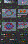
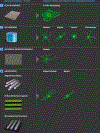
Over several decades, biomaterial scientists have developed materials to spur axonal regeneration and limit secondary injury and tested these materials within preclinical animal models. Rarely, though, are astrocytes examined comprehensively when biomaterials are placed into the injury site. Astrocytes support neuronal function in the central nervous system. Following an injury, astrocytes undergo reactive gliosis and create a glial scar. The astrocytic glial scar forms a dense barrier which restricts the extension of regenerating axons through the injury site. However, there are several beneficial effects of the glial scar, including helping to reform the blood-brain barrier, limiting the extent of secondary injury, and supporting the health of regenerating axons near the injury site. This review provides a brief introduction to the role of astrocytes in the spinal cord, discusses astrocyte phenotypic changes that occur following injury, and highlights studies that explored astrocyte changes in response to biomaterials tested within in vitro or in vivo environments. Overall, we suggest that in order to improve biomaterial designs for spinal cord injury applications, investigators should more thoroughly consider the astrocyte response to such designs.
Keywords: Astrocytes; Biomaterials; Reactive gliosis; Spinal cord injury.
© 2018 S. Karger AG, Basel.
Conflict of interest statement
4.2. Statement of Ethics
The authors have no ethical conflicts to disclose.
4.3. Disclosure Statement
The authors have no conflicts of interest to declare.
Figures


Similar articles
-
A new in vitro model of the glial scar inhibits axon growth.Glia. 2008 Nov 15;56(15):1691-709. doi: 10.1002/glia.20721. Glia. 2008. PMID: 18618667 Free PMC article.
-
Glial scar and axonal regeneration in the CNS: lessons from GFAP and vimentin transgenic mice.Acta Neurochir Suppl. 2004;89:87-92. doi: 10.1007/978-3-7091-0603-7_12. Acta Neurochir Suppl. 2004. PMID: 15335106
-
Astrocyte reactivity and astrogliosis after spinal cord injury.Neurosci Res. 2018 Jan;126:39-43. doi: 10.1016/j.neures.2017.10.004. Epub 2017 Oct 17. Neurosci Res. 2018. PMID: 29054466 Review.
-
Toll-like receptor 9 antagonism modulates astrocyte function and preserves proximal axons following spinal cord injury.Brain Behav Immun. 2019 Aug;80:328-343. doi: 10.1016/j.bbi.2019.04.010. Epub 2019 Apr 3. Brain Behav Immun. 2019. PMID: 30953770
-
Biomaterials and strategies for repairing spinal cord lesions.Neurochem Int. 2021 Mar;144:104973. doi: 10.1016/j.neuint.2021.104973. Epub 2021 Jan 23. Neurochem Int. 2021. PMID: 33497713 Review.
Cited by
-
Modulating neuroinflammation through molecular, cellular and biomaterial-based approaches to treat spinal cord injury.Bioeng Transl Med. 2022 Aug 31;8(2):e10389. doi: 10.1002/btm2.10389. eCollection 2023 Mar. Bioeng Transl Med. 2022. PMID: 36925680 Free PMC article. Review.
-
Selenium Nanoparticles in Protecting the Brain from Stroke: Possible Signaling and Metabolic Mechanisms.Nanomaterials (Basel). 2024 Jan 11;14(2):160. doi: 10.3390/nano14020160. Nanomaterials (Basel). 2024. PMID: 38251125 Free PMC article. Review.
-
Injectable hydrogels in central nervous system: Unique and novel platforms for promoting extracellular matrix remodeling and tissue engineering.Mater Today Bio. 2023 Mar 22;20:100614. doi: 10.1016/j.mtbio.2023.100614. eCollection 2023 Jun. Mater Today Bio. 2023. PMID: 37008830 Free PMC article. Review.
-
A narrative review of current therapies in unilateral recurrent laryngeal nerve injury caused by thyroid surgery.Gland Surg. 2022 Jan;11(1):270-278. doi: 10.21037/gs-21-708. Gland Surg. 2022. PMID: 35242688 Free PMC article. Review.
-
Controlled assembly of retinal cells on fractal and Euclidean electrodes.PLoS One. 2022 Apr 6;17(4):e0265685. doi: 10.1371/journal.pone.0265685. eCollection 2022. PLoS One. 2022. PMID: 35385490 Free PMC article.
References
Publication types
MeSH terms
Substances
Grants and funding
LinkOut - more resources
Full Text Sources
Medical

