

Cell Wall Epitopes and Endoploidy as Reporters of Embryogenic Potential in Brachypodium Distachyon Callus Culture
- PMID: 30501101
- PMCID: PMC6321580
- DOI: 10.3390/ijms19123811
Cell Wall Epitopes and Endoploidy as Reporters of Embryogenic Potential in Brachypodium Distachyon Callus Culture
Abstract
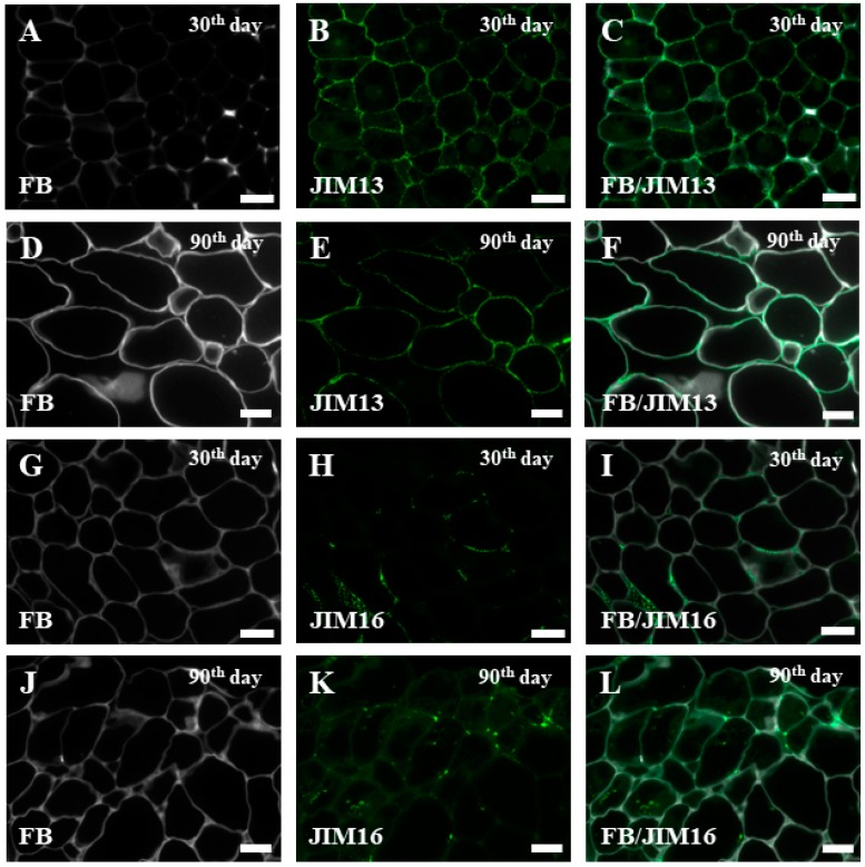
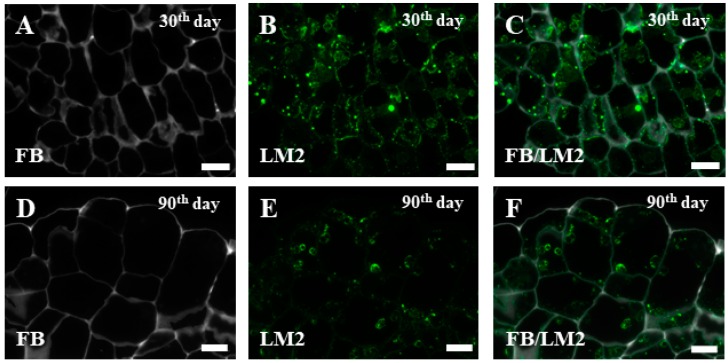
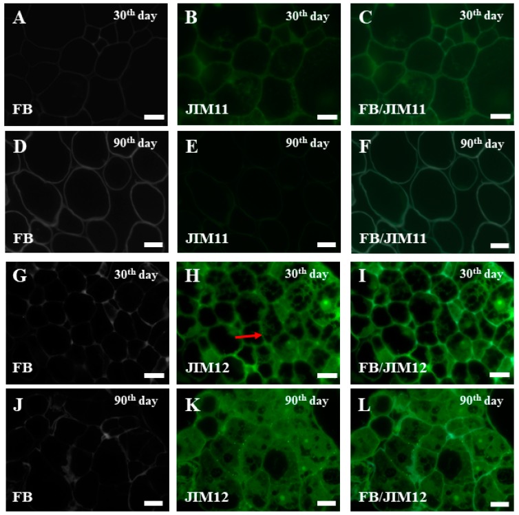
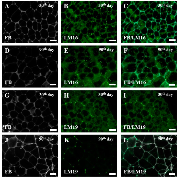
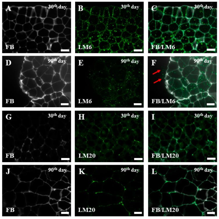
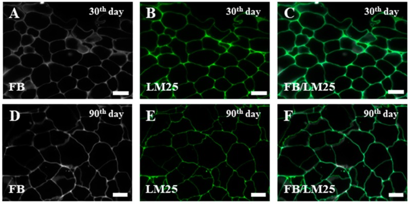
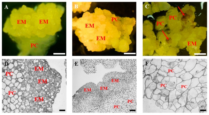
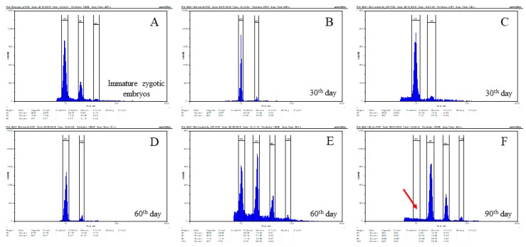
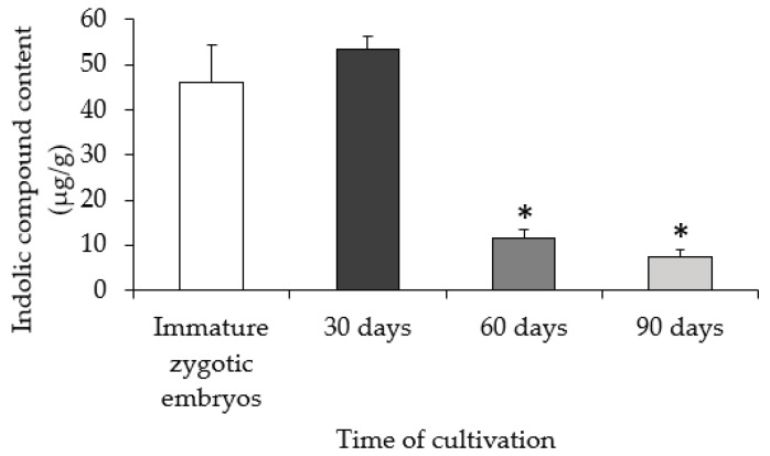
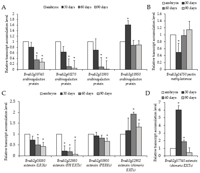
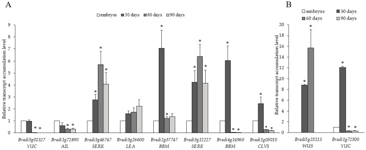
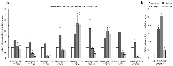
Effective regeneration of callus tissue into embryos and then into whole plants is essential for plant biotechnology. The embryonic potential is often low and can further decrease with time in culture, which limits the utilisation of calli for transformation procedures and in vitro propagation. In this study, we show that the loss of embryogenic potential in callus cultures of Brachypodium distachyon is progressive over time. Flow cytometry analyses indicated endoploidy levels increased in 60- and 90-day-old calli with effective loss of the 2C DNA content peak in the latter. Analysis of indolic compounds content revealed a decrease in 60- and 90-day-old calli compared to either freshly isolated explants or 30-day-old calli. Immunohistochemical analysis revealed a decrease in arabinogalactan proteins (AGP) signal with the time of culture, but extensin (EXT) epitopes either increased (JIM12 epitopes) or decreased (JIM11 epitopes). The transcript accumulation levels of AGPs and EXTs confirmed these results, with most of AGP and EXT transcripts gradually decreasing. Some chimeric EXT transcripts significantly increased on the 30th day of culture, perhaps because of an increased embryogenic potential. Selected somatic embryogenesis-related genes and cyclins demonstrated a gradual decrease of transcript accumulation for YUCCA (YUC), AINTEGUMENTA-LIKE (AIL), BABY BOOM (BBM), and CLAVATA (CLV3) genes, as well as for most of the cyclins, starting from the 30th day of culture. Notably, WUSCHEL (WUS) transcript was detectable only on the 30th and 60th day and was not detectable in the zygotic embryos and in 90-day-old calli.
Keywords: Brachypodium distachyon; arabinogalactan proteins; cell wall; cyclins; extensins; pectins; ploidy instability; somaclonal variation; somatic embryogenesis; transcript accumulation.
Conflict of interest statement
The authors declare no conflict of interest.
Figures
Similar articles
-
Spatial Distribution of Selected Chemical Cell Wall Components in the Embryogenic Callus of Brachypodium distachyon.PLoS One. 2016 Nov 28;11(11):e0167426. doi: 10.1371/journal.pone.0167426. eCollection 2016. PLoS One. 2016. PMID: 27893856 Free PMC article.
-
Hydroxyproline-Rich Glycoproteins as Markers of Temperature Stress in the Leaves of Brachypodium distachyon.Int J Mol Sci. 2019 May 25;20(10):2571. doi: 10.3390/ijms20102571. Int J Mol Sci. 2019. PMID: 31130622 Free PMC article.
-
Regenerative potential, metabolic profile, and genetic stability of Brachypodium distachyon embryogenic calli as affected by successive subcultures.Protoplasma. 2018 Mar;255(2):655-667. doi: 10.1007/s00709-017-1177-x. Epub 2017 Oct 28. Protoplasma. 2018. PMID: 29080994
-
In Vitro Tissue Culture in Brachypodium: Applications and Challenges.Int J Mol Sci. 2020 Feb 4;21(3):1037. doi: 10.3390/ijms21031037. Int J Mol Sci. 2020. PMID: 32033195 Free PMC article. Review.
-
Plant Cell Wall Proteomics: A Focus on Monocot Species, Brachypodium distachyon, Saccharum spp. and Oryza sativa.Int J Mol Sci. 2019 Apr 23;20(8):1975. doi: 10.3390/ijms20081975. Int J Mol Sci. 2019. PMID: 31018495 Free PMC article. Review.
Cited by
-
One-Week Scutellar Somatic Embryogenesis in the Monocot Brachypodium distachyon.Plants (Basel). 2022 Apr 14;11(8):1068. doi: 10.3390/plants11081068. Plants (Basel). 2022. PMID: 35448796 Free PMC article.
-
The callus formation capacity of strawberry leaf explant is modulated by DNA methylation.Hortic Res. 2022 Jan 19;9:uhab073. doi: 10.1093/hr/uhab073. Online ahead of print. Hortic Res. 2022. PMID: 35043170 Free PMC article.
-
3,4-Dehydro-L-proline Induces Programmed Cell Death in the Roots of Brachypodium distachyon.Int J Mol Sci. 2021 Jul 14;22(14):7548. doi: 10.3390/ijms22147548. Int J Mol Sci. 2021. PMID: 34299166 Free PMC article.
-
H3K4me3 changes occur in cell wall genes during the development of Fagopyrum tataricum morphogenic and non-morphogenic calli.Front Plant Sci. 2024 Sep 25;15:1465514. doi: 10.3389/fpls.2024.1465514. eCollection 2024. Front Plant Sci. 2024. PMID: 39385990 Free PMC article.
-
Apoplastic and Symplasmic Markers of Somatic Embryogenesis.Plants (Basel). 2023 May 11;12(10):1951. doi: 10.3390/plants12101951. Plants (Basel). 2023. PMID: 37653868 Free PMC article. Review.
References
-
- Mujib A., Samaj J. Somatic Embryogenesis. Springer; Berlin, Germany: New York, NY, USA: 2006. 357p
-
- Yang X., Zhang X. Regulation of somatic embryogenesis in higher plants. Crit. Rev. Plant Sci. 2010;29:36–57. doi: 10.1080/07352680903436291. - DOI
MeSH terms
Substances
Grants and funding
- BBS/E/J/000CA348/BB_/Biotechnology and Biological Sciences Research Council/United Kingdom
- DEC-2014/14/M/NZ2/00519/Narodowym Centrum Nauki
- 10754/The Leverhulme Trust
- BBS/E/J/000CA364/BB_/Biotechnology and Biological Sciences Research Council/United Kingdom
- BBS/E/J/000CA387/BB_/Biotechnology and Biological Sciences Research Council/United Kingdom
LinkOut - more resources
Full Text Sources
