. 2019 Feb;179(2):601-615.
doi: 10.1104/pp.18.01286. Epub 2018 Nov 28.

OXA2b is Crucial for Proper Membrane Insertion of COX2 during Biogenesis of Complex IV in Plant Mitochondria

Affiliations

OXA2b is Crucial for Proper Membrane Insertion of COX2 during Biogenesis of Complex IV in Plant Mitochondria

Renuka Kolli et al. Plant Physiol. 2019 Feb.

Abstract

The evolutionarily conserved YidC/Oxa1/Alb3 proteins are involved in the insertion of membrane proteins in all domains of life. In plant mitochondria, individual knockouts of OXA1a, OXA2a, and OXA2b are embryo-lethal. In contrast to other members of the protein family, OXA2a and OXA2b contain a tetratricopeptide repeat (TPR) domain at the C-terminus. Here, the role of Arabidopsis (Arabidopsis thaliana) OXA2b was determined by using viable mutant plants that were generated by complementing homozygous lethal OXA2b T-DNA insertional mutants with a C-terminally truncated OXA2b lacking the TPR domain. The truncated-OXA2b-complemented plants displayed severe growth retardation due to a strong reduction in the steady-state abundance and enzyme activity of the mitochondrial respiratory chain complex IV. The TPR domain of OXA2b directly interacts with cytochrome c oxidase subunit 2, aiding in efficient membrane insertion and translocation of its C-terminus. Thus, OXA2b is crucial for the biogenesis of complex IV in plant mitochondria.


Figures

Figure 1.
Figure 1.
Conserved membrane topology of the Oxa1 superfamily. Conserved membrane topology and conserved structural features of the members of the Oxa1 superfamily are shown. The three highly conserved transmembrane helices are colored orange, red, and blue. The extra two transmembrane domains of the Oxa1/YidC/Alb3 proteins are colored two different shades of green. The coiled-coil region located between transmembrane helices 1 and 2 is colored light brown. S. mutans, Streptococus mutans; TPR, tetratricopeptide repeat; N, N terminus; C, C terminus.
Figure 2.
Figure 2.
Phylogeny of the Oxa1 superfamily. A maximum-likelihood phylogenetic tree of Oxa1/YidC/Alb3 proteins is shown. Numbers represent ultrafast bootstrap values from IQTREE. Only the main branches bootstrap values are shown for better visibility. Species and sequences can be found in Supplemental Dataset S1.
Figure 3.
Figure 3.
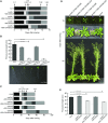
Phenotypes of oxa2b complementation plants. A, Plate-based growth progression analysis. Arrows indicate the time taken by wild-type plants to reach the developmental stages: 0.1: Imbibition; 0.5: Radicle emergence; 0.7: Hypocotyl and cotyledon emergence; 1.0: Cotyledons fully open; 1.02: two rosette leaves > 1 mm in length; 1.04: four rosette leaves > 1 mm in length. The boxes represent the time between the growth stages. Data are given as averages for 100 plants. B, Primary root length of plants grown vertically for 14 d. Data are given as averages ± se. n = 12, 20, 16, 17, and 9 for Col-0, oxa2b-1+35S:OXA2B, oxa2b-1+35S:OXA2B∆235, oxa2b-2+35S:OXA2B, and oxa2b-2+35S:OXA2B∆235, respectively. Statistical significance based on Student’s t test is indicated by * with a specified P value. C, Soil-based growth progression analysis. Developmental stages: 1.10: 10 rosette leaves > 1 mm in length; 5.10: First flower buds visible; 6.00: First flower open; 6.90: Flowering complete. Data are given as averages for 24 plants. D, Representative pictures of plants grown for the soil-based phenotyping. Pictures were taken after the indicated days of growth. E, Plant height measured at stage 6.90. Data are given as averages ± se for 24 plants. Statistical significance based on Student's t test is indicated by * with a specified P value.
Figure 4.
Figure 4.
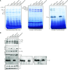
Analysis of mitochondrial complexes in oxa2b complementation plants. A, BN-PAGE analysis of mitochondrial complexes. The gel on the left was Coomassie-stained, the gel in the middle was stained for NADH dehydrogenase activity (Complex I), and the gel on the right was stained for cytochrome c oxidase activity (complex IV). Complexes and super complexes are indicated where appropriate. B, Immunoblot analysis of mitochondrial complexes after BN-PAGE with the following antibodies: carbonic anhydrase2 (complex I), succinate dehydrogenase subunit4 (complex II), Rieske iron-sulfur subunit of cytochrome c reductase (complex III), cytochrome c oxidase subunits 1, 2, and 3 (COX1, 2 and 3, complex IV), and subunits alpha and beta of ATP synthase (complex V). I, complex I; V, complex V; III2, dimeric complex III; F1, F1 part of complex V; I+III2, supercomplex composed of complex I and dimeric complex III; I2+III4, supercomplex composed of two complex I monomers and two copies of dimeric complex III; I+III2+IV, super complex composed of complex I, dimeric complex III, and complex IV. CA2, carbonic anhydrase2; SDH4, succinate dehydrogenase subunit4; RISP, Rieske iron-sulfur subunit of cytochrome c reductase; ATPα/β, subunits alpha and beta of ATP synthase.
Figure 5.
Figure 5.
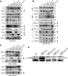
Analysis of mitochondrial proteins in oxa2b complementation plants. A, Immunoblot analysis of the indicated proteins involved in the respiratory chain. B, Immunoblot analysis of the indicated proteins involved in the import of proteins into mitochondria. C, Immunoblot analysis of a selection of other mitochondrial proteins. D, Immunoblot for OXA2b. Because OXA2b is a low abundance protein, 100 µg mitochondrial protein was loaded per lane, instead of 30 µg. In all panels, the antibody used is on the left of the gel and the molecular mass in kD is located on the right. The correct band is indicated with a small arrow head on the left-hand side. In some cases, more than one isoform is detected. Bands not indicated are nonspecific reactions of the antibody. Where protein abundance is notably different, it is indicated on the right side by either an up (up-regulated) or a down (down-regulated) arrow. For full list of antibodies, see Supplemental Table S2. M, matrix; IM, inner membrane; R, ribosomal proteins.
Figure 6.
Figure 6.
Translation and assembly of mitochondrial-encoded proteins in oxa2b-1 complementation plants. A, Autoradiogram of proteins synthesized in organello for 10, 30, and 60 min in the mitochondria isolated from the indicated plants after separation by SDS-PAGE. The proteins identified in Supplemental Fig. S5 are indicated on the left. The lane labeled “control” is a bacterial contamination control where sodium acetate was used as an energy source. The large smear at the bottom of the gel is ATP9. Due to its high hydrophobicity, ATP9 runs aberrantly on SDS gels. B, The same treatment as in (A), but separated by BN-PAGE to show the incorporation of newly translated proteins into complexes. The respective respiratory complexes are indicated on the right. The question mark indicates a complex of unknown origin. C, Potential topologies of COX2 in mitoplasts before and after PK treatment. Before PK treatment, both forms of COX2 would have identical sizes. After PK treatment, if COX2 is partially inserted and only the N terminus is in the IMS, it has a predicted size of 24.8 kD, whereas if COX2 is fully inserted and has both the N- and C terminus in the IMS, it has a predicted size of 7.3 kD. D, Autoradiogram of a 60 min in-organello translation reaction that was split in half, with one half mock-treated (−) and the other half PK-treated (+) after preparing mitoplasts. The major proteins are indicated on the left. The asterisk indicates a band that is produced only when treated with PK in the truncated-OXA2b-complemented plant mitochondria. The full gel is shown on the left and the region containing COX2 is enlarged on the right. E, The same treatments as in (D), except that another complex IV mutant (rpoTmp) was included for comparison. Again, the full gel is shown on the left and the region containing COX2 is enlarged on the right.
Figure 7.
Figure 7.
The TPR domain of OXA2b interacts with newly synthesized COX2. A, Mitochondria isolated from Col-0 plants were used for in-organello translation reactions, lysed with Triton X-100 and incubated with recombinant GST or GST fused to OXA2bTPR, prebound to glutathione beads. After washing, the bound material was eluted and proteins were analyzed by SDS-PAGE and autoradiography was recorded. Lysate (8.3% of mitochondrial lysate) was used for binding. Mitochondrial translation products are shown with arrows on the left and molecular weight markers are indicated on the right. B, In vitro translated COX2 (34 kD) and COX2Ct (17 kD) were incubated with recombinant GST or GST fused to OXA2bTPR, prebound to glutathione beads. After washing, the bound material was eluted, proteins were analyzed by SDS-PAGE, and autoradiography was recorded. For translation, 20% of translation reaction was used for binding.

Similar articles

Cited by

References

    1. Altschul SF, Gish W, Miller W, Myers EW, Lipman DJ (1990) Basic local alignment search tool. J Mol Biol 215: 403–410 - PubMed
    1. Anghel SA, McGilvray PT, Hegde RS, Keenan RJ (2017) Identification of Oxa1 homologs operating in the eukaryotic endoplasmic reticulum. Cell Reports 21: 3708–3716 - PMC - PubMed
    1. Benz M, Soll J, Ankele E (2013) Arabidopsis thaliana Oxa proteins locate to mitochondria and fulfill essential roles during embryo development. Planta 237: 573–588 - PubMed
    1. Blatch GL, Lässle M (1999) The tetratricopeptide repeat: A structural motif mediating protein-protein interactions. BioEssays 21: 932–939 - PubMed
    1. Bonnefoy N, Fiumera HL, Dujardin G, Fox TD (2009) Roles of Oxa1-related inner-membrane translocases in assembly of respiratory chain complexes. Biochim Biophys Acta 1793: 60–70 - PMC - PubMed

Publication types

MeSH terms