. 2018 Nov 26;6(4):107.
doi: 10.3390/medsci6040107.

OK-432 Administration Inhibits Murine Allergic Rhinitis at the Induction Phase, through the Macrophage Activation with TLR2 Signaling Pathway

Affiliations

OK-432 Administration Inhibits Murine Allergic Rhinitis at the Induction Phase, through the Macrophage Activation with TLR2 Signaling Pathway

Noriaki Aoi et al. Med Sci (Basel). .

Abstract

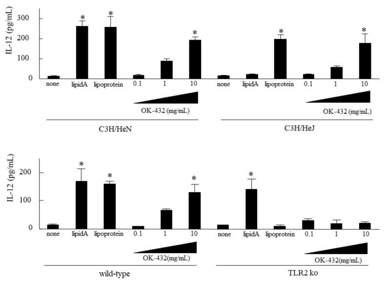
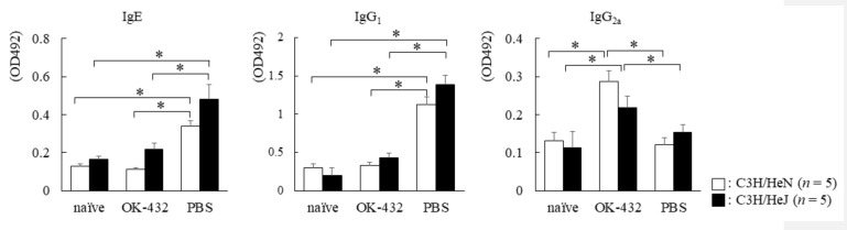
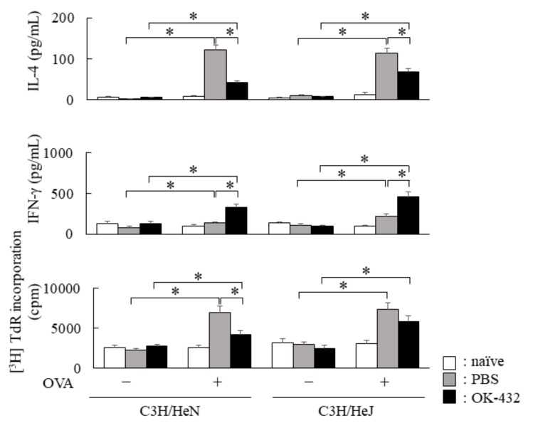
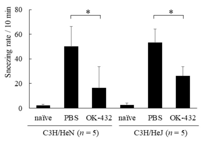
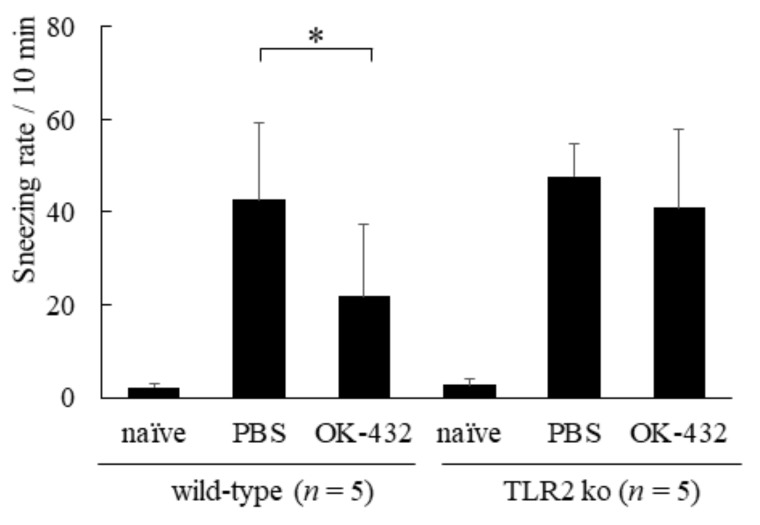
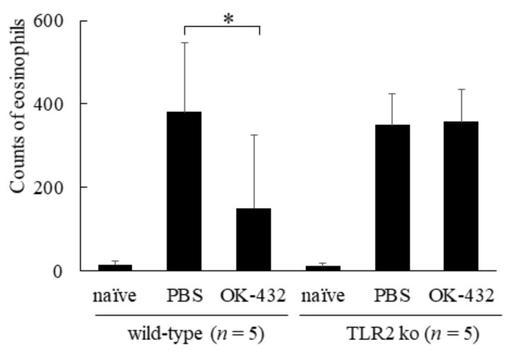
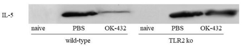
OK-432, a preparation of a low-virulence strain (Su) of Streptococcus pyogenes (Group A) killed by a penicillin and lyophilized, is a stiff inducer of Th1 cytokines, and exerts anti-cancer effects in tumor-bearing mice. OK-432 has been reported to consist of many bacterial components, such as peptidoglycan, M-protein, etc. However, it is yet to be ascertained which bacterial component induces T helper 1 (Th1) responses. For the last decade, Toll-like receptor (TLR) family proteins are well elucidated to play a role in recognizing bacterial components and inducing interleukin (IL)-12 from macrophages. Above all, peptidoglycan seems to be the agonist of TLR2 rather than the obverse. In our present study, the role of TLR2 for the recognition of OK-432 by macrophages and the effects of OK-432 are examined on murine allergic rhinitis model. Interestingly, results show IL-12 production by macrophages derived from TLR2 knock-out (ko) mice was significantly decreased, in comparison with that of macrophages derived from wild-type mice. Moreover, in TLR2 ko mice, no regulatory effect of OK-432 was observed on an allergic rhinitis model. These data indicate that TLR2 signaling is involved in regulating OK-432-induced anti-T helper 2 (Th2) immunity, and may offer a new prophylactic and therapeutic approach using OK-432 to downregulate allergic disorders, such as allergic rhinitis.

Keywords: OK-432; TLR2; allergic rhinitis; macrophage.


Conflict of interest statement

We have no conflict of interest to submit this manuscript.

Figures

Figure 1
Figure 1
IL-12 production by splenic macrophages from C3H/HeN, C3H/HeJ, wild-type or TLR2 ko mice in response to OK-432. The macrophages from the spleen of C3H/HeN, C3H/HeJ, wild-type or TLR2 knock-out mice were cultured in the presence of lipid A, lipoprotein or OK-432 for 48 h at 37 °C. IL -12 production by macrophages was measured by an ELISA. The data are representative of four independent experiments using pooled cells from five mice and are shown as the mean of triplicate determinations ± standard deviation (SD). *, p < 0.005.
Figure 2
Figure 2
IL-12 production by peritoneal macrophages from intraperitoneally PBS or OK-432-treated mice. The peritoneal macrophages from phosphate-buffered saline (PBS)-treated (white bars) or OK-432-treated (black bars) mice sensitized with OVA were cultured in the presence of OVA for 48 h at 37°C. IL-12 production by murine peritoneal macrophages were measured by an ELISA. Data were obtained from three independent experiments and are expressed as the mean of triplicate determinations ± SD. Per experimental group, five mice were used. *, p < 0.005.
Figure 3
Figure 3
Serum levels of OVA-specific Igs in C3H/HeN (white bars) or C3H/HeJ (black bars) mice sensitized with OVA. Individual levels of OVA-specific Igs were determined by an ELISA in C3H/HeN or C3H/HeJ mice on day 14 after i.p. injections with OVA/ALUM on day 0 and day 7, or naive C3H/HeN or C3H/HeJ mice without sensitization of OVA. PBS-treated mice were intraperitoneal (i.p.) injected with PBS on day 0 and day 7. OK-432-treated mice were i.p. injected with 100 mg OK-432 on day 0 and day 7. Data were obtained from three independent experiments and are expressed as the mean of triplicate determinations ± SD. Per experimental group, five mice were used. *, p < 0.005.
Figure 4
Figure 4
Cytokine production by spleen T cells from OK-432-treated or PBS-treated mice at the induction phase. The enriched T cells (5 × 10⁵ cells) from the spleen of OK-432-treated (black bars) or PBS-treated (hatched bars) mice sensitized with OVA or naive mice (white bars) were cultured with OVA in the presence of MMC-treated spleen cells (5 × 10⁵ cells) for 48 h at 37 °C. All mice were immunized with OVA/ALUM. The proliferative responses of splenic T cells were measured by an incorporation of [3H]TdR, and data was represented by CPM (counts per minute). IL-4 and interferon (IFN)-γ production by T cells was measured by an ELISA. The data are representative of four independent experiments using pooled cells from five mice and are shown as the mean of triplicate determinations ± SD. *, p < 0.005.
Figure 5
Figure 5
Sneezing rate in a murine model of C3H/HeN or C3H/HeJ mice systemically sensitized with OVA and receiving subsequent intranasal challenge of OVA. Naive mice underwent no systemic sensitization. PBS-treated mice were i.p. injected with PBS on day 0 and day 7. OK-432-treated mice were i.p. injected with 100 mg OK-432 on day 0 and day 7. Data were obtained and expressed as the mean of triplicate determinations ± SD. Per experimental group, five mice were used. *, p < 0.005.
Figure 6
Figure 6
Eosinophil infiltration in nasal mucosae in a murine model of C3H/HeN or C3H/HeJ mice systemically sensitized with OVA and receiving subsequent intranasal challenge of OVA. Naive mice underwent no systemic sensitization. PBS-treated mice were i.p. injected with PBS on day 0 and day 7. OK-432-treated mice were i.p. injected with 100 mg OK-432 on day 0 and day 7. Data were obtained and expressed as the mean of triplicate determinations ± SD. Per experimental group, five mice were used. *, p < 0.005.
Figure 7
Figure 7
IL-5 expressions of nasal mucosae assessed by Western blot assay, in a murine model of C3H/HeN or C3H/HeJ mice systemically sensitized with OVA and receiving subsequent intranasal challenge of OVA. Naive mice underwent no systemic sensitization. PBS-treated mice were i.p. injected with PBS on day 0 and day 7. OK-432-treated mice were i.p. injected with 100 mg OK-432 on day 0 and day 7. IL-5 expressions were detected in PBS-treated C3H/HeN and C3H/HeJ mice, but not found in OK-432-treated-mice of both strains.
Figure 8
Figure 8
Serum levels of OVA-specific Igs in wild-type (white bars) or TLR2 knock-out (black bars) mice sensitized with OVA. Individual levels of OVA-specific Igs were determined by an ELISA in wild-type or TLR2 knock-out mice on day 14 after i.p. injections with OVA/ALUM on day 0 and day 7, or naive wild-type or TLR2 knock-out mice without sensitization of OVA. PBS-treated mice were i.p. injected with PBS on day 0 and day 7. OK-432-treated mice were i.p. injected with 100 mg OK-432 on day 0 and day 7. Data were obtained from three independent experiments and are expressed as the means of triplicate determinations ± SD. Per experimental group, five mice were used. *, p < 0.005.
Figure 9
Figure 9
Cytokine production by splenic T cells from OK-432-treated or PBS-treated mice at the induction phase. The enriched T cells (5 × 10⁵ cells) from the spleen of OK-432-treated (black bars) or PBS-treated (hatched bars) mice sensitized with OVA or naive mice (white bars) were cultured with OVA in the presence of MMC-treated spleen cells (5 × 10⁵ cells) for 48 h at 37 °C. All mice were immunized with OVA/ALUM. The proliferative responses of splenic T cells was measured by an incorporation of [3H]TdR. IL-4 and IFN-γ production by the splenic T cells was measured by an ELISA. The data are representative of four independent experiments using pooled cells from five mice and are shown as mean of triplicate determinations ± SD. *, p < 0.005.
Figure 10
Figure 10
Sneezing rate in a murine model of wild-type and TLR2 knock-out mice systemically sensitized with OVA and receiving subsequent intranasal challenge of OVA. Naive mice underwent no systemic sensitization. PBS-treated mice were i.p. injected with PBS on day 0 and day 7. OK-432-treated mice were i.p. injected with 100 mg OK-432 on day 0 and day 7. Data were obtained and expressed as the mean of triplicate determinations ± SD. Per experimental group, five mice were used. *, p < 0.005.
Figure 11
Figure 11
Eosinophil infiltration in nasal mucosae in a murine model of wild-type and TLR2 knock-out mice systemically sensitized with OVA and receiving subsequent intranasal challenge of OVA. Naive mice underwent no systemic sensitization. PBS-treated mice were i.p. injected with PBS on day 0 and day 7. OK-432-treated mice were i.p. injected with 100 mg OK-432 on day 0 and day 7. Data were obtained and expressed as the mean of triplicate determinations ± SD. Per experimental group, five mice were used. *, p < 0.005.
Figure 12
Figure 12
IL-5 expressions of nasal mucosae assessed by western blot assay, in a murine model of wild-type and TLR2 knock-out mice systemically sensitized with OVA and receiving subsequent intranasal challenge of OVA. Naive mice underwent no systemic sensitization. PBS-treated mice were i.p. injected with PBS on day 0 and day 7. OK-432-treated mice were i.p. injected with 100 mg OK-432 on day 0 and day 7. IL-5 expression was detected in PBS-treated wild type of mice and TLR 2 knock-out mice. Interestingly, it was also detected in OK-432-treated TLR2 knock-out mice, whereas it was downregulated in PBS-treated wild-type mice.

Similar articles

Cited by

References

    1. Baraniuk J.N. Mechanisms of allergic rhinitis. Curr. Allergy Asthma Rep. 2001;1:207–217. doi: 10.1007/s11882-001-0007-5. - DOI - PubMed
    1. Campbell J.D., HayGlass K.T. T cell chemokine receptor expression in human Th1- and Th2-associated diseases. Arch. Immunol. Ther. Exp. 2000;48:451–456. - PubMed
    1. Pearlman D.S. Pathophysiology of the inflammatory response. Pt 1J. Allergy Clin. Immunol. 1999;104:S132–S137. doi: 10.1016/S0091-6749(99)70308-8. - DOI - PubMed
    1. Durham S.R. Mechanisms of mucosal inflammation in the nose and lungs. Clin Exp Allergy. 1998;28:11–16. - PubMed
    1. Baraniuk J.N. Pathogenesis of allergic rhinitis. J. Allergy Clin. Immunol. 1997;99:S763–S772. doi: 10.1016/S0091-6749(97)70125-8. - DOI - PubMed