Review
. 2019 Jan;19(1):3-14.
doi: 10.3892/mmr.2018.9679. Epub 2018 Nov 20.

Roles of the inflammasome in the gut‑liver axis (Review)

Affiliations
Review

Roles of the inflammasome in the gut‑liver axis (Review)

Junfeng Wang et al. Mol Med Rep. 2019 Jan.

Abstract

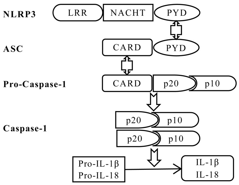
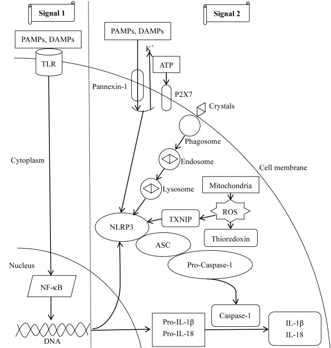
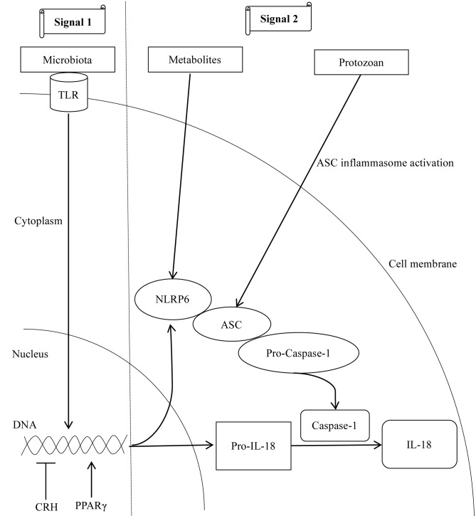
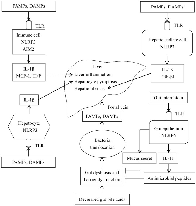
The gut‑liver axis connects the liver with the intestine via bile acid metabolism. Bile acid dysregulation leads to intestinal dysbiosis, that allows enterogenous pathogenic bacteria, including Gram‑negative bacteria and their products lipopolysaccharide (LPS), into the liver via the portal vein, triggering inflammation in the liver. The inflammasome serves as an intracellular pattern recognition receptor that detects pathogens or danger signals and mediates innate immunity in the liver or gut. Specifically, the NACHT, LRR and PYD domains‑containing protein (NLRP)6 inflammasome maintains intestinal microbial balance, by promoting interleukin (IL)‑18‑dependent antimicrobial peptide synthesis and mucus secretion from goblet cells. The NLRP3 inflammasome, in contrast, primarily induces IL‑1β and aggravates inflammatory liver injury. Furthermore, the NLRP3 inflammasome affects the epithelial integrity of cholangiocytes by inducing the production of pro‑inflammatory cytokines. In addition, bile acids, including deoxycholic acid and chenodeoxycholic acid, are able to activate the NLRP3 inflammasome in macrophages; however, bile acids have the potential to exert the opposite role by interacting with the membrane‑bound Takeda G‑protein receptor 5 or by activating nuclear farnesoid‑X receptor. Therefore, further investigation of the molecular mechanisms underlying the inflammasome, involved in the gut‑liver axis, may provide important insights into the identification of a potential therapeutic target for the treatment of liver and gut diseases. The present review discusses the roles of the inflammasome in the gut‑liver axis, and the emerging associations between the inflammasome and the intestinal microbiota or the bile acids in the gut‑liver axis.

Keywords: bile acid; gut-liver axis; inflammation; intestinal microbiota; NLRP3 inflammasome; NLRP6 inflammasome.


Figures

Figure 1.
Figure 1.
Schematic diagram of NLRP3 inflammasome assembly. The NLRP3 inflammasome is assembled by NLRP3, the ASC adaptor and pro-caspase-1. Upon NLRP3 activation, NLRP3 interacts with ASC via PYDs, and the CARD domain of ASC recruits the CARD of pro-caspase-1, leading to autocleavage of the inactive CARD domain from pro-caspase-1. This cleavage allows the formation of the active caspase-1 p10/p20 tetramer, which cleaves cytokine precursors to produce and release mature IL-1β and IL-18. IL, interleukin; CARD, caspase recruitment domain; LRR, leucine-rich repeat; NACHT, nucleotide-binding and oligomerization domain; PYD, pyrin domain; NLRP3, NACHT, LRR and PYD domains-containing protein 3; ASC, apoptotic speck-like protein containing a CARD.
Figure 2.
Figure 2.
Mechanisms involved in NLRP3 inflammasome activation. Two signals are required for the activation of the NLRP3 inflammasome. For signal 1, PAMPs and DAMPs combine with TLRs on the cell membrane to activate NF-κB-dependent transcription and translation of NLRP3, pro-IL-1β and pro-IL-18. For signal 2, three mechanisms have been described. In the first, ATP interacts with P2X7, leading to intracellular K+-depletion and opening of a large-pored pannexin-1 channel, through which PAMPs and DAMPs enter the cell and activate the NLRP3 inflammasome. Furthermore, endocytosis of large molecules, including crystals, results in lysosomal disruption, leading to the release of its components and activation of the NLRP3 inflammasome. Additionally, mitochondria-derived ROS detach TXNIP from thioredoxin and enable activation of the NLRP3 inflammasome. The second signal results in caspase-1 activation, and cleavage of pro-IL-1β and pro-IL-18 into mature IL-1β and IL-18. ASC, apoptosis-associated speck-like protein containing a CARD; DAMPs, danger-associated molecular patterns; NF-κB, nuclear factor-κB; P2X7, P2X purinoceptor 7; PAMPs, pathogen-associated molecular patterns; ROS, reactive oxygen species; TLR, Toll-like receptor; TXNIP, thioredoxin-interacting protein; IL, interleukin; NLRP3, NACHT, LRR and PYD domains-containing protein 3; ATP, adenosine 5′-triphosphate.
Figure 3.
Figure 3.
Mechanisms involved in NLRP6 inflammasome activation. Intestinal microbiota initiate two signals for the activation of the NLRP6 inflammasome. In the first signal, the commensal microbiota serve as a TLR ligand and promotes the transcription of NLRP6 and pro-IL-18. For the second signal, microbial metabolites, including taurine, promote the multiprotein complex assembly to activate the NLRP6 inflammasome. In particular, commensal protozoans promote epithelial IL-18 secretion via activation of the ASC inflammasome. In addition to the microbial roles, CRH inhibits the transcription of NLRP6 inflammasome components, whereas, the nuclear transcription factor PPAR-γ activates NLRP6 by binding to its promoter region. Arrows indicate ‘promotion’, whereas, the symbol ‘┴’ indicates ‘inhibition’. CRH, corticotrophin-releasing hormone; PPAR-γ, peroxisome proliferator-activated receptor-γ; NLRP6, NACHT, LRR and PYD domains-containing protein 6; IL-18, interleukin 18; ASC, apoptosis-associated speck-like protein containing a CARD; TLR, Toll-like receptor.
Figure 4.
Figure 4.
Effect of BA on the NLRP3 inflammasome in the macrophage. Elevated intracellular BAs, including deoxycholic acid and chenodeoxycholic acid, directly activate the NLRP3 inflammasome in macrophages. However, the BA nuclear receptor FXR interacts with NLRP3 to prevent the assembly of NLRP3 inflammasome components, thereby repressing its activation. Furthermore, the BA membrane receptor TGR5 may negatively regulate NLRP3 inflammasome activation by TGR5-cAMP-PKA axis-dependent NLRP3 phosphorylation and ubiquitination. However, due to the limited expression of FXR and TGR5 under cholestasis conditions, the aforementioned protective mechanisms fail to counteract the cytotoxic effects of BAs. Black arrows indicate ‘promotion’, whereas, purple arrow indicates ‘inhibition’. BA, bile acid; cAMP, cylic adenosine monophosphate; FXR, farnesoid-X receptor; PKA, protein kinase A; TGR5, Takeda G-protein receptor 5; NLRP3, NACHT, LRR, and PYD domains-containing protein 3.
Figure 5.
Figure 5.
Roles of the inflammasome in the gut-liver axis. Decreased intestinal BAs induce intestinal flora disorders, increase intestinal permeability, and impair intestinal barrier function. The NLRP6 inflammasome in the intestinal epithelium physiologically induces IL-18 synthesis, and promotes the production of antimicrobial peptides and mucus secretion by goblet cells, which eventually inhibits intestinal barrier disruption and maintains intestinal microbial balance. However, in gut-liver dysfunction, protective effects of the NLRP6 inflammasome may be inhibited, resulting in bacterial translocation, and the transfer of PAMPs and DAMPs to the liver via the portal vein. In the liver, accumulative PAMPs and DAMPs act on TLRs on the cell membrane to activate the intracellular inflammasome. Furthermore, in immune cells, NLRP3 and AIM2 inflammasome activation induces the synthesis of IL-1β, which primarily mediates inflammation in the liver, and increases the expression of MCP-1 and TNF-α. In hepatocytes, MCP-1 further aggravates hepatocyte steatosis. Furthermore, IL-1β sensitizes hepatocytes to TNF-mediated cellular toxicity and induces pyroptosis of hepatocytes in combination with TNF-α. In hepatic stellate cells, NLRP3 inflammasome activation upregulates the expression level of TGF-β1 and promotes hepatic fibrosis. Arrows indicate ‘promotion’, whereas, the symbol ‘┴’ indicates ‘inhibition’. BA, bile acid; DAMPs, danger-associated molecular patterns; MCP-1, monocyte chemoattractant protein 1; PAMPs, pathogen-associated molecular patterns; TLR, Toll-like receptor; IL, interleukin; NLRP6, NACHT, LRR and PYD domains-containing protein 6; AIM, interferon-inducible protein AIM2; TNF, tumor necrosis factor; TGF-β1, transforming growth factor β1.

Similar articles

Cited by

References

    1. Visschers RG, Luyer MD, Schaap FG, Olde Damink SW, Soeters PB. The gut-liver axis. Curr Opin Clin Nutr Metab Care. 2013;16:576–581. doi: 10.1097/MCO.0b013e32836410a4. - DOI - PubMed
    1. Islam KB, Fukiya S, Hagio M, Fujii N, Ishizuka S, Ooka T, Ogura Y, Hayashi T, Yokota A. Bile acid is a host factor that regulates the composition of the cecal microbiota in rats. Gastroenterology. 2011;141:1773–1781. doi: 10.1053/j.gastro.2011.07.046. - DOI - PubMed
    1. Yokota A, Fukiya S, Islam KB, Ooka T, Ogura Y, Hayashi T, Hagio M, Ishizuka S. Is bile acid a determinant of the gut microbiota on a high-fat diet? Gut Microbes. 2012;3:455–459. doi: 10.4161/gmic.21216. - DOI - PubMed
    1. Cai SY, Boyer JL. Studies on the mechanisms of bile acid initiated hepatic inflammation in cholestatic liver injury. Inflamm Cell Signal. 2017;4:e1561. - PMC - PubMed
    1. Cai SY, Ouyang X, Chen Y, Soroka CJ, Wang J, Mennone A, Wang Y, Mehal WZ, Jain D, Boyer JL. Bile acids initiate cholestatic liver injury by triggering a hepatocyte-specific inflammatory response. JCI Insight. 2017;2:e90780. doi: 10.1172/jci.insight.90780. - DOI - PMC - PubMed