Review
. 2018 Dec:53:187-198.
doi: 10.1016/j.sbi.2018.11.002. Epub 2018 Nov 24.

PARP family enzymes: regulation and catalysis of the poly(ADP-ribose) posttranslational modification

Affiliations
Review

PARP family enzymes: regulation and catalysis of the poly(ADP-ribose) posttranslational modification

Marie-France Langelier et al. Curr Opin Struct Biol. 2018 Dec.

Abstract

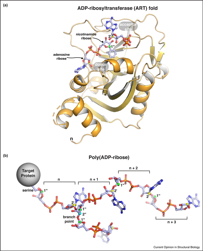
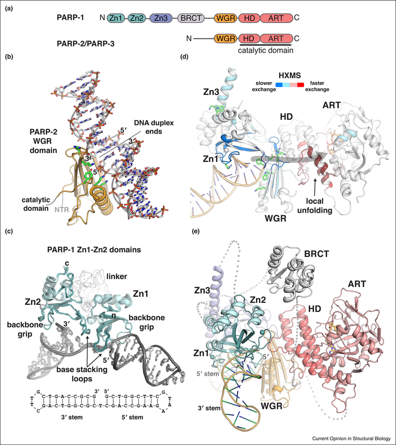
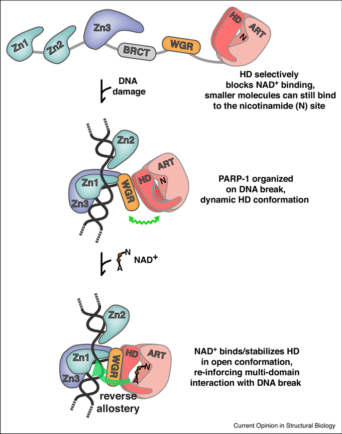
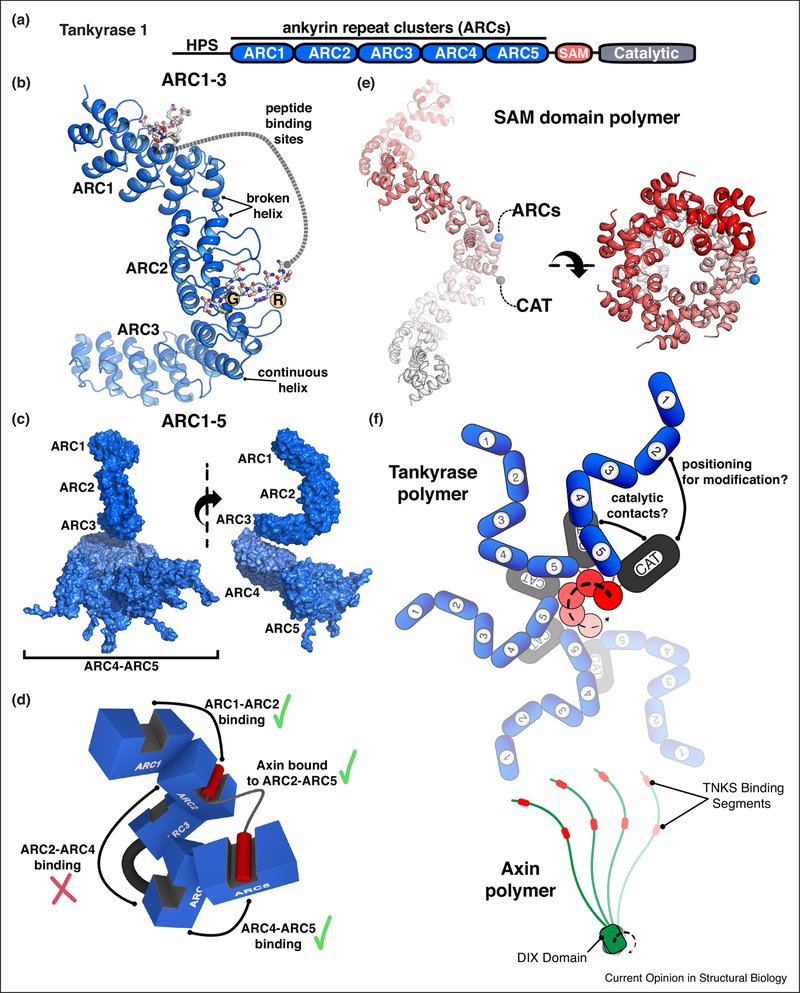
Poly(ADP-ribose) is a posttranslational modification and signaling molecule that regulates many aspects of human cell biology, and it is synthesized by enzymes known as poly(ADP-ribose) polymerases, or PARPs. A diverse collection of domain structures dictates the different cellular roles of PARP enzymes and regulates the production of poly(ADP-ribose). Here we primarily review recent structural insights into the regulation and catalysis of two family members: PARP-1 and Tankyrase. PARP-1 has multiple roles in the cellular response to DNA damage and the regulation of gene transcription, and Tankyrase regulates a diverse set of target proteins involved in cellular processes such as mitosis, genome integrity, and cell signaling. Both enzymes offer interesting modes of regulating the production and the target site selectivity of the poly(ADP-ribose) modification.


Conflict of interest statement

Conflict of interest

None declared.

Figures

Figure 1.
Figure 1.. Poly(ADP-ribose) and the ADP-ribosyltransferase (ART) fold.
(a) The ADP-ribosyltransferase (ART) fold of PARP family enzymes. The minimal catalytic region of human PARP-1 is shown bound to a non-hydrolyzable NAD+ analog (PDB code 6bhv; [32]). Substrate NAD+ binds to the “donor site.” The adenosine ribose and the nicotinamide ribose are indicated. Adenosime ribose atoms are labeled as single prime (‘) and nicotinamde ribose atoms are labeled as double prime (“). Protein side chains to be modified with ADP-ribose attack the 1 ” carbon of nicotinamide ribose (labeled and colored green), thus releasing nicotinamide. The current model for the extension of the primary ADP-ribose group is based on an “acceptor site” identified in the crystal structure of chicken PARP-1 (PDB code la26; [76]). The ADP group from the chicken PARP-1 structure was overlayed on the structure of human PARP-1. Extension of the ADP-ribose chain is proposed to arise from the acceptor site adenosine ribose 2’ hydroxyl (2’ carbon is labeled and colored yellow) attacking the donor site NAD+ on the nicotinamide ribose 1” carbon (creating a 1” to 2’ linkage; see panel b). Branches are proposed to arise from an ADP-ribose that binds in a reversed orientation in the “acceptor site,” thus instead placing the nicotinamide ribose 2” hydroxyl in position to attack the donor site NAD+ on the nicotinamide ribose 1” carbon (creating a 1” to 2” linkage; see panel b). Structures that capture reaction intermediates are required to fully understand the catalysis of poly(ADP-ribose). The termini of the ART fold are labeled (n and c). (b) The poly(ADP-ribose) posttranslational modification. Four linear units of ADP-ribose (denoted n, n+1, …) are shown attached to a serine side chain on a target protein. A single branch point is shown extending from the n + 1 ADP-ribose of the linear chain. The serine sidechain oxygen is attached to the 1” carbon of the nicotinamide ribose (colored green as above). Linear chains of ADP-ribose units are formed through a ribose-ribose linkage: nicotinamide ribose 1” carbon linked to the adenosine ribose 2’ carbon (colored yellow as above). A branch in the poly(ADP-ribose) chain forms a different ribose-ribose linkage: nicotinamide ribose 1” carbon linked to a nicotinamide ribose 2” carbon (colored cyan on the n + 1 ADP-ribose unit).
Figure 2.
Figure 2.. Structural insights into PARP-1 and PARP-2 DNA damage detection.
(a) Schematic representation of human PARP-1, PARP-2, and PARP-3 domain organization. Zinc fingers: Zn1, Zn2, and Zn3; BRCT – BRCA C-terminus; WGR: Trp-Gly-Arg domain; HD –helical domain; ART – ADP-ribosyltransferase. (b) Structure of the human PARP-2 WGR domain bound to 5’ phosphorylated DNA (PDB code 6f5b; [18]). The crystal asymmetric unit contains two DNA duplexes. The WGR binds where the two DNA ends meet, which mimics a DNA strand break. Thus, the WGR spans the DNA break, engaging the 5’ phosphorylated end of one DNA duplex (labeled 5’), and the 3ΌH terminated end of the second duplex (labeled 3’). Some of the key esidues involved in binding the 5’ phosphate, or the second duplex, are shown as sticks and colored green. A second WGR domain is present in the crystal asymmetric unit, but has not been illustrated for clarity. The second WGR domain is bound to the opposite face of the DNA in the same manner as shown. (c) Structure of the Zn1-Zn2 fragment of human PARP-1 detecting a DNA single-strand break (PDB code 2n8a; [21]). A schematic of the DNA is drawn below the complex structure, illustrating the single strand break with a one nucleotide gap. The two sides of the DNA break are denoted 3’ stem and 5’ stem, based on the nature of the DNA terminus. Zn1 engages nucleotide bases on the 5’ stem, and Zn2 engages nucleotide bases on the 3’ stem. The N-terminus of Zn1 is labeled (n), and the C-terminus of Zn2 is labeled (c). The extended linker region connecting Zn1 to Zn2 adopts multiple conformations, and is likely to allow the two zinc fingers to engage a variety of damaged DNA structures in which the relative positioning of the ends could vary [21]. (c) Changes in PARP-1 dynamics upon detecting DNA damage were revealed by HXMS [31]. PARP-1 peptides experiencing slower amide hydrogen exchange in the presence of a DNA strand break are indicative of newly formed protein-DNA contacts and/or domain-domain contacts, and the peptides exhibiting slower exchange (colored blue) are consistent with contacts observed in the PARP-1 complex with a DNA double-strand break (shown in cartoon with domains labeled) [13]. Remarkably, several PARP-1 peptides grouped in the HD experienced much faster amide hydrogen exchange in the presence of DNA (colored red). The rate of exchange was much faster than possible for amide hydrogens involved in forming a helix, thus these helical regions of the HD were proposed to unfold in the presence of DNA, or to rapidly sample the unfolded state [31]. Several of the key PARP-1 residues essential for DNA-dependent poly(ADP-ribose) catalysis [13,19,20,29] are drawn as sticks, and they highlight the allosteric network that connects PARP-1 DNA damage detection to the catalytic active site (indicated by grey arrow). (d) A composite model for full-length PARP-1 detecting a single-strand DNA break. The crystal structure of Zn1-Zn3-WGR-CAT was aligned to the NMR structure of the Zn1-Zn2 fragment bound to a single-strand break [13,21]. The BRCT domain was manually positioned between the C-terminus of the Zn3 domain and the N-terminus of the WGR domain. The linker residues connecting PARP-1 domains are shown as grey spheres, with each sphere representing an amino acid residue. The DNA break site is noted by the labeling of the 5’ terminus adjacent to the WGR domain.
Figure 3.
Figure 3.. Allosteric regulation of PARP-1.
Regulation of NAD+ access and reverse allostery. The HD regulates PARP-1 catalytic activity through a substrate-blocking mechanism [32]. In the absence of DNA, the domains of PARP-1 exist in an open configuration. In this state, small molecules can bind to the nicotinamide site (N) of the ART; however, NAD+ binding is completely blocked. Upon detecting DNA damage, PARP-1 domains are organized around the DNA break, and an allosteric network of contacts destabilizes the HD, leading to a dynamic HD structure, illustrated as multiple potential HD conformations, and accented by a wavy green line that indicates a flexible conformation. NAD+ is now able to access the catalytic active site, engaging the nicotinamide site (N) and the adenosine site (A) that was previously blocked by the HD. NAD+ binding pushes the distribution of HD conformations toward the unfolded state, thus promoting the PARP-1 assembly of domains on DNA. Thus, NAD+ binding can influence PARP-1 interaction with DNA through a reverse allostery mechanism from catalytic active site to DNA binding domain (large green arrow).
Figure 4.
Figure 4.. Structural biology of Tankyrase regulation.
(a) Schematic representation of human Tankyrase-1 domain architecture. ARC – ankyrin repeat cluster (ARC); SAM – sterile alpha motif; HPS – histidine/proline/serine-rich region of unknown function. Human Tankyrase-2 has the same domain architecture, but lacks the HPS region. (b) Crystal structure of the ARC1–ARC2–ARC3 fragment of human Tankyrase-1 bound to a peptide derived from IRAP (insulin regulated aminopeptidase) [59]. Peptide bound to ARC1 and ARC2 are drawn as sticks. The peptide bound to ARC2 indicates the essential contact points – an Arg (R) at position 1 and a Gly (G) at position 6. ARC3 lacks the amino acids necessary for peptide binding. The structure indicated two types of transitions between consecutive ARCs: a “broken helix” connecting ARC1 to ARC2, and a “continuous helix” connecting ARC2 to ARC3. The relative positioning of the two peptide binding sites is fixed. The ARC1–ARC2–ARC3 conformation was confirmed by SAXS analysis [59]. (c) SAXS analysis of the entire ankyrin repeat region, ARC1–5. In contrast to the rigid conformation of ARC1–3, ARC4 and ARC5 are more flexibly connected. An ensemble of structures was used to model the SAXS data [59], and a selection of ARC4-ARC5 conformations are shown. (d) Axin interaction with ARC1–5. A cartoon representation of ARC1–5 based on SAXS-based modeling in panel (c). Axin contains two peptide regions that bind to Tankyrase [60] (shown here as red cylinders). Combinatorial mutagenesis of ARC peptide binding sites and Axin binding affinity analysis indicated that specific ARC pairs can function together to bind Axin: ARC1:ARC2, ARC4:ARC5, ARC2:ARC5 (shown as green check mark). Non-binding ARC3 separates ARC2 and ARC4 and prevents them from simultaneously engaging Axin (red check mark. (e) Tankyrase SAM domain polymer. The crystal structure of the wild-type human Tankyrase-1 SAM domain is shown from two views related by a 90° rotation (PDB code 5kni [69]; see also [68]). For one SAM domain in the polymer, the N-terminus (blue sphere) and C-terminus (grey sphere) are indicated to highlight that the ARCs and the catalytic domain (CAT) will extend from the outside surface of the SAM polymer. (f) A model for the polymeric from of Tankyrase based on current structural information. A juxtaposed cartoon model of the Axin polymer [72] illustrates the potential for avidity-dependent interaction of these proteins [51,68].

Similar articles

Cited by

References

    1. Palazzo L, Mikoč A, Ahel I: ADP-ribosylation: new facets of an ancient modification. FEBS J 2017, doi:10.1111/febs.14078. - DOI - PMC - PubMed
    1. Gupte R Liu Z, Kraus WL: PARPs and ADP-ribosylation: recent advances linking molecular functions to biological outcomes. Genes Dev 2017, 31:101–126. - PMC - PubMed
    1. Vyas S, Chang P: New PARP targets for cancer therapy. Nat Rev Cancer 2014, 14:502–509. - PMC - PubMed
    1. Hottiger MO, Hassa PO, Lüscher B, Schüler H, Koch-Nolte F: Toward a unified nomenclature for mammalian ADP-ribosyltransferases. Trends Biochem Sci 2010, 35:208–219. - PubMed
    1. Vyas S, Matic I, Uchima L, Rood J, Zaja R Hay RT, Ahel I, Chang P: Family-wide analysis of poly(ADP-ribose) polymerase activity. 2014, 5:4426. - PMC - PubMed

Publication types

MeSH terms

LinkOut - more resources