. 2018 Nov 26;8(1):17375.
doi: 10.1038/s41598-018-35745-1.

HIF-2α, but not HIF-1α, mediates hypoxia-induced up-regulation of Flt-1 gene expression in placental trophoblasts

Affiliations

HIF-2α, but not HIF-1α, mediates hypoxia-induced up-regulation of Flt-1 gene expression in placental trophoblasts

Tadashi Sasagawa et al. Sci Rep. .

Abstract

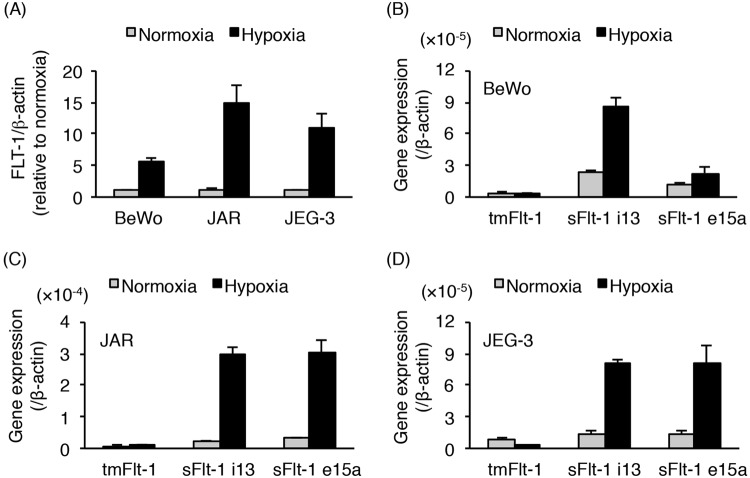
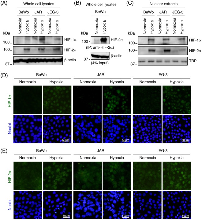
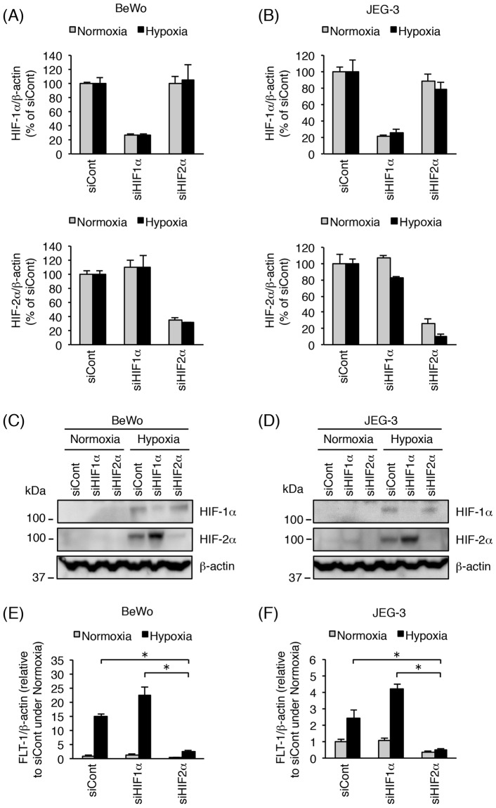
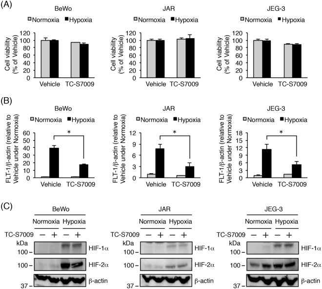
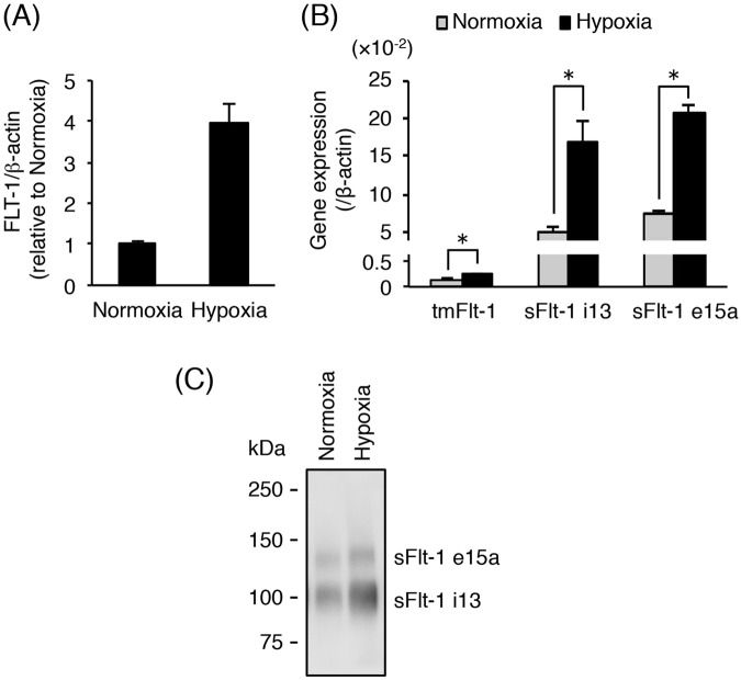
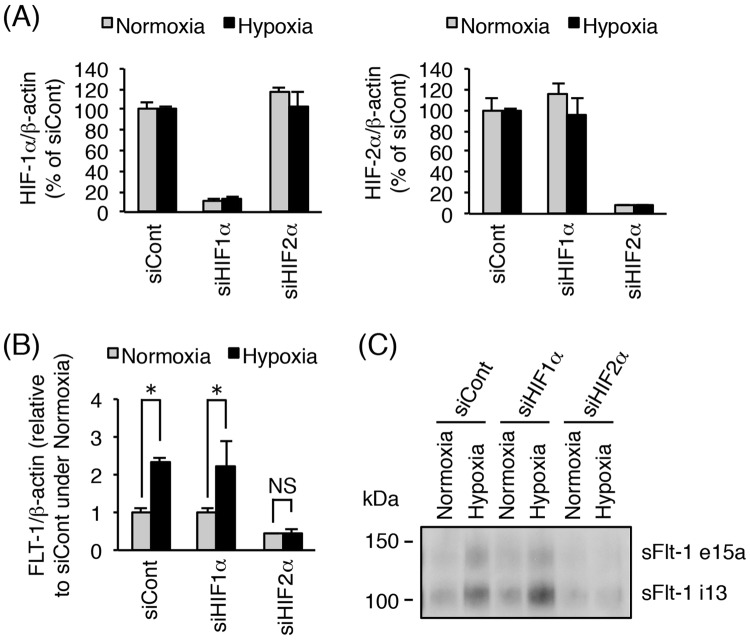
Placental hypoxia and elevated levels of circulating soluble Fms-like tyrosine kinase-1 (sFlt-1), an anti-angiogenic factor, are closely related to the pathogenesis of preeclampsia. Although sFlt-1 secretion from the placental trophoblasts is increased under hypoxic conditions, the underlying molecular mechanism remains unclear. Previously, an authentic hypoxia response element in the Flt-1 gene promoter was shown to be a potential binding site for hypoxia-inducible factors (HIFs). Here, we investigated the roles of HIF-1α and HIF-2α in Flt-1 gene expression in trophoblast-derived choriocarcinoma cell lines and cytotrophoblasts exposed to hypoxic conditions. In the cell lines, increased expression of sFlt-1 splice variants and nuclear accumulation of HIF-1α and HIF-2α were observed after hypoxic stimulation. A specific small interfering RNA or an inhibitor molecule targeting HIF-2α decreased hypoxia-induced up-regulation of Flt-1 gene expression. Moreover, in cytotrophoblasts, increased sFlt-1 mRNA expression and elevated sFlt-1 production were induced by hypoxic stimulation. Notably, hypoxia-induced elevation of sFlt-1 secretion from the cytotrophoblasts was inhibited by silencing the HIF-2α, but not HIF-1α mRNA. These findings suggest that hypoxia-induced activation of HIF-2α is essential for the increased production of sFlt-1 proteins in trophoblasts. Targeting the HIF-2α may be a novel strategy for the treatment of preeclampsia.


Conflict of interest statement

The authors declare no competing interests.

Figures

Figure 1
Figure 1
Hypoxia exposure up-regulates the expression of sFlt-1 variants in human trophoblast-derived choriocarcinoma cell lines. BeWo, JAR, and JEG-3 cells were incubated for 24 h under normoxic or hypoxic conditions. The mRNA expression levels of all Flt-1 transcript variants (FLT-1) and three splice variants of the Flt-1 gene in these cells were determined by quantitative real-time PCR analysis using β-actin mRNA as a reference. (A) FLT-1 mRNA expression level in the three trophoblast-derived choriocarcinoma cell lines. Results are expressed as a fold change relative to the cells under normoxic conditions. (BD) The mRNA expression levels of three Flt-1 splice variants (tmFlt-1, sFlt-1 i13, and sFlt-1 e15a) in BeWo (B), JAR (C), and JEG-3 (D) cells. Results are represented as a ratio relative to the expression of β-actin mRNA. All values are represented as the means ± SD (n = 3).
Figure 2
Figure 2
Hypoxia-induced activation of HIF-1α and HIF-2α in trophoblast-derived choriocarcinoma cell lines. Cells were incubated for 24 h under normoxic or hypoxic condition, and then whole cell lysates or nuclear extracts were prepared. (A) HIF-1α and HIF-2α expression in whole cell lysates derived from the three choriocarcinoma cell lines was assessed by Western blot analysis. β-actin was used as a loading control. (B) Immunoprecipitation (IP) of HIF-2α protein from the whole cell lysates of BeWo cells. The immunoprecipitates and whole cell lysates (input) were subjected to Western blot analysis with anti- HIF-2α and anti-β-actin antibodies, respectively. Four percent of the whole cell lysates were used as the input. (C) HIF-1α and HIF-2α expression in nuclear extracts derived from the three choriocarcinoma cell lines. The nuclear protein TBP served as a loading control. Uncropped images of Western blots are presented in Supplementary Fig. S5. (D,E) Cellular localization of HIF-1α (D) or HIF-2α (E) in the three choriocarcinoma cell lines was determined by immunofluorescence staining.
Figure 3
Figure 3
Inhibition of HIF-2α by siRNA transfection decreases hypoxia-induced up-regulation of Flt-1 gene expression in BeWo and JEG-3 cells. BeWo and JEG-3 cells were transfected with 10 nM of control siRNA, HIF-1α siRNA, or HIF-2α siRNA. Seventy-two hours after transfection, cells were incubated for 24 h under normoxic or hypoxic conditions. (A,B) Evaluation of siRNA-mediated HIF-1α/2α mRNA knockdown in BeWo (A) and JEG-3 (B) cells. HIF-1α/2α mRNA expression levels were measured by quantitative real-time PCR analysis using β-actin mRNA as a reference. Results are expressed as a percentage relative to control siRNA (siCont)-transfected cells under normoxic or hypoxic conditions. (C,D) HIF-1α/2α protein expression was assessed in BeWo (C) and JEG-3 (D) cells by Western blot analysis. β-actin was used as a loading control. Uncropped images of Western blots are presented in Supplementary Fig. S5. (E, F) The mRNA expression of all Flt-1 transcript variants (FLT-1) was measured in BeWo (E) and JEG-3 (F) cells by quantitative real-time PCR analysis. FLT-1 mRNA expression level was calculated by normalizing to β-actin mRNA and represented as a fold change relative to control siRNA (siCont)-transfected cells under normoxic conditions. All values are represented as the means ± SD (n = 3). Asterisks indicate a significant difference (P < 0.05).
Figure 4
Figure 4
Effects of the HIF-2α inhibitor, TC-S7009, on hypoxia-induced up-regulation of Flt-1 gene expression in trophoblast-derived choriocarcinoma cell lines. Three cell lines were cultured for 24 h under normoxic or hypoxic conditions in the presence of 0.1% DMSO (vehicle control) or 30 µM HIF-2α-specific inhibitor TC-S7009. (A) Cell viability was assessed to determine the potential toxicity of TC-S7009. Results are expressed as a percentage relative to vehicle-treated cells under normoxic or hypoxic conditions. (B) Quantitative real-time PCR analysis of the mRNA expression level of all Flt-1 transcript variants (FLT-1). FLT-1 mRNA expression level was calculated by normalizing to β-actin mRNA and represented as a fold change relative to vehicle-treated cells under normoxic conditions. (C) Western blot analysis of HIF-1α/2α protein expression levels in whole cell lysates derived from three choriocarcinoma cell lines. β-actin was used as a loading control. Uncropped images of Western blots are presented in Supplementary Fig. S5. All values are represented as the means ± SD (n = 3). Asterisks indicate a significant difference (P < 0.05).
Figure 5
Figure 5
Hypoxia-induced up-regulation of sFlt-1 secretion in primary cytotrophoblasts. Thawed primary cytotrophoblasts were cultured for 16 h and then incubated for 24 h under normoxic or hypoxic conditions. Conditioned media were collected and analyzed for secretion of sFlt-1 proteins by Western blot analysis. (A) The mRNA expression of all Flt-1 transcripts (FLT-1) in cytotrophoblasts was assessed by quantitative real-time PCR analysis using β-actin mRNA as a reference. Results are represented as a fold change relative to cells under normoxic conditions. (B) The mRNA expression levels of three Flt-1 splice variants (tmFlt-1, sFlt-1 i13, and sFlt-1 e15a) in cytotrophoblasts. Results are represented as a ratio relative to the expression of β-actin mRNA. (C) Secreted sFlt-1 proteins in conditioned media were concentrated by heparin-sepharose beads and then subjected to Western blot analysis with anti-human Flt-1 N-terminal antibody. Uncropped image of Western blot is presented in Supplementary Fig. S5. All values are represented as the means ± SD (n = 3). Asterisks indicate a significant difference (P < 0.05).
Figure 6
Figure 6
Silencing of HIF-2α, but not HIF-1α, inhibits hypoxia-induced increase in sFlt-1 secretion in primary cytotrophoblasts. Thawed primary cytotrophoblasts were cultured for 16 h and then transfected with 10 nM of control siRNA, HIF-1α siRNA, or HIF-2α siRNA. Forty-eight hours after transfection, cells were incubated for 24 h under normoxic or hypoxic conditions. Conditioned media were collected and analyzed for secretion of sFlt-1 proteins by Western blot analysis. (A) Evaluation of HIF-1α/2α mRNA knockdown by siRNA transfection. HIF-1α/2α mRNA expression levels were measured by quantitative real-time PCR analysis using β-actin mRNA as a reference. Results are expressed as a percentage relative to control siRNA (siCont)-transfected cells under normoxic or hypoxic condition. (B) Quantitative real-time PCR analysis of mRNA expression of all Flt-1 transcript variants (FLT1). FLT-1 mRNA expression level was calculated by normalizing to β-actin mRNA and represented as a fold change relative to control siRNA (siCont)-transfected cells under normoxic conditions. (C) Western blot analysis of sFlt-1 proteins secreted into the conditioned media. sFlt-1 proteins in the media were harvested and concentrated by heparin-sepharose beads. Uncropped image of Western blot is presented in Supplementary Fig. S5. All values are represented as the means ± SD (n = 3). Asterisks indicate a significant difference (P < 0.05). NS: No significance.

Similar articles

Cited by

References

    1. Wang A, Rana S, Karumanchi SA. Preeclampsia: the role of angiogenic factors in its pathogenesis. Physiology (Bethesda) 2009;24:147–158. - PubMed
    1. Young BC, Levine RJ, Karumanchi SA. Pathogenesis of preeclampsia. Annu. Rev. Pathol. 2010;5:173–192. doi: 10.1146/annurev-pathol-121808-102149. - DOI - PubMed
    1. Maynard SE, et al. Excess placental soluble fms-like tyrosine kinase 1 (sFlt1) may contribute to endothelial dysfunction, hypertension, and proteinuria in preeclampsia. J. Clin. Invest. 2003;111:649–658. doi: 10.1172/JCI17189. - DOI - PMC - PubMed
    1. Koga K, et al. Elevated serum soluble vascular endothelial growth factor receptor 1 (sVEGFR-1) levels in women with preeclampsia. J. Clin. Endocrinol. Metab. 2003;88:2348–2351. doi: 10.1210/jc.2002-021942. - DOI - PubMed
    1. Levine RJ, et al. Circulating angiogenic factors and the risk of preeclampsia. N. Engl. J. Med. 2004;350:672–683. doi: 10.1056/NEJMoa031884. - DOI - PubMed

Publication types

MeSH terms

Substances