. 2019 Jan 18;294(3):1019-1034.
doi: 10.1074/jbc.RA118.003769. Epub 2018 Nov 26.

Kinetic and catalytic properties of M.HpyAXVII, a phase-variable DNA methyltransferase from Helicobacter pylori

Affiliations

Kinetic and catalytic properties of M.HpyAXVII, a phase-variable DNA methyltransferase from Helicobacter pylori

Yedu Prasad et al. J Biol Chem. .

Erratum in

Abstract

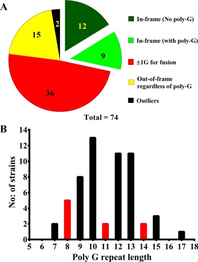
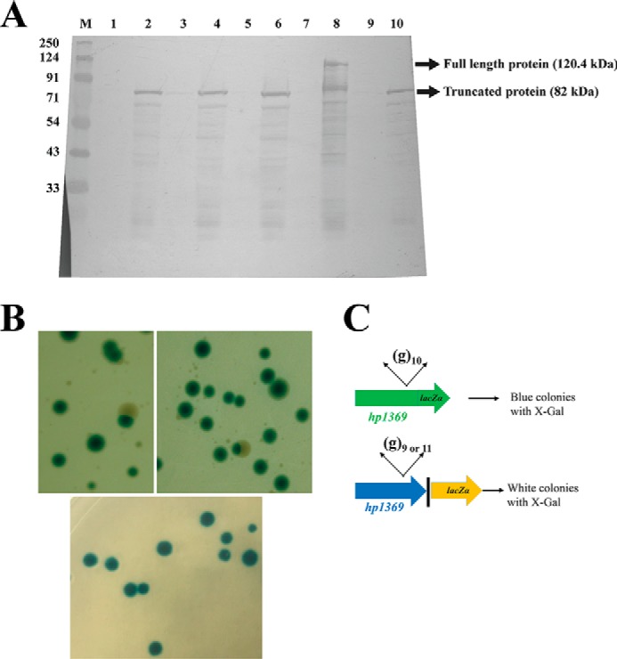
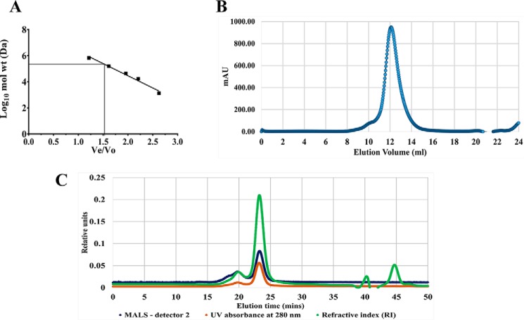
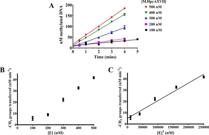
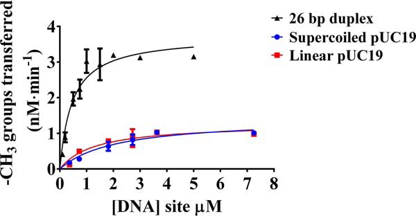
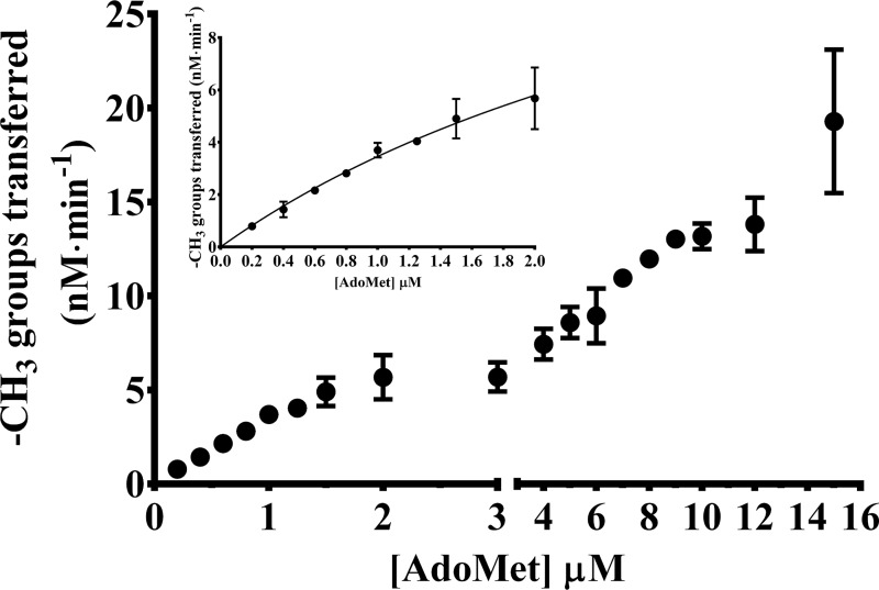
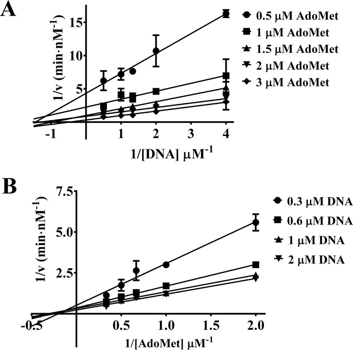
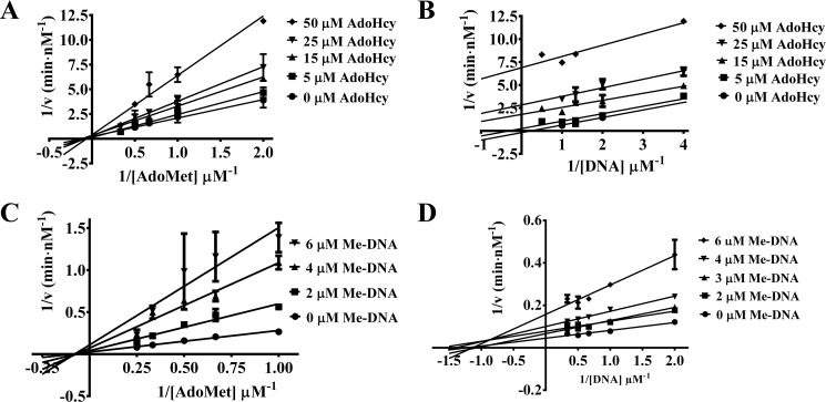
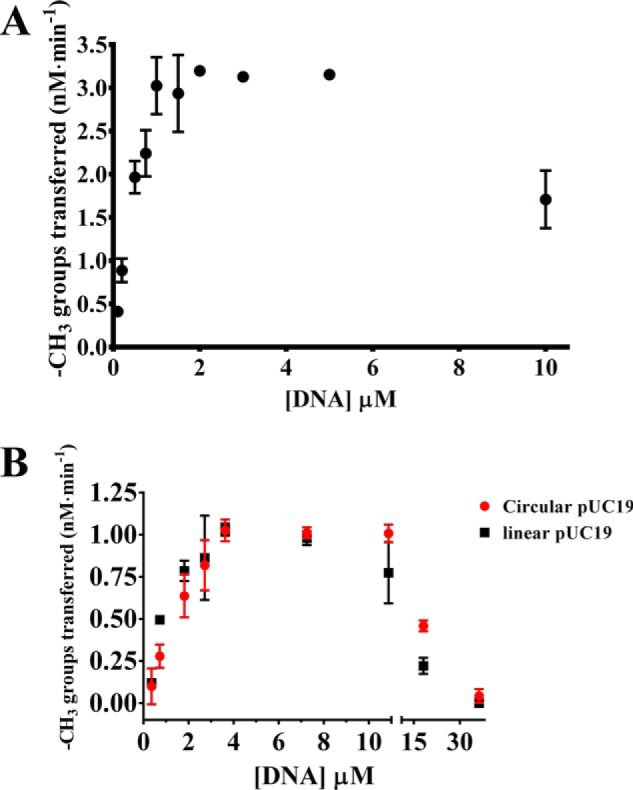
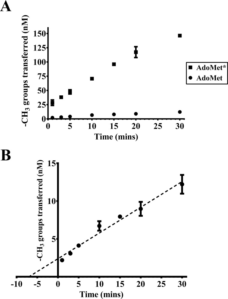
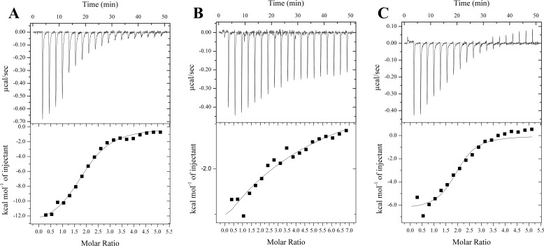
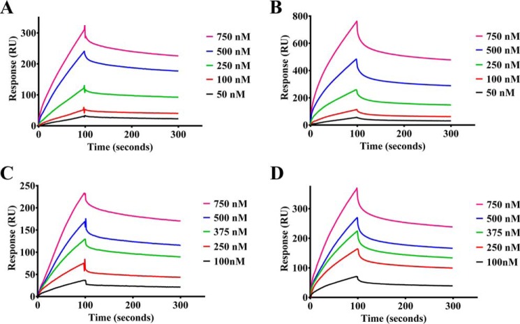
The bacterium Helicobacter pylori is one of the most common infectious agents found in the human stomach. H. pylori has an unusually large number of DNA methyltransferases (MTases), prompting speculation that they may be involved in the cancerization of epithelial cells. The mod-4a/4b locus, consisting of the hp1369 and hp1370 ORFs, encodes for a truncated and inactive MTase in H. pylori strain 26695. However, slipped-strand synthesis within the phase-variable polyguanine tract in hp1369 results in expression of an active HP1369-1370 fusion N6-adenine methyltransferase, designated M.HpyAXVII. Sequence analysis of the mod-4a/4b locus across 74 H. pylori strain genomes has provided insights into the regulation of M.HpyAXVII expression. To better understand the role of M.HpyAXVII in the H. pylori biology, here we cloned and overexpressed the hp1369-70 fusion construct in Escherichia coli BL21(DE3) cells. Results from size-exclusion chromatography and multi-angle light scattering (MALS) analyses suggested that M.HpyAXVII exists as a dimer in solution. Kinetic studies, including product and substrate inhibition analyses, initial velocity dependence between substrates, and isotope partitioning, suggested that M.HpyAXVII catalyzes DNA methylation in an ordered Bi Bi mechanism in which the AdoMet binding precedes DNA binding and AdoMet's methyl group is then transferred to an adenine within the DNA recognition sequence. Altering the highly conserved catalytic motif (DPP(Y/F)) as well as the AdoMet-binding motif (FXGXG) by site-directed mutagenesis abolished the catalytic activity of M.HpyAXVII. These results provide insights into the enzyme kinetic mechanism of M.HpyAXVII. We propose that AdoMet binding conformationally "primes" the enzyme for DNA binding.

Keywords: DNA binding; DNA methyltransferase; M.HpyAXVII; S-adenosylmethionine (AdoMet); allosteric regulation; bioinformatics; enzyme kinetics; enzyme mechanism; ordered Bi Bi mechanism; phase variation.


Conflict of interest statement

The authors declare that they have no conflicts of interest with the contents of this article

Figures

Figure 1.
Figure 1.
The poly-G tract length is variable across H. pylori strains. A, pie chart depicting the distribution of hpyaxvii gene status across 74 fully genome-sequenced H. pylori strains. B, histogram depicting the frequency of poly-G tract lengths found across 74 fully sequenced H. pylori strain genomes. The red columns distinguish the poly-G tract lengths (8, 11, and 14 nucleotides) at which M.HpyAXVII is expressed as a full-length, possibly functional, MTase.
Figure 2.
Figure 2.
hp1369-hp1370 is a phase-variable locus. A, Western blotting of pGEX4T2-hp1369-hp1370 induction profile using anti-GST antibodies. Lane M, prestained protein ladder (sizes of proteins in the ladder, in kDa, are marked on the left). Lanes 1, 3, 5, 7, and 9, uninduced samples. Lanes 2, 4, 6, 8, and 10, IPTG-induced samples. B (top), E. coli DH5α cells transformed with hp1369-lacZ construct and grown on lysogeny broth agar medium supplemented with X-Gal. The sectored colonies can be seen along with the predominant blue colonies. Bottom, the all-blue control colonies obtained from transforming a nonphase-variable lacZ construct. C, schematic representation of the hp1369-lacZ fusion construct. The change in repeats within the poly-G tract causes random switching ON/OFF of the lacZ gene.
Figure 3.
Figure 3.
M.HpyAXVII molecular mass determination. A, standard curve depicting the relationship between Ve/Vo and log10 molecular mass of five standard proteins (Bio-Rad gel filtration standard). Ve = peak elution volume of individual components. Vo = void volume of the column calculated by injecting blue dextran (2 MDa). Ve/Vo of M.HpyAXVII is interpolated into the standard curve to obtain a corresponding log10 molecular mass value. B, elution profile of 750 μg of M.HpyAXVII injected into a Sephadex S200 column as measured by absorbance at 280 nm. C, elution profile of 500 μg of M.HyAXVII injected into a Sephadex S200 gel filtration column (GE Healthcare) as measured by MALS (blue), absorbance at 280 nm (brown), and refractive index (green) at a flow rate of 0.5 ml/min. A and B are from independent experiments performed on an FPLC system that was not integrated with a MALS detector array.
Figure 4.
Figure 4.
Rate of methylation versus enzyme concentration. In vitro methylation of 26-bp duplex DNA containing a single recognition site was performed in standard reaction buffer at 37 °C. The reactions were followed as time courses of product formation at different concentrations of M.HpyAXVII. 4 μm DNA and 6 μm [3H]AdoMet were used in each case. 10-μl aliquots were taken at fixed time points, and the samples were processed as described under “Experimental procedures.” A, the progress of the reaction in first 5 min after the addition of M.HpyAXVII is shown. B, initial velocity versus enzyme concentrations. C, initial velocity versus square of enzyme concentrations. Error bars, S.D.
Figure 5.
Figure 5.
Determination of kinetic parameters (DNA substrates): Determination of KmDNA. M.HpyAXVII-dependent methylation was assayed with various DNA substrates. The reactions were performed in standard reaction buffer at 37 °C for 30 min. Enzyme concentration in all cases was 200 nm. AdoMet concentration in all cases was 2 μm. Error bars, S.D.
Figure 6.
Figure 6.
Determination of kinetic parameters (AdoMet): Determination of KmAdoMet. M.HpyAXVII-dependent methylation was assayed at increasing concentrations of AdoMet ranging from 0.2 to 15 μm. The reactions were performed in standard reaction buffer at 37 °C for 30 min. Enzyme concentration in all cases was 200 nm. DNA concentration in all cases was 4 μm. Inset, the Michaelis–Menten fit for the initial region of the curve (up to 2 μm AdoMet) is depicted. Error bars, S.D.
Figure 7.
Figure 7.
Double-reciprocal or Lineweaver–Burk plots of initial velocity versus substrate concentration. A, Lineweaver–Burk plots of rates of methylation versus DNA concentration. B, Lineweaver–Burk plots of rates of methylation versus AdoMet concentration. Error bars, S.D.
Figure 8.
Figure 8.
Product inhibition analysis. Methylation reactions were performed at the indicated concentrations of reaction products. Lineweaver–Burk plots are shown of rates of methylation versus AdoMet concentrations at various constant concentrations of AdoHcy (A), rates of methylation versus DNA concentrations at various constant concentrations of AdoHcy (B), rates of methylation versus AdoMet concentrations at various constant concentrations of Me-DNA (C), and rates of methylation versus DNA concentrations at various constant concentrations of Me-DNA (D). Error bars, S.D.
Figure 9.
Figure 9.
Substrate inhibition of M.HpyAXVII. Methylation was monitored against DNA concentration. AdoMet was kept constant at 2 μm in each reaction. 200 nm enzyme was used in each case. Reactions were performed in standard reaction buffer at 37 °C, and samples were processed according to the protocol described under “Experimental procedures”. A, rate of methylation versus single-site duplex DNA concentration. B, rate of methylation versus pUC19 plasmid DNA (supercoiled and linear) concentration normalized for number of sites. Error bars, S.D.
Figure 10.
Figure 10.
Isotope partitioning. A, methylation reactions were performed in standard reaction buffer at 37 °C. 200 nm enzyme was preincubated with 3 μm [3H]AdoMet at 37 °C for 3 min. The presaturated enzyme–AdoMet mix was split into two. ■, profile of product formation when the reaction was initiated by diluting the presaturated mix into DNA (2 μm) and [3H]AdoMet. ●, profile product formation when the reaction was initiated by diluting the presaturated mix into DNA (2 μm) and unlabeled AdoMet. B, a profile of the initiation of reaction by the addition of unlabeled AdoMet and DNA is depicted separately to highlight the positive y intercept value, which translates to the burst amplitude. Error bars, S.D.
Figure 11.
Figure 11.
Isothermal titration calorimetry to measure KdAdoMet. The calorimetric titration of AdoMet with M.HpyAXVII WT (A), AdoMet with M.HpyAXVII F658C (B), and AdoMet with M.HpyAXVII Y407V (C) is depicted. The top panels in A–C represent the raw heat change measured during 15–20 injections of 500 μm AdoMet (AdoMet = 1.5 mm in B) into 280 μl of 20 μm protein. Bottom panels, integrated heat values derived from raw data after factoring in dilution enthalpy of protein. Curves were fitted using a “one-set binding model,” as elaborated under “Experimental procedures.”
Figure 12.
Figure 12.
Surface plasmon resonance analyses to measure DNA binding. The indicated protein concentrations were injected onto the streptavidin chip containing bound biotinylated single-site duplex DNA for 100 s followed by buffer wash for 300 s. The binding isotherms are depicted in the panels. A, M.HpyAXVII WT binding in the absence of sinefungin; B, M.HpyAXVII WT binding in the presence of 40 μm sinefungin; C, M.HpyAXVII F658C binding in the absence of sinefungin; D, M.HpyAXVII F658C binding in the presence of 40 μm sinefungin. KdDNA was calculated as Koff/Kon.

Similar articles

Cited by

References

    1. Ahmad I., and Rao D. N. (1996) Chemistry and biology of DNA methyltransferases. Crit. Rev. Biochem. Mol. Biol. 31, 361–380 10.1074/jbc.RA117.001068 - DOI - PubMed
    1. Bheemanaik S., Reddy Y. V. R., and Rao D. N. (2006) Structure, function and mechanism of exocyclic DNA methyltransferases. Biochem. J. 399, 177–190 10.1074/jbc.RA117.001068 - DOI - PMC - PubMed
    1. Adhikari S., and Curtis P. D. (2016) DNA methyltransferases and epigenetic regulation in bacteria. FEMS Microbiol. Rev. 40, 575–591 10.1074/jbc.RA117.001068 - DOI - PubMed
    1. Jeltsch A. (2002) Beyond Watson and Crick: DNA methylation and molecular enzymology of DNA methyltransferases. ChemBioChem 3, 274–293 10.1074/jbc.RA117.001068 - DOI - PubMed
    1. Kumar R., Sabareesh V., Mukhopadhyay A. K., and Rao D. N. (2012) Mutations in hpyAVIBM, C5 cytosine DNA methyltransferase from Helicobacter pylori result in relaxed specificity. FEBS J. 279, 1080–1092 10.1074/jbc.RA117.001068 - DOI - PubMed

Publication types

MeSH terms

Substances

LinkOut - more resources