. 2019 Jan 3;24(1):153-165.e7.
doi: 10.1016/j.stem.2018.10.016. Epub 2018 Nov 21.

IKZF2 Drives Leukemia Stem Cell Self-Renewal and Inhibits Myeloid Differentiation

Affiliations

IKZF2 Drives Leukemia Stem Cell Self-Renewal and Inhibits Myeloid Differentiation

Sun-Mi Park et al. Cell Stem Cell. .

Abstract

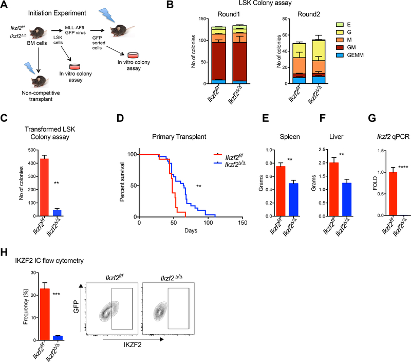
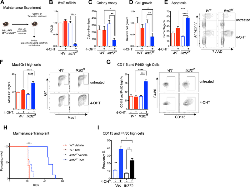
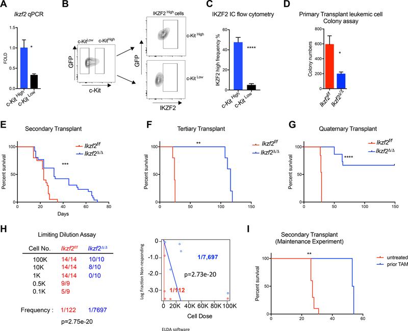
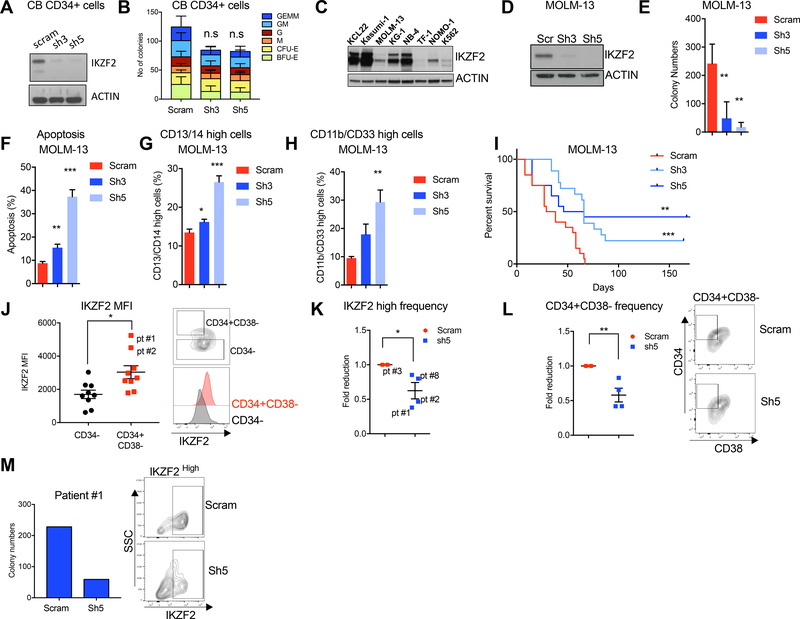
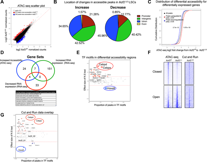
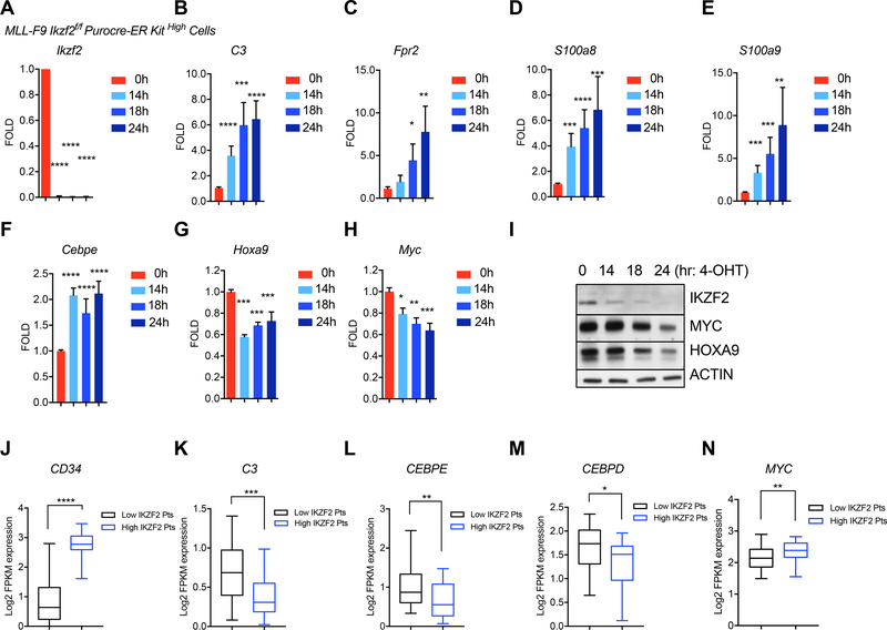
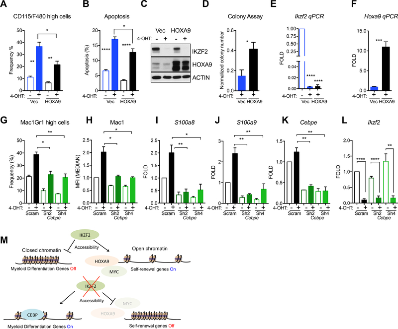
Leukemias exhibit a dysregulated developmental program mediated through both genetic and epigenetic mechanisms. Although IKZF2 is expressed in hematopoietic stem cells (HSCs), we found that it is dispensable for mouse and human HSC function. In contrast to its role as a tumor suppressor in hypodiploid B-acute lymphoblastic leukemia, we found that IKZF2 is required for myeloid leukemia. IKZF2 is highly expressed in leukemic stem cells (LSCs), and its deficiency results in defective LSC function. IKZF2 depletion in acute myeloid leukemia (AML) cells reduced colony formation, increased differentiation and apoptosis, and delayed leukemogenesis. Gene expression, chromatin accessibility, and direct IKZF2 binding in MLL-AF9 LSCs demonstrate that IKZF2 regulates a HOXA9 self-renewal gene expression program and inhibits a C/EBP-driven differentiation program. Ectopic HOXA9 expression and CEBPE depletion rescued the effects of IKZF2 depletion. Thus, our study shows that IKZF2 regulates the AML LSC program and provides a rationale to therapeutically target IKZF2 in myeloid leukemia.

Keywords: C/EBP; HOXA9; IKZF2; leukemic stem cells.


Conflict of interest statement

Declaration of Interests

S.A.A. consults for Epizyme Inc, Imago Biosciences, Cyteir Therapeutics, C4 Therapeutics, Syros Pharmaceuticals and Accent Therapeutics. S.A.A. receives research support from Janssen, Novartis, and AstraZeneca.

Figures

Figure 1.
Figure 1.. IKZF2 is required for leukemogenesis but dispensable for normal hematopoiesis.
(A) Experimental scheme for investigating normal and transformed LSK cells. Leukemia initiation experiment was performed using the murine MLL-AF9 model. (B) Ikzf2 is dispensable for colony-forming ability of normal stem cells. Colony Assay was performed with LSK cells from Ikzf2f/f n=4 and Ikzf2Δ/Δ n=4 mice (C) Ikzf2 deletion in MLL-AF9 transformed LSK cells reduced colony formation. MLL-AF9 transformed LSK cells from Ikzf2f/f n=3 and Ikzf2Δ/Δ n=3 mice were used for colony assay. Mean +/− S.E.M of three independent experiments, Student’s t test **, p<0.01. (D) Deletion of Ikzf2 delays leukemia progression in MLL-AF9 model. Survival analysis is from the result of three combined transplants with genotypes Ikzf2f/f n=13 and Ikzf2Δ/Δ n=28 mice. **, p<0.01 logrank test. (E,F) Ikzf2Δ/Δ deleted leukemic mice have reduced disease burden. (E) Spleen and (F) liver weights of moribund Ikzf2f/f and Ikzf2Δ/Δ mice with proper deletion. Result is from Ikzf2f/f n=12 and Ikzf2Δ/Δ n=8 mice. **P < 0.01, unpaired Student’s t test. (G) QPCR of Ikzf2 in bone marrow leukemic cells from Ikzf2f/f n=12 and Ikzf2Δ/Δ n=8 mice. ****p<0.0001. unpaired Student’s t test. (H) Bone marrow leukemic cells from Ikzf2Δ/Δ mice have reduced IKZF2 expression. Frequency of IKZF2High cells in primary leukemic mice was examined by intracellular flow cytometry for IKZF2. Result from Ikzf2f/f n=12 and Ikzf2Δ/Δ n=10 mice is shown. ***, p<0.001. unpaired Student’s t test.
Figure 2.
Figure 2.. IKZF2 is required for leukemia maintenance in vitro and in vivo
(A) Experimental scheme for investigating IKZF2 role in leukemia maintenance in vitro and in vivo. (B) QPCR analysis showing Ikzf2 is acutely deleted by 4-OHT treatment in MLL-AF9 Ikzf2f/f cre-ER cells. (C) Ikzf2 deletion reduces colony forming ability in MLL-AF9 Ikzf2f/f cre-ER cells. (D) Ikzf2 deletion reduces cell growth. (E) Deletion of Ikzf2 increases apoptosis. Left, the percentage of apoptotic cells was determined at 24hr after 4-OHT treatment. Cells were stained for Annexin V and 7-AAD, and quantified by flow cytometry. Right, representative cytometric flow plot showing Annexin V and 7-AAD staining. (F-G) Myeloid differentiation is increased in Ikzf2 deleted leukemic cells. Frequency of population with high myeloid differentiation markers, (F) Mac-1/Gr-1 and (G) CD115 and F4/80 was measured by flow cytometry. Representative flow plots for these myeloid markers are shown in panels (F, Right and G, Right). Data shown in (B, D-G) were analyzed 24 hrs after MLL-AF9 WT or Ikzf2f/f cre-ER cells were treated with10 nM 4-OHT. Results (shown in (B - G) were combined from at least three independent experiments, using one MLL-AF9 WT cre-ER line and three MLL-AF9 Ikzf2f/f cre-ER lines. Mean +/− SEM Student’s t test *p<0.05, ** p<0.01, *** p<0.001, **** p<0.0001 (H) Ikzf2 deletion after leukemic cells are transplanted in mice, delays progression. Survival analysis of mice transplanted with MLL-AF9 WT or Ikzf2f/f cre-ER leukemic cells, treated with corn oil (vehicle) or 160mg/kg Tamoxifen. Data shown are combined from two independent transplants. Indicated groups WT,WT+TAM, Ikzf2f/f and Ikzf2f/f TAM have n=5, n=5, n=9, n=9 mice respectively. ****, p<0.0001 log-rank test. (I) Overexpression of Flag-CBP-IKZF2 partially rescued the differentiation phenotype of MLL-AF9 Ikzf2f/f leukemic cells deleted of endogenous Ikzf2. FACS analysis of F480 and CD115 expression in MLL-AF9 Ikzf2f/f leukemic cells treated with or without 20 nM 4-OHT for 24 hrs. n = 3 independent experiments; error bars, S.E.M *P < 0.05, **P < 0.01, two-tailed t test.
Figure 3.
Figure 3.. IKZF2 is highly expressed in LSCs and is required for maintaining LSC activity.
(A) Ikzf2 mRNA is highly expressed in LSCs, cells enriched in c-KitHigh cells compared to c-KitLow cells. Ikzf2 qPCR was performed in sorted c-KitHigh and c-KitLow cells from bone marrow of MLL-AF9 Ikzf2f/f leukemic mice n=4, Student’s t test * p<0.05. (B) Representative flow plot showing the gating for c-KitHigh (top 15%) and c-KitLow (bottom 40%) cells, and also the gating for IKZF2 High cells shown in (C). (C) c-KitHigh cells have higher expression of IKZF2 compared to c-KitLow cells. Frequency of IKZF2High population was measured in c-KitHigh and c-KitLow cells of MLL-AF9 leukemic mice. Mean and S.E.M of experiments from leukemic bone marrow cells isolated from Ikzf2f/f n=15 mice ***, Student’s t test p<0.001. (D) Colony Assay was performed with bone marrow leukemic cells from primary transplanted Ikzf2f/f n=3 and Ikzf2Δ/Δ n=3 mice. Student’s t test. *p<0.05. (E) Delay in survival between Ikzf2f/f and Ikzf2Δ/Δ mice is increased in secondary transplant of cells from (Fig 1D), compared to primary transplant. Result is from two combined transplants using four Ikzf2f/f and five Ikzf2Δ/Δ donors with recipients Ikzf2f/f n=19 and Ikzf2Δ/Δ n=26 mice. ***p<0.001. log-rank test. (F) Survival Curve of tertiary transplant of leukemic bone marrow cells from (E) shows worse LSC function when Ikzf2 is deleted. Transplant is shown with Ikzf2f/f n=5 and Ikzf2Δ/Δ n=5 mice ***p<0.001. log-rank test. (G) Survival Curve of quarternary transplant exhibits exhaustion of LSCs in Ikzf2Δ/Δ mice. Three donors of each genotype from (F) were transplanted into recipients n=13 and n=15 mice for Ikzf2f/f and Ikzf2Δ/Δ, respectively. **p<0.01. log-rank test. (H) Ikzf2 deleted leukemic mice have reduced LSC frequency. Limiting dilution experiment assay performed with bone marrow cells from primary leukemic mice is shown. Left panel, table shows different number of cells and mice used for the transplant.. Right panel, graph showing the frequency of LSCs in Ikzf2f/f and Ikzf2Δ/Δ leukemic mice. ELDA software was used to calculate the frequency of the LSCs (Hu and Smyth, 2009). (I) Survival analysis for secondary transplant of the maintenance experiment is shown. Result is from transplanting Ikzf2f/f nonTAM=10 and Ikzf2 f/f prior TAM n=10 mice, with two and one donors respectively. **p<0.01.
Figure 4.
Figure 4.. IKZF2 is required for human leukemia cell survival
(A) Western blot analysis showing depletion of IKZF2 in human cord blood CD34+ (HSPCs) at day 4 post transduction with lentivirus expressing either Scramble control or two independent IKZF2 shRNAs. (B) Colony assay was performed with HSPCs in (A). 104 cells were plated at day 4 post transduction and different colonies were counted two weeks later. Result is from n=3 independent experiments. (C) Western blot of IKZF2 was performed in whole cell lysates from eight cell lines, using ACTIN as loading control. (D) Western blot analysis showing IKZF2 depletion in MOLM-13 cells after four days post-transduction with lentivirus expressing IKZF2 shRNAs. ACTIN was used as loading control. (E) Depletion of IKZF2 in MOLM-13 cells reduced colony formation. Colonies were measured a week after plating shRNA transduced-MOLM-13 cells. (F) IKZF2 knockdown increases apoptosis in MOLM-13 cells. Apoptosis was measured by flow cytometry using Annexin V-PE and 7-AAD staining at day 7 post-transduction. (G-H) IKZF2 knockdown leads to increased differentiation. Flow cytometry measuring myeloid markers (G) CD13/CD14 and (H) CD11b/CD33 in MOLM-13 cells at day 7 post-transduction. Results in (B-H) are from more than three independent experiments using triplicates are shown. Mean +/− S.E.M Student’s t test p value. *p<0.05, **p<0.01 and ***p<0.001. (I) IKZF2 knockdown prolongs leukemia-free survival in mice. MOLM-13 cells transduced with scramble or IKZF2 shRNA virus were counted at day 4 post-transduction and transplanted into sub-lethally irradiated NSG mice. Results from four combined transplants with total scram n=20, Ikzf2 shRNA-3 n=18, and Ikzf2 shRNA-5 n=20 mice are shown. log-rank test. **p<0.01 and ***p<0.001. (J) IKZF2 is highly expressed in CD34+CD38 population in AML patients. Left, IKZF2 intracellular flow cytometry data showing IKZF2 expression in CD34 and CD34+CD38 populations in nine AML patients. Right, representative flow plot showing gating of CD34 and CD34+CD38 populations. (K) IKZF2 Intracellular flow cytometry data in AML patients cells transduced with lentivirus expressing Scram control or IKZF2 shRNA at day 4 post transduction. Results are from experiments using cells from four AML patients. Mean +/− S.E.M Student’s t test p value. *p<0.05, (L) CD34+CD38 population is reduced when IKZF2 is knocked down in AML patient cells. Leukemic stem cell population was analyzed at day 4 post transduction with Scram or IKZF2 shRNA. Results are from four AML patients. Mean +/− S.E.M Student’s t test p value. *p<0.05. (M) IKZF2 depletion reduces colony formation in AML patient cells. Cells from Patient #1 were transduced with lentivirus expressing Scramble or IKZF2 shRNA and sorted for GFP at day 4 post transduction. Left, graph showing number of colonies scored 2 weeks after plating. Right, flow plot of intracellular IKZF2 staining in patient cells.
Figure 5.
Figure 5.. IKZF2 loss leads to increased accessibility of differentiation genes and decreased accessibility of self-renewal genes in LSCs.
(A) Scatterplot to show differentially accessible peaks from ATAC-seq analysis on Ikzf2f/f n=2 and Ikzf2Δ/Δ n=2 LSCs (c-Kit High) sorted from bone marrow of primary leukemic mice, under the Benjamini-Hochberg adjusted q-value threshold of 0.2. Red dots represent more accessible and blue represents less accessible peaks. (B) Location of increased accessible (left panel) and decreased accessible (right) ATAC-seq peaks in Ikzf2Δ/Δ LSCs. (C) Chromatin accessibility has positive correlation with gene expression. Cumulative distribution function (CDF) graph shows enrichment of accessibility in upregulated genes (shown in red) from the RNA sequencing data of Ikzf2f/f and Ikzf2Δ/ Δ LSCs. Loss of accessibility is found in downregulated genes (shown in blue). (D) Ikzf2 deletion results in increased accessibility and expression of myeloid differentiation program. Overlap of gene sets from genes with increased accessibility (ATAC-seq data), increased and decreased RNA expression (RNA-seq data) leading to seven gene sets including myeloid development, C/EBPα network, neutrophil maturation and gene set that is increased when Hoxa9 and Meis1 is upregulated. (E) TF motifs enriched in differentially accessible regions in Ikzf2Δ/Δ LSCs. C/EBPε and C/EBPδ motifs are the most enriched motifs in the increased accessible regions whereas HOXA9 motif is the top TF motif for loss of enrichment in decreased accessible regions in Ikzf2 deleted LSCs. Further information is found in STAR methods. (F) Tornado plot of differentially accessible ATAC-seq peaks (left) selected by nominal p ≤ 0.05. 1191 opened and 1523 closed peaks are shown. The genomic domain is defined as the summit ± 3 kb. To show the native binding of IKZF2, the signal of CUT&RUN sequencing at the same loci (right) is shown in parallel. (G) Data analyzed in (E) was further queried by additionally overlapping with genomic regions bound by IKZF2. TF motifs enriched in differentially accessible regions by the nominal p-value cutoff of 0.05 and showing averaged signal of IKZF2 CUT&RUN above 0.5 TPM over the region of summit ± 250 bp, encompassing 9259 peaks. TFs with the K-S test effect size above 0.05 and the odds ratio above 1.3 are highlighted with red (opened) and blue (closed) colors, respectively. The two IKZF2 motifs from TRANSFAC are also highlighted.
Figure 6.
Figure 6.. IKZF2 represses the expression of differentiation transcription factor, C/EBP and maintains expression of self-renewal transcription factors, HOXA9 and C-MYC.
(A) QPCR analysis of Ikzf2 mRNA in sorted MLL-AF9 Ikzf2f/f cre-ER c-Kit High cells treated with 4-OHT for different time points. (B-E) Acute deletion of Ikzf2 in LSCs increases genes data showing that acute deletion of Ikzf2 reduces Hoxa9 and c-Myc mRNA levels. (A-H) Results shown are from four different experiments. Student’s t test *p<0.05, ** p<0.01, *** p<0.001, **** p<0.0001. (I) Western blot analysis showing reduction of IKZF2, HOXA9 and C-MYC after Ikzf2 is acutely deleted in MLL-AF9 Ikzf2f/f cre-ER High cells. (J-N) Box and whiskers plot showing correlation of the indicated genes in patients with high (n=24) versus low IKZF2 (n=45) expression in AML patients from TCGA dataset. (J) CD34 (K) C3 (L) CEBPE (M) CEBPD (N) MYC .*P < 0.05, **P < 0.01, ***P < 0.001, two-tailed t test.
Figure 7.
Figure 7.. CEBPE and HOXA9 can partially rescue phenotypes of Ikzf2 loss
(A) Differentiation measured by CD115/F480 and (B) apoptosis were partially rescued when Ikzf2 was deleted in HOXA9 overexpressing MLL-AF9 Ikzf2f/f cre-ER cells. n=3 independent experiments. Student’s t test *p<0.05, ** p<0.01, **** p<0.0001. (C) Western blot analysis demonstrating efficient deletion of IKZF2 and overexpression of HOXA9. (D) Colony assay showing rescue of Ikzf2 deletion by HOXA9 overexpression in 4-OHT treated MLL-AF9 Ikzf2f/f cre-ER cells. (E) QPCR showing complete deletion of Ikzf2 in MLL-AF9 Ikzf2f/f cre-ER vector control and HOXA9 overexpressing cells after 4-OHT treatment. (F) QPCR showing expression of Hoxa9 in the vector control and HOXA9 expressing cells after 4-OHT treatment in the MLL-AF9 Ikzf2f/f cre-ER cells. (D-F) All data represent the mean +S.E.M of at least three independent replicates. * p < 0.05, ***p < 0.001, ****p<0.0001 Student’s t test. (G-J) CEBPE depleted cells lacks differentiation induction and IKZF2 targets caused by IKZF2 loss. (G) Differentiation state was measured by flow cytometry using Mac1 and Gr1 as markers in the MLL-AF9 Ikzf2f/f cre-ER cells transduced with lentivirus expressing scramble or Cebpe shRNA and were treated with 4-OHT or left alone. (H) Median Fluorescent Intensity of Mac1 was measured in same experiment in (G). (I) S100a8 (J) S100a9, targets of IKZF2 were measured using qPCR in experiments in (G). (K) Cebpe and (L) Ikzf2 mRNAs were measured to check for depletion in experiment mentioned in (G). (G-L) All data represent the mean +S.E.M of at least three independent replicates. * p < 0.05, **p < 0.01, ****p<0.0001 Student’s t test. (M) Model showing the role of IKZF2 in regulating chromatin accessibility of differentiation and self-renewal program in LSCs. IKZF2 loss leads to increased C/EBPε and chromatin accessibility of regions in differentiation genes containing C/EBP motifs, thereby turning on myeloid genes. HOXA9 expression and chromatin accessibility of HOXA9 motifs are reduced, turning off the self-renewal genes.

Comment in

Similar articles

Cited by

References

    1. Asanuma S, Yamagishi M, Kawanami K, Nakano K, Sato-Otsubo A, Muto S, Sanada M, Yamochi T, Kobayashi S, Utsunomiya A, et al. (2013). Adult T-cell leukemia cells are characterized by abnormalities of Helios expression that promote T cell growth. Cancer science 104, 1097–1106. - PMC - PubMed
    1. Buenrostro JD, Giresi PG, Zaba LC, Chang HY, and Greenleaf WJ (2013). Transposition of native chromatin for fast and sensitive epigenomic profiling of open chromatin, DNA-binding proteins and nucleosome position. Nature methods 10, 1213–1218. - PMC - PubMed
    1. Collins CT, and Hess JL (2016). Role of HOXA9 in leukemia: dysregulation, cofactors and essential targets. Oncogene 35, 1090–1098. - PMC - PubMed
    1. Dobin A, Davis CA, Schlesinger F, Drenkow J, Zaleski C, Jha S, Batut P, Chaisson M, and Gingeras TR (2013). STAR: ultrafast universal RNA-seq aligner. Bioinformatics 29, 15–21. - PMC - PubMed
    1. Ee LS, McCannell KN, Tang Y, Fernandes N, Hardy WR, Green MR, Chu F, and Fazzio TG (2017). An Embryonic Stem Cell-Specific NuRD Complex Functions through Interaction with WDR5. Stem cell reports 8, 1488–1496. - PMC - PubMed

Publication types

MeSH terms