

Dynamic Notch signalling regulates neural stem cell state progression in the Drosophila optic lobe
- PMID: 30466475
- PMCID: PMC6251220
- DOI: 10.1186/s13064-018-0123-8
Dynamic Notch signalling regulates neural stem cell state progression in the Drosophila optic lobe
Abstract
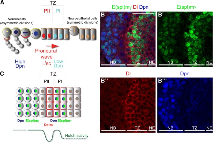
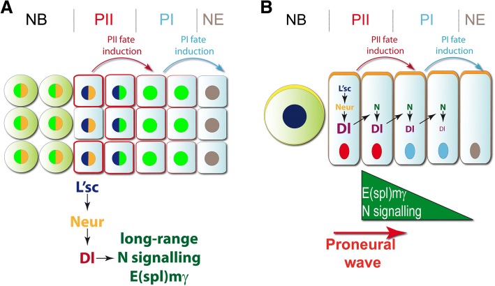
Background: Neural stem cells generate all of the neurons and glial cells in the central nervous system, both during development and in the adult to maintain homeostasis. In the Drosophila optic lobe, neuroepithelial cells progress through two transient progenitor states, PI and PII, before transforming into neuroblasts. Here we analyse the role of Notch signalling in the transition from neuroepithelial cells to neuroblasts.
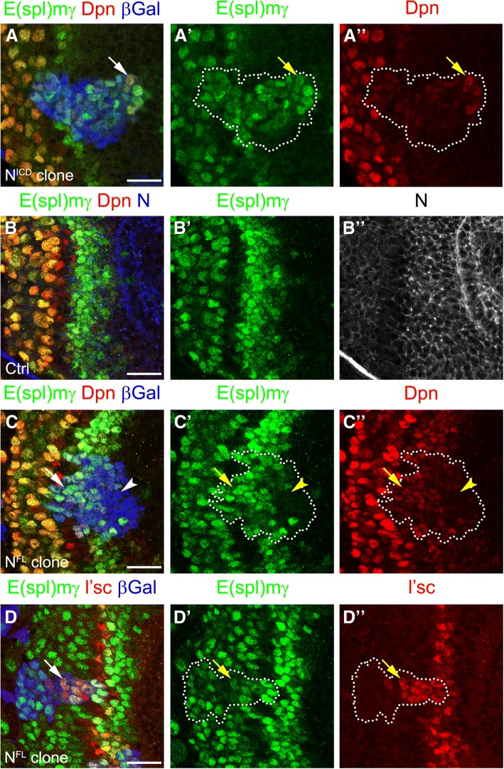
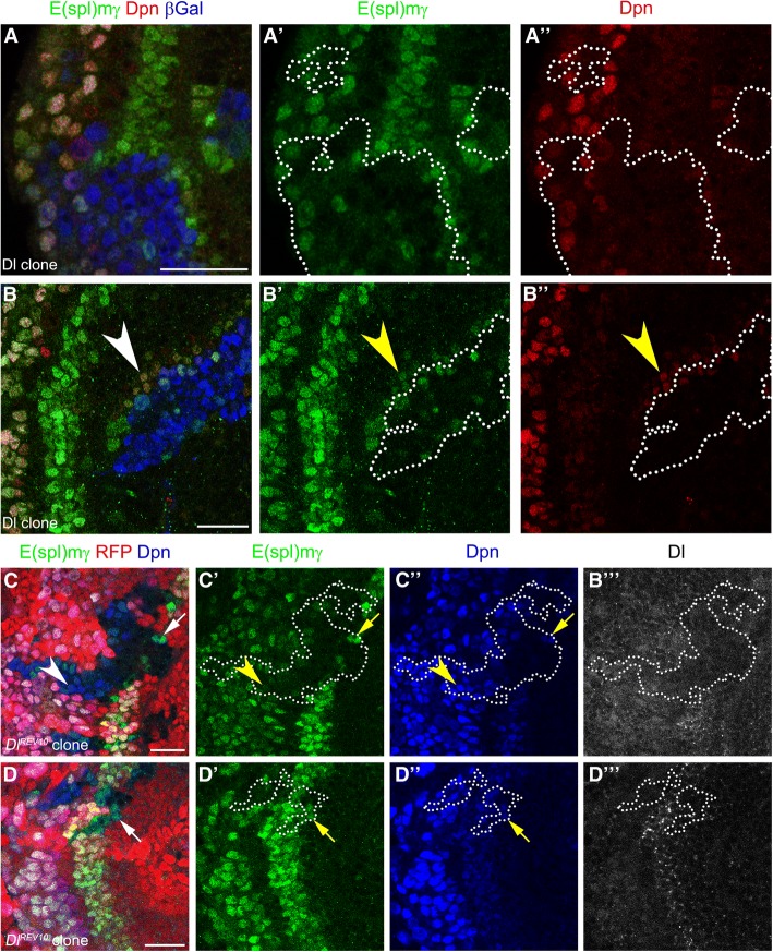
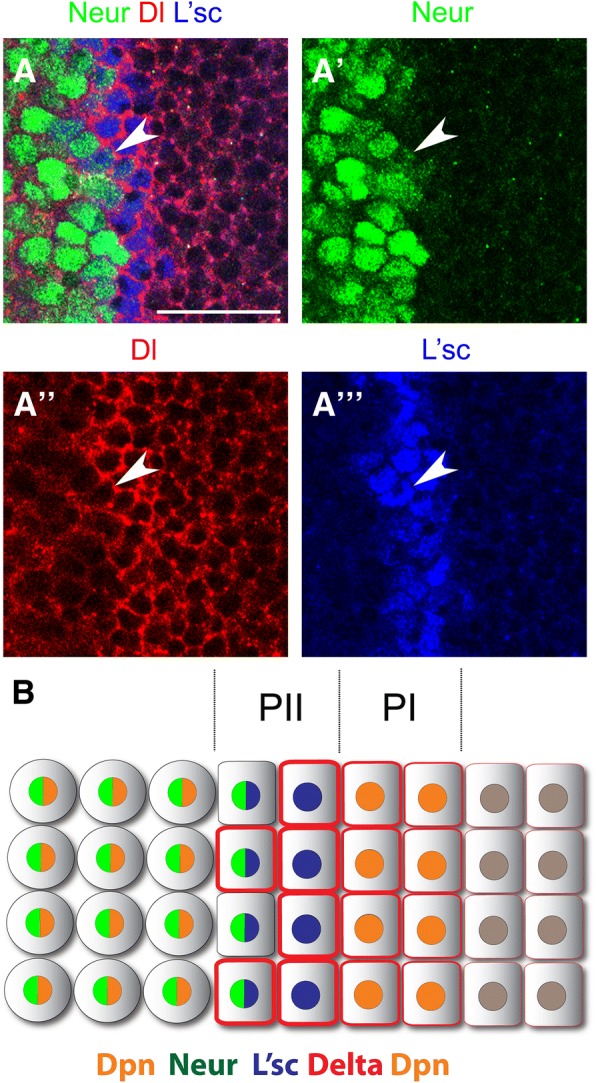
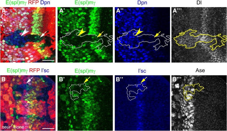
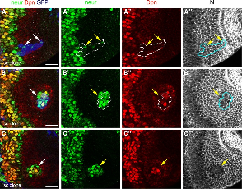
Results: We observed dynamic regulation of Notch signalling: strong activity in PI progenitors, low signalling in PII progenitors, and increased activity after neuroblast transformation. Ectopic expression of the Notch ligand Delta induced the formation of ectopic PI progenitors. Interestingly, we show that the E3 ubiquitin ligase, Neuralized, regulates Delta levels and Notch signalling activity at the transition zone. We demonstrate that the proneural transcription factor, Lethal of scute, is essential to induce expression of Neuralized and promote the transition from the PI progenitor to the PII progenitor state.
Conclusions: Our results show dynamic regulation of Notch signalling activity in the transition from neuroepithelial cells to neuroblasts. We propose a model in which Lethal of scute activates Notch signalling in a non-cell autonomous manner by regulating the expression of Neuralized, thereby promoting the progression between different neural stem cell states.
Keywords: Neural stem cell; Neuralized; Notch; Optic lobe.
Conflict of interest statement
Ethics approval and consent to participate
Not applicable.
Consent for publication
Not applicable.
Competing interests
The authors declare that they have no competing interests.
Publisher’s Note
Springer Nature remains neutral with regard to jurisdictional claims in published maps and institutional affiliations.
Figures
Similar articles
-
Notch signaling regulates neuroepithelial stem cell maintenance and neuroblast formation in Drosophila optic lobe development.Dev Biol. 2011 Feb 15;350(2):414-28. doi: 10.1016/j.ydbio.2010.12.002. Epub 2010 Dec 10. Dev Biol. 2011. PMID: 21146517
-
A Serrate-Notch-Canoe complex mediates essential interactions between glia and neuroepithelial cells during Drosophila optic lobe development.J Cell Sci. 2013 Nov 1;126(Pt 21):4873-84. doi: 10.1242/jcs.125617. Epub 2013 Aug 22. J Cell Sci. 2013. PMID: 23970418
-
Coordinated sequential action of EGFR and Notch signaling pathways regulates proneural wave progression in the Drosophila optic lobe.Development. 2010 Oct;137(19):3193-203. doi: 10.1242/dev.048058. Epub 2010 Aug 19. Development. 2010. PMID: 20724446
-
A challenge of numbers and diversity: neurogenesis in the Drosophila optic lobe.J Neurogenet. 2014 Sep-Dec;28(3-4):233-49. doi: 10.3109/01677063.2014.922558. Epub 2014 Jul 8. J Neurogenet. 2014. PMID: 24912777 Review.
-
Regulating the balance between symmetric and asymmetric stem cell division in the developing brain.Fly (Austin). 2011 Jul-Sep;5(3):237-41. doi: 10.4161/fly.5.3.15640. Epub 2011 Jul 1. Fly (Austin). 2011. PMID: 21502820 Review.
Cited by
-
Intracellular trafficking of Notch orchestrates temporal dynamics of Notch activity in the fly brain.Nat Commun. 2021 Apr 7;12(1):2083. doi: 10.1038/s41467-021-22442-3. Nat Commun. 2021. PMID: 33828096 Free PMC article.
-
Novel Strategies for the Generation of Neuronal Diversity: Lessons From the Fly Visual System.Front Mol Neurosci. 2019 May 31;12:140. doi: 10.3389/fnmol.2019.00140. eCollection 2019. Front Mol Neurosci. 2019. PMID: 31213980 Free PMC article. Review.
-
Different modes of Notch activation and strength regulation in the spermathecal secretory lineage.Development. 2020 Feb 7;147(3):dev184390. doi: 10.1242/dev.184390. Development. 2020. PMID: 31988187 Free PMC article.
-
Mathematical modeling of Notch dynamics in Drosophila neural development.Fly (Austin). 2022 Dec;16(1):24-36. doi: 10.1080/19336934.2021.1953363. Fly (Austin). 2022. PMID: 34609265 Free PMC article. Review.
-
Separable Roles for Neur and Ubiquitin in Delta Signalling in the Drosophila CNS Lineages.Cells. 2023 Dec 14;12(24):2833. doi: 10.3390/cells12242833. Cells. 2023. PMID: 38132160 Free PMC article.
References
Publication types
MeSH terms
Substances
Grants and funding
LinkOut - more resources
Full Text Sources
Molecular Biology Databases
Research Materials
Miscellaneous
