

Preparation of Sliver and Selenium Nanoparticles and Its Characterization by Dynamic Light Scattering and Scanning Electron Microscopy
- PMID: 30464890
- PMCID: PMC6206752
- DOI: 10.4103/JMAU.JMAU_3_18
Preparation of Sliver and Selenium Nanoparticles and Its Characterization by Dynamic Light Scattering and Scanning Electron Microscopy
Abstract
Aim: In our study, two different methods were used to determine the size and size distribution of the sliver and selenium nanoparticles via dynamic light scattering (DLS) and scanning electron microscopy (SEM).
Background: Nanotechnology dealing with metal and metalloid nanoparticles has been usually applied in nearly each field of science, engineering, and technology including biology and medicine etc due to presence of size and shape dependent unusual physical and chemical properties. In the most recent decade, numerous groups including appreciably developed metal and metalloid nanoparticles based theranostic approaches for the treatment of almost human diseases. Amongst many nanoparticles, recently silver and selenium nanoparticles have been broadly used in the antimicrobial coatings, textiles, paints, keyboards, engineering, food industry, electronics, cosmetics, bio-sensing, wound dressings, and even in biomedical devices.
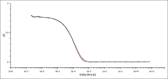
Methods: In our study, silver nanoparticles were prepared by using the chemical reduction method. Selenium nanoparticles (SeNPs) were synthesized by the chemical reduction of sodium selenite by glutathione (reduced form) and stabilized by bovine serum albumin (BSA). Characterization of silver and selenium nanoparticles samples were analyzed by dynamic light scattering (DLS) and Scanning Electron Microscopy (SEM).
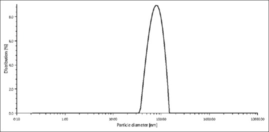
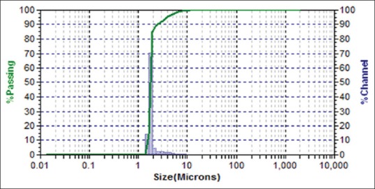
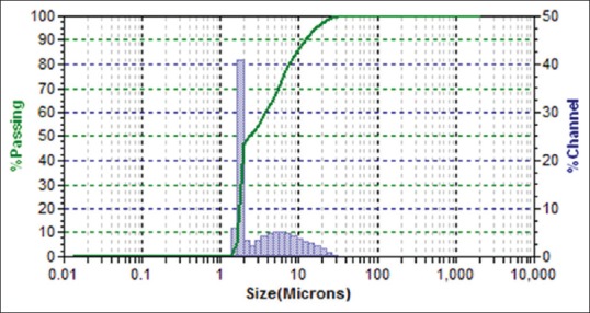
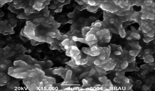
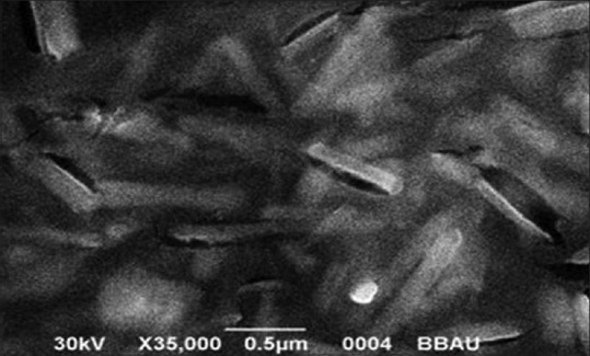
Conclusions: Due to characterization by DLS technique, nanoparticles size was found the range of 79.22 nm and 178 nm for Sliver and Selenium Nanoparticles respectively. Sliver nanoparticles shown morphological average size and shape with SEM reveals spherical shape particles with the size of 80.32 nm whereas Selenium nanoparticles shown rod shape particles with the size of 74.29 nm.
Keywords: Biomedical devices; dynamic light scattering; microbial infections; nanotechnology; scanning electron microscopy; selenium nanoparticles.
Conflict of interest statement
There are no conflicts of interest.
Figures
Similar articles
-
A novel one-pot green synthesis of selenium nanoparticles and evaluation of its toxicity in zebrafish embryos.Artif Cells Nanomed Biotechnol. 2016;44(2):471-7. doi: 10.3109/21691401.2014.962744. Epub 2014 Oct 7. Artif Cells Nanomed Biotechnol. 2016. PMID: 25287880
-
Determination of size and mass-and number-based concentration of biogenic SeNPs synthesized by lactic acid bacteria by using a multimethod approach.Anal Chim Acta. 2017 Nov 1;992:34-41. doi: 10.1016/j.aca.2017.09.033. Epub 2017 Sep 28. Anal Chim Acta. 2017. PMID: 29054148
-
Hybrid of niosomes and bio-synthesized selenium nanoparticles as a novel approach in drug delivery for cancer treatment.Mol Biol Rep. 2020 Sep;47(9):6517-6529. doi: 10.1007/s11033-020-05704-z. Epub 2020 Aug 7. Mol Biol Rep. 2020. PMID: 32767222
-
A review on plant-mediated selenium nanoparticles and its applications.J Popul Ther Clin Pharmacol. 2022 Jan 6;28(2):e29-e40. doi: 10.47750/jptcp.2022.870. eCollection 2022. J Popul Ther Clin Pharmacol. 2022. PMID: 35016267 Review.
-
Role of nano-selenium in health and environment.J Biotechnol. 2021 Jan 10;325:152-163. doi: 10.1016/j.jbiotec.2020.11.004. Epub 2020 Nov 4. J Biotechnol. 2021. PMID: 33157197 Review.
Cited by
-
Synthesis Monitoring, Characterization and Cleanup of Ag-Polydopamine Nanoparticles Used as Antibacterial Agents with Field-Flow Fractionation.Antibiotics (Basel). 2022 Mar 8;11(3):358. doi: 10.3390/antibiotics11030358. Antibiotics (Basel). 2022. PMID: 35326821 Free PMC article.
-
The Impact of Berberine Loaded Selenium Nanoparticles on K. pneumoniae and Candida albicans Antibiotics Resistance Isolates.Arch Razi Inst. 2023 Jun 30;78(3):1005-1015. doi: 10.22092/ARI.2022.359898.2509. eCollection 2023 Jun. Arch Razi Inst. 2023. PMID: 38028848 Free PMC article.
-
Anticancer efficacy of biosynthesized silver nanoparticles loaded with recombinant truncated parasporin-2 protein.Sci Rep. 2024 Jul 5;14(1):15544. doi: 10.1038/s41598-024-66650-5. Sci Rep. 2024. PMID: 38969695 Free PMC article.
-
Green Synthesis, Characterization and Application of Silver Nanoparticles Using Bioflocculant: A Review.Bioengineering (Basel). 2024 May 15;11(5):492. doi: 10.3390/bioengineering11050492. Bioengineering (Basel). 2024. PMID: 38790359 Free PMC article. Review.
-
Biosynthesized Silver Nanoparticles Using Morus alba (White Mulberry) Leaf Extract as Potential Antibacterial and Anticancer Agents.Molecules. 2023 Jan 26;28(3):1213. doi: 10.3390/molecules28031213. Molecules. 2023. PMID: 36770881 Free PMC article.
References
-
- Brambilla G, Masri D E, Pierno M, Berthier L, Cipelletti L, Petekidis G, Schofield AB. Probing the equilibrium dynamics of colloidal hard spheres above the mode-coupling glass transition Phys.Rev. Lett. 2009;102:1–4. - PubMed
-
- Iker MB, Dorota W, Patrick H, Jonathan S, Iseult L, Kenneth D. Characterisation of nanoparticle size and state prior to nanotoxicological studies J.Nanopart. Res. 2010;12:47–53.
-
- Madrid AP, Lapas LC, Rubi JM. Heat exchange between two interacting nanoparticles beyond the fluctuation-dissipation regime Phys.Rev. Lett. 2009;103:1–4. - PubMed
-
- Schoch RB, Han J, Renaud P. Transport phenomena in nanofluidics Rev.Mod. Phys. 2008;80:839–883.
-
- Klasen HJ. Historical review of the use of silver in the treatment of burns. I. Early uses. Burns. 2000;26:117–130. - PubMed
LinkOut - more resources
Full Text Sources