. 2019 Feb 5;93(4):e01916-18.
doi: 10.1128/JVI.01916-18. Print 2019 Feb 15.

Efficient Inhibition of Avian and Seasonal Influenza A Viruses by a Virus-Specific Dicer-Substrate Small Interfering RNA Swarm in Human Monocyte-Derived Macrophages and Dendritic Cells

Affiliations

Efficient Inhibition of Avian and Seasonal Influenza A Viruses by a Virus-Specific Dicer-Substrate Small Interfering RNA Swarm in Human Monocyte-Derived Macrophages and Dendritic Cells

Miao Jiang et al. J Virol. .

Abstract

Influenza A viruses (IAVs) are viral pathogens that cause epidemics and occasional pandemics of significant mortality. The generation of efficacious vaccines and antiviral drugs remains a challenge due to the rapid appearance of new influenza virus types and antigenic variants. Consequently, novel strategies for the prevention and treatment of IAV infections are needed, given the limitations of the presently available antivirals. Here, we used enzymatically produced IAV-specific double-stranded RNA (dsRNA) molecules and Giardia intestinalis Dicer for the generation of a swarm of small interfering RNA (siRNA) molecules. The siRNAs target multiple conserved genomic regions of the IAVs. In mammalian cells, the produced 25- to 27-nucleotide-long siRNA molecules are processed by endogenous Dicer into 21-nucleotide siRNAs and are thus designated Dicer-substrate siRNAs (DsiRNAs). We evaluated the efficacy of the above DsiRNA swarm at preventing IAV infections in human primary monocyte-derived macrophages and dendritic cells. The replication of different IAV strains, including avian influenza H5N1 and H7N9 viruses, was significantly inhibited by pretransfection of the cells with the IAV-specific DsiRNA swarm. Up to 7 orders of magnitude inhibition of viral RNA expression was observed, which led to a dramatic inhibition of IAV protein synthesis and virus production. The IAV-specific DsiRNA swarm inhibited virus replication directly through the RNA interference pathway although a weak induction of innate interferon responses was detected. Our results provide direct evidence for the feasibility of the siRNA strategy and the potency of DsiRNA swarms in the prevention and treatment of influenza, including the highly pathogenic avian influenza viruses.IMPORTANCE In spite of the enormous amount of research, influenza virus is still one of the major challenges for medical virology due to its capacity to generate new variants, which potentially lead to severe epidemics and pandemics. We demonstrated here that a swarm of small interfering RNA (siRNA) molecules, including more than 100 different antiviral RNA molecules targeting the most conserved regions of the influenza A virus genome, could efficiently inhibit the replication of all tested avian and seasonal influenza A variants in human primary monocyte-derived macrophages and dendritic cells. The wide antiviral spectrum makes the virus-specific siRNA swarm a potentially efficient treatment modality against both avian and seasonal influenza viruses.

Keywords: Dicer-substrate siRNA; DsiRNA; IAV; IFN; RNA interference; avian influenza virus; gene silencing; human macrophage; human moDC; influenza A virus; interferon response; siRNA swarm; viral replication.


Figures

FIG 1
FIG 1
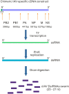
Schematic representation of enzymatic in vitro synthesis of DsiRNA molecules of a chimeric IAV construct. A chimeric cDNA construct containing selected conserved regions of the PB1, PB2, PA, NP, M, and NS genes of the A/wild duck/Hunan/211/2005 (H5N1) IAV strain and the promoter sequence of the T7 polymerase is shown. The sizes of the sequences derived from the different IAV genes are indicated. The ssRNA molecules are transcribed from the chimeric cDNA construct using bacteriophage T7 polymerase. The ssRNA molecules are used as templates for the bacteriophage φ6 RNA-dependent RNA polymerase to produce corresponding dsRNA molecules. Produced dsRNA molecules are then digested into a swarm of DsiRNA molecules (cIAV DsiRNA swarm) using recombinant Giardia intestinalis Dicer. The chimeric IAV sequence is shown at the top with the T7 promoter indicated in green. Other bars represent the ssRNA and dsRNA synthesis intermediate products and the cIAV siRNA swarm, as indicated.
FIG 2
FIG 2
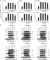
Inhibition of avian IAV replication in human macrophages and moDCs by pretransfection with cIAV siRNAs. Human primary macrophages or moDCs obtained from four different blood donors were separately mock transfected (control, UV IAV, or no-siRNA bars) or pretransfected with the indicated siRNA or DsiRNAs (10 nM) for 21 h. Cells were then infected with live or UV-irradiated H5N1 or H7N9 virus at an MOI of 1. Macrophages were washed twice with PBS at 1 h p.i. and then maintained in macrophage medium. Input virus was retained in moDC cultures (A) Cells from four different blood donors were subsequently collected at 7 and 24 h p.i. and were pooled; then IAV M1 RNA expression was determined by qRT-PCR from isolated total cellular RNA samples. The values were normalized against β-actin gene-specific mRNA, and relative IAV M1 RNA levels were calculated by the ΔΔCT method using untreated cellular RNA as a calibrator. The means (±SD) of three parallel analyses are shown. Data are representative of three individual experiments. Statistical significance was determined against results from samples of nontransfected cells (boxed bars). *; P < 0.05, **, P < 0.01. (B) Western blot analysis for the expression of viral PB1, NP, M1, and NS1 proteins and β-actin and GAPDH proteins in siRNA/DsiRNA transfected human macrophages and moDCs. Cells were collected at 24 h after avian H5N1 or H7N9 IAV infection, and whole-cell lysates were prepared. Cellular proteins (30 μg/lane) were separated by 10% SDS-PAGE, followed by electrophoretic transfer of the proteins onto polyvinylidene difluoride membranes and visualization of the transferred proteins by protein-specific antibodies, as indicated. The data of one representative experiment of three independent experiments is shown.
FIG 3
FIG 3
Inhibition of the productivity of H5N1 infection by pretransfection with cIAV DsiRNA swarm in human macrophage and moDCs. Human primary macrophages or moDCs obtained from four different blood donors (A, B, C, and D) were left nontransfected (control, UV IAV, or no siRNA) or separately pretransfected with the indicated siRNA or DsiRNAs (10 nM) for 21 h. Cells were then infected with live or UV-irradiated H5N1 viruses at an MOI of 1. To remove the input virus, macrophages were washed twice with PBS at 1 h p.i. and then maintained in a macrophage medium. MoDC cultures were not washed, and therefore the H5N1 virus titers in supernatant samples at 1 h p.i. represented the input amounts of virus. (A) The infective viral titers produced from macrophages and moDCs were determined by plaque assay in Madin-Darby canine kidney cells. Statistical significance was determined against results from samples of nontransfected H5N1 virus-infected cells (no siRNA; boxed bars). *, P < 0.05; **, P < 0.01. (B) The RNA was isolated from the supernatant samples from macrophages and moDCs, and the viral M1 gene-specific RNA levels were detected by qRT-PCR. The viral RNA expression was calculated relative to the level in UV-irradiated samples with the ΔΔCT method. Statistical significance was determined against results from samples of nontransfected H5N1 virus-infected cells (no siRNA; boxed bars). *, P < 0.05.
FIG 4
FIG 4
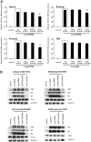
Inhibition of seasonal IAV replication in human moDCs by pretransfection with a cIAV DsiRNA swarm. Human moDCs from four different blood donors were separately mock transfected (control, UV IAV, or no-siRNA bars) or pretransfected with the indicated siRNAs (10 nM) for 21 h. Cells were subsequently infected with the indicated live or with UV-irradiated seasonal IAVs at an MOI of 1. (A) At 24 h p.i. cells from different blood donors were pooled, and the IAV M1 RNA expression was determined by qRT-PCR from isolated total cellular RNA samples. The values were normalized against β-actin gene-specific mRNA, and relative IAV M1 RNA levels were calculated by the ΔΔCT method using untreated cells as a calibrator. The means (±SD) of three parallel analyses are shown. Data are representative of three individual experiments. Statistical significance was determined against results from samples of nontransfected cells (no siRNA; boxed bars). *, P < 0.05; **, P < 0.01. (B) Western blot analysis for the expression of viral PB1, NP, M1, and NS1 proteins and β-actin in siRNA/DsiRNA transfected moDCs infected with the indicated seasonal IAVs. Cells were collected at 24 h p.i., and whole-cell lysates were prepared. Cellular proteins (30 μg/lane) were separated by 10% SDS-PAGE, followed by Western blot analysis with the indicated antibodies. One representative experiment of three independent experiments is shown.
FIG 5
FIG 5
Antiviral specificity of cIAV DsiRNAs in Calu-3 cells. Human lung cancer Calu-3 cells (in 12-well plates; 5 × 105 cells/well) were mock transfected (control or no-siRNA bars) or pretransfected with the indicated control and IAV-specific siRNA/DsiRNAs (10 nM). After 21 h of incubation, cells were infected with the indicated IAVs or influenza B virus (IBV) at an MOI of 1 for an additional 24 h. Cells were then collected for RNA isolation and quantitative RT-PCR analysis and for Western blot analysis. (A) The values of RT-PCR analyses were normalized against β-actin gene-specific mRNA, and relative IAV M1 RNA or IBV NP RNA levels were calculated by the ΔΔCT method using untreated control cells as a calibrator. The means (±SD) of three parallel analyses are shown. Data are representative of three individual experiments. Statistical significance was determined against results from samples of nontransfected cells (no siRNA; boxed bars). *, P < 0.05. (B) Western blot analysis for the expression of IAV proteins PB1, NP, and M1, IBV proteins NP and M1, and cellular β-actin and GAPDH proteins in siRNA/DsiRNA transfected moDCs after the infection of indicated IAVs or IBV. Cells were collected at 24 h after infection, and whole-cell lysates were prepared. Cellular proteins (30 μg/lane) were separated by 10% SDS-PAGE, followed by Western blot analysis with the indicated antibodies. One representative experiment of three independent experiments is shown.
FIG 6
FIG 6
Inhibition of seasonal IAV replication by pretransfection with a cIAV DsiRNA swarm in A549 cells. Human lung epithelial A549 cells (in 12-well plates; 5 × 105 cells/well) were mock transfected (control or no-siRNA bars) or pretransfected with the indicated control and IAV-specific siRNA/DsiRNAs (10 nM). After 21 h of incubation, cells were infected with the indicated IAVs at an MOI of 1 for an additional 24 h. Cellular or the supernatant samples from A549 cells were then collected for RNA isolation and quantitative RT-PCR analysis. (A) The values of RT-PCR analyses were normalized against β-actin gene-specific mRNA, and the relative cellular IAV M1 mRNA level was calculated by the ΔΔCT method using untreated cells as a calibrator. The means (±SD) of three parallel analyses are shown. Data are representative of three individual experiments. Statistical significance was determined against results from samples of nontransfected cells (boxed bars). *, P < 0.05. (B) Cell culture supernatant viral RNA levels from isolated total RNA were calculated relative to the level in untreated control cell supernatants with the ΔΔCT method.
FIG 7
FIG 7
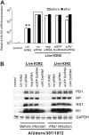
Difference between antiviral effects of cIAV DsiRNAs in moDCs transfected before and after IAV infection. Human moDCs from four different blood donors were separately mock transfected (control, UV IAV, or no-siRNA bars) or pretransfected with the indicated siRNAs (10 nM) either 21 h before or 1 h after A/Udorn/307/72 (H3N2) virus infection. Cells were infected with live or UV-irradiated A/Udorn/307/72 IAV at an MOI of 1. At 24 h p.i. cells from four different blood donors were pooled, and IAV M1 RNA expression was determined by qRT-PCR from isolated total cellular RNA or by Western blotting from cell lysates. (A) Relative IAV M1 RNA expression in cells transfected 21 h before infection or 1 h after infection. The values were normalized against β-actin gene-specific mRNA, and relative IAV M1 RNA levels were calculated by the ΔΔCT method using untreated cells as a calibrator. The means (±SD) of three parallel determinations are shown. Data are representative of three individual experiments. Statistical significance was determined against results from samples of nontransfected cells (boxed bars). *, P < 0.05. (B) Western blot analysis for the expression of viral PB1, NP, M1, and NS1 proteins and GAPDH protein in siRNA/DsiRNA transfected moDCs after A/Udorn/72 IAV infection. Cells were collected at 24 h p.i., and whole-cell lysates were prepared. Cellular proteins (30 μg/lane) were separated by 10% SDS-PAGE, followed by Western blot analysis with the indicated antibodies. One representative experiment of three independent experiments is shown.
FIG 8
FIG 8
Induction of IFN gene expression and inhibition of IAV replication by siRNA, DsiRNA swarms, and an 88-bp dsRNA in human macrophages and moDCs. Human macrophages or moDCs were left nontransfected (control, UV IAV, or no siRNA) or separately transfected with the indicated siRNA or DsiRNA swarms or 88-bp dsRNA (10 nM) for 21 h. (A) qRT-PCR analysis of IFN gene expression. IFN-β and IFN-λ1 mRNA expression is shown. Values are the means (±SD) of three parallel analyses. Data are representative of three individual experiments. The statistical significance of results with siRNA/DsiRNA swarm pretransfected cellular samples was determined against results with s 88-bp dsRNA pretransfected cellular samples (positive control, boxed bars). *, P < 0.05. (B) Western blot analysis for the expression of IFN-α/β/λ-inducible MxA protein and GAPDH in siRNA/DsiRNA/88-bp dsRNA transfected macrophages or moDCs. Cells were collected at 21 h after transfection, and whole-cell lysates were prepared. Cellular proteins (30 μg/lane) were separated by 10% SDS-PAGE, followed by Western blot analysis with anti-MxA and anti-GAPDH antibodies. One representative experiment of three independent experiments is shown. (C) Transfected macrophages or moDCs were infected with the indicated live or UV-irradiated seasonal IAV at an MOI of 1. Macrophages were washed twice with PBS at 1 h p.i. and subsequently maintained in a macrophage medium, while the input virus remained in moDCs throughout the experiment. At 24 h after infection cells from different blood donors were pooled, and IAV M1 mRNA expression was determined by qRT-PCR from isolated total cellular RNA. The values were normalized against β-actin gene-specific mRNA, and relative IAV M1 mRNA levels were calculated with the ΔΔCT method using untreated control cells as a calibrator. The means (±SD) of three parallel analyses are shown. Data are representative of three individual experiments. Statistical significance was determined against results of mock transfected cellular samples (no-siRNA boxed bars). *, P < 0.05; **, P < 0.01.
FIG 9
FIG 9
Inhibition of H3N2 IAV replication by pretransfection with cIAV DsiRNA swarm in mouse KO cells. Mouse wt cells, IRF3/IRF7 double-KO cells (IRF3/7 KO cells), NF-κB RelA/c-Rel/Nfkb1 triple-KO cells (NF-κB KO cells), and IFN-α/β receptor 1 KO cells (IFNAR1 KO cells) (in 12-well plates; 5 × 105 cells/well) were mock transfected (control or no-siRNA bars) or pretransfected with the indicated control and IAV-specific siRNA/DsiRNAs (10 nM). After 21 h of incubation, cells were infected with H3N2 IAV (A/Udorn/307/1972) at an MOI of 1 for an additional 24 h. Cells were then collected for RNA isolation and quantitative RT-PCR analysis and for Western blot analysis. (A) The values of RT-PCR analyses were normalized against β-actin gene-specific mRNA, and the relative IAV M1 mRNA level was calculated by the ΔΔCT method using untreated control cells as a calibrator. The means (±SD) of three parallel analyses are shown. Data are representative of three individual experiments. Statistical significance was determined against results of samples of nontransfected cells (boxed bars). *, P < 0.05. (B) Western blot analysis for the expression of IAV proteins PB1 and NP and cellular β-actin in siRNA/DsiRNA transfected cells after the infection of H3N2 IAV. Cells were collected at 24 h after infection, and whole-cell lysates were prepared. Cellular proteins (30 μg/lane) were separated by 10% SDS-PAGE, followed by Western blot analysis with the indicated antibodies. One representative experiment of three independent experiments is shown.

Similar articles

Cited by

References

    1. Taubenberger JK, Reid AH, Fanning TG. 2000. The 1918 influenza virus: a killer comes into view. Virology 274:241–245. doi:10.1006/viro.2000.0495. - DOI - PubMed
    1. Smith GJ, Vijaykrishna D, Bahl J, Lycett SJ, Worobey M, Pybus OG, Ma SK, Cheung CL, Raghwani J, Bhatt S, Peiris JS, Guan Y, Rambaut A. 2009. Origins and evolutionary genomics of the 2009 swine-origin H1N1 influenza A epidemic. Nature 459:1122–1125. doi:10.1038/nature08182. - DOI - PubMed
    1. Lindstrom SE, Cox NJ, Klimov A. 2004. Genetic analysis of human H2N2 and early H3N2 influenza viruses, 1957–1972: evidence for genetic divergence and multiple reassortment events. Virology 328:101–119. doi:10.1016/j.virol.2004.06.009. - DOI - PubMed
    1. Palese P, Tumpey TM, Garcia-Sastre A. 2006. What can we learn from reconstructing the extinct 1918 pandemic influenza virus? Immunity 24:121–124. doi:10.1016/j.immuni.2006.01.007. - DOI - PubMed
    1. Chan PK. 2002. Outbreak of avian influenza A(H5N1) virus infection in Hong Kong in 1997. Clin Infect Dis 34(Suppl 2):S58–S64. doi:10.1086/338820. - DOI - PubMed

Publication types

MeSH terms

LinkOut - more resources