. 2019 Jan 18;431(2):368-390.
doi: 10.1016/j.jmb.2018.11.015. Epub 2018 Nov 17.

Clustering, Spatial Distribution, and Phosphorylation of Discoidin Domain Receptors 1 and 2 in Response to Soluble Collagen I

Affiliations

Clustering, Spatial Distribution, and Phosphorylation of Discoidin Domain Receptors 1 and 2 in Response to Soluble Collagen I

David A Yeung et al. J Mol Biol. .

Abstract

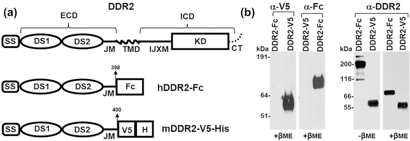
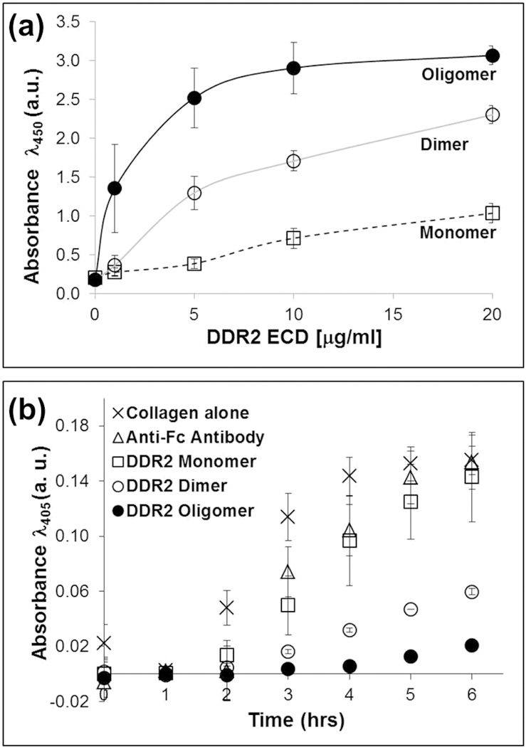
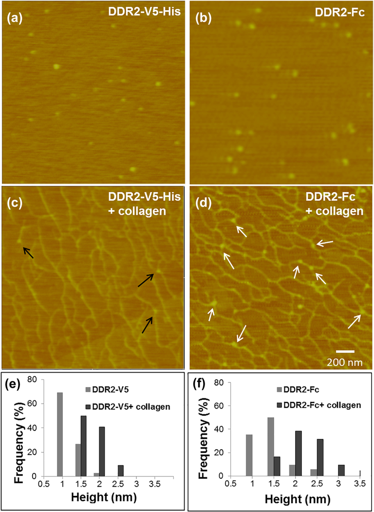
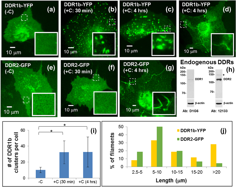
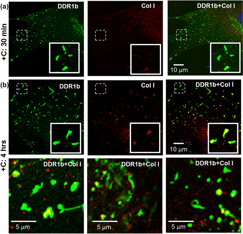
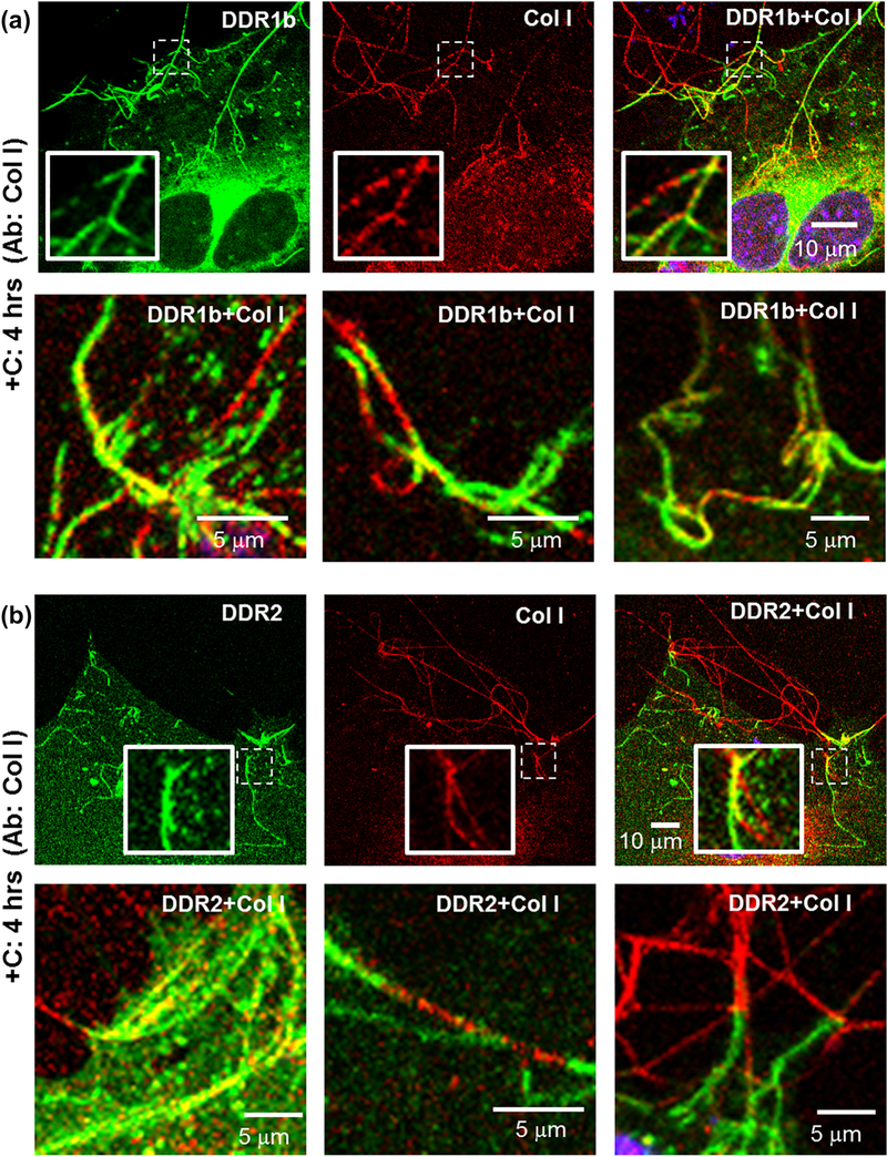
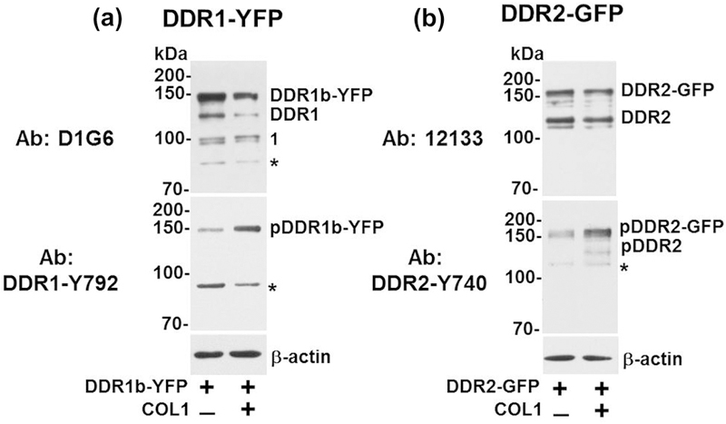
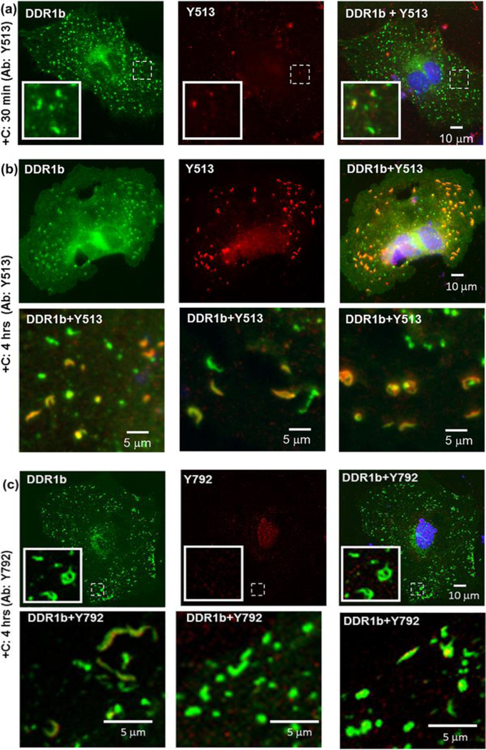
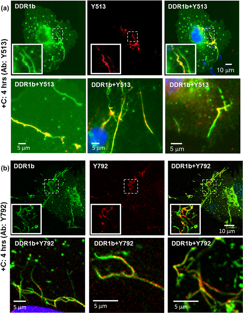
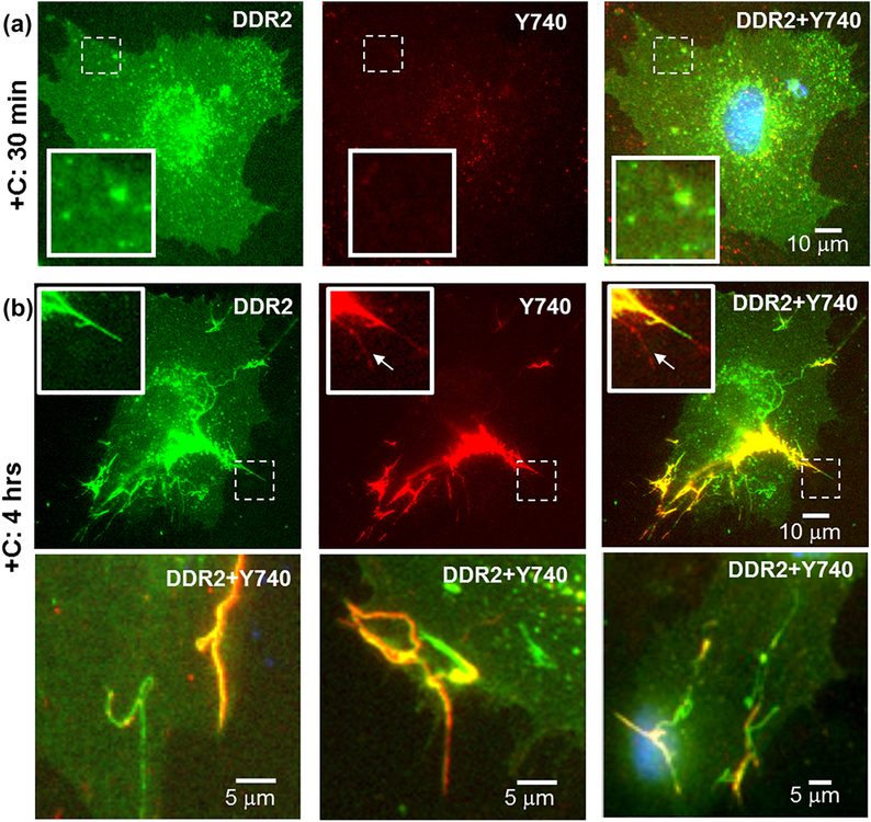
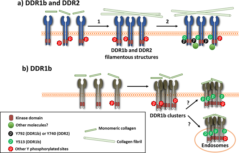
Discoidin domain receptors (DDR1 and DDR2) are receptor tyrosine kinases that signal in response to collagen. We had previously shown that collagen binding leads to clustering of DDR1b, a process partly mediated by its extracellular domain (ECD). In this study, we investigated (i) the impact of the oligomeric state of DDR2 ECD on collagen binding and fibrillogenesis, (ii) the effect of collagen binding on DDR2 clustering, and (iii) the spatial distribution and phosphorylation status of DDR1b and DDR2 after collagen stimulation. Studies were conducted using purified recombinant DDR2 ECD proteins in monomeric, dimeric or oligomeric state, and MC3T3-E1 cells expressing full-length DDR2-GFP or DDR1b-YFP. We show that the oligomeric form of DDR2 ECD displayed enhanced binding to collagen and inhibition of fibrillogenesis. Using atomic force and fluorescence microscopy, we demonstrate that unlike DDR1b, DDR2 ECD and DDR2-GFP do not undergo collagen-induced receptor clustering. However, after prolonged collagen stimulation, both DDR1b-YFP and DDR2-GFP formed filamentous structures consistent with spatial re-distribution of DDRs in cells. Immunocytochemistry revealed that while DDR1b clusters co-localized with non-fibrillar collagen, DDR1b/DDR2 filamentous structures associated with collagen fibrils. Antibodies against a tyrosine phosphorylation site in the intracellular juxtamembrane region of DDR1b displayed positive signals in both DDR1b clusters and filamentous structures. However, only the filamentous structures of both DDR1b and DDR2 co-localized with antibodies directed against tyrosine phosphorylation sites within the receptor kinase domain. Our results uncover key differences and similarities in the clustering abilities and spatial distribution of DDR1b and DDR2 and their impact on receptor phosphorylation.

Keywords: atomic force microscopy; fibrillogenesis; fluorescence microscopy; oligomer; receptor tyrosine kinase.


Figures

Figure 1:
Figure 1:
(a) Schematic diagram showing the structure of full length DDR2, human DDR2-Fc and mouse DDR2-V5-His constructs. SS: signal sequence; DS: discoidin domain; ECD: extracellular domain; IJXM: intracellular juxtamembrane region; TMD: transmembrane domain; ICD: intracellular domain; KD: kinase domain (b) Purified recombinant DDR2-V5-His and DDR2-Fc proteins (20 ng/lane) were resolved by SDS-PAGE under reducing conditions (+βME), with either 4–12% (w/v) Bis-Tris Gels (left panel) or under reducing and non-reducing (-βME, 100 ng/lane)) conditions with 10% SDS-PAGE (right panel). The separated protein was detected by immunoblotting using anti-epitope or anti-DDR2 antibodies as indicated.
Figure 2:
Figure 2:
(a) Solid phase binding of DDR2 ECD proteins to immobilized bovine-dermal collagen I as indicated. Binding was detected using antibodies against DDR2 ECD. (b) Inhibition of fibrillogenesis of bovine-dermal collagen I assessed using turbidity measurements. DDR2 ECD proteins (40 μg/ml) as indicated were incubated with 200 μg/ml of neutralized collagen I in 96-well plates at 37°C.
Figure 3:
Figure 3:
AFM height images of monomeric DDR2-V5-His and dimeric DDR2-Fc before and after binding to bovine-dermal collagen I as indicated (a-d). DDR2-V5-His and DDR2-Fc particles bound to collagen are indicated by black and white arrows respectively. Particle size distribution and average sizes are indicated in the accompanying histograms in (e) and (f) and in Table 1.
Figure 4:
Figure 4:
Live cell imaging of DDR1b-YFP- (a-d) and DDR2-GFP- (e-g) expressing MC3T3-E1 cells using wide-field fluorescence microscopy, before and after addition of collagen ‘C’ as indicated. Insets (in a-g) show selected regions which have been magnified from the corresponding images to visualize receptor assemblies. The location of these selected regions on the cell surface is indicated by dashed boxes. Western blotting of endogenous DDR expression in MC3T3-E1 cells is shown in (h). Fluorescence microscopy images (a-c) show that DDR1b-YFP exhibits a uniform distribution on the cell surface before collagen stimulation and results in cluster formation upon collagen stimulation. Quantitative analysis (i) indicates that the number of punctuate structures in cells significantly increases upon collagen stimulation (*p-value <0.05) and persist at 4 hrs. After 4 hrs of collagen stimulation, a subpopulation of DDR1b-YFP expressing cells also exhibits the presence of long, filamentous structures (d). DDR2-GFP exhibits a uniform distribution on the cell surface before collagen stimulation (e) and does not result in cluster formation upon collagen stimulation (f). However, at 4 hrs post collagen administration, filamentous structures were also observed in DDR2-GFP-expressing cells (g). The distribution of uninterrupted contour length of filamentous structures formed in DDR1b-YFP- and DDR2-GFP- expressing cells after 4 hrs of collagen stimulation is shown in (j).
Figure 5:
Figure 5:
Immunocytochemistry (ICC) performed on non-permeabilized cells for evaluating the association of collagen I with DDR1b-YFP clusters. Total receptor is indicated in YFP channel (green) while staining with collagen I antibodies is shown by TRITC (red). Co-localization YFP and TRITC is shown in yellow. Blue represents nuclear (DAPI) staining. Insets (in first row of each panel) show selected regions which have been magnified form the corresponding images. (a) Little to no signal for collagen staining was detected on cells stimulated with collagen for 30 min, despite the presence of DDR1b clusters. (b) At prolonged collagen stimulation (4 hrs), a number of DDR1b clusters co-localized with signals for collagen I antibodies. The second row consists of selected regions from three different cells showing co-localization of DDR1b clusters and collagen signal. The collagen associated with DDR1b clusters was non-fibrillar in morphology.
Figure 6:
Figure 6:
Immunocytochemistry (ICC) performed on non-permeabilized cells for evaluating the association of collagen I with filamentous structures formed in (a) DDR1b-YFP- and (b) DDR2-GFP- expressing cells after 4 hrs of collagen stimulation. Total receptor is indicated in YFP channel (green) while staining with collagen antibodies is shown by TRITC (red). Co-localization YFP and TRITC is shown in yellow. Blue represents nuclear (DAPI) staining. Insets (in first row of each panel) show selected regions which have been magnified from the corresponding images. Selected regions from three different cells are shown in the second row in each panel. YFP/GFP positive filamentous structures had a very similar morphology as collagen fibrils and were observed to anchor the fibrils at cell edges. Collagen staining was intermittently present on or interspersed with YFP/GFP positive filamentous structures.
Figure 7:
Figure 7:
Western blotting of DDR expression and phosphorylation in MC3T3 cells. MC3T3-E1 cells were transiently transfected with (a) DDR1b-YFP or (b) DDR2-GFP expression vectors and stimulated with 20 µg/ml collagen I (+), as described in Materials and Methods. After 4 hrs of collagen stimulation, the cells were lysed in RIPA buffer and equal protein concentrations (25 μg/lane) were resolved by reducing 7.5% SDS-PAGE followed by immunoblot analyses using the indicated antibodies to DDR1 or DDR2. β-actin was used as loading control. Number 1 in panel b indicates an additional band detected with D1G6 antibodies (discussed in the Results section). Asterisks (*) show non-specific bands.
Figure 8:
Figure 8:
Immunocytochemistry (ICC) performed on permeabilized cells for evaluating receptor phosphorylation associated with DDR1b clusters by using pDDR1 antibodies Y513 and Y792, as indicated. Total receptor is indicated in YFP channel (green) while staining with Y513 or Y792 antibodies is shown by TRITC (red). Co-localization YFP and TRITC is shown in yellow. Blue represents nuclear (DAPI) staining. Insets (in first row in panels a and c) show selected regions which have been magnified from the corresponding images. (a) Little to no Y513 signal was detected in cells, 30 min after collagen stimulation despite the presence of DDR1b clusters. (b) At prolonged collagen stimulation (4 hrs), a number of DDR1b clusters co-localized with antibodies to Y513. The second row consists of selected regions from three different cells showing co-localization of DDR1b clusters with Y513 signal. (c) Little to no Y792 antibody signal was detected in cells even after 4 hrs of collagen stimulation despite the presence of DDR1b clusters. The second row consists of selected regions from three different cells showing the presence of DDR1b clusters with little to no co-localization with Y792 signal.
Figure 9:
Figure 9:
Immunocytochemistry (ICC) performed on permeabilized cells for evaluating receptor phosphorylation associated with filamentous structures formed in DDR1b-YFP- expressing cells after 4 hrs of collagen stimulation, using pDDR1 antibodies (a) Y513 and (b) Y792 as indicated. Total receptor is indicated in YFP channel (green) while staining with Y513 and Y792 antibodies is shown by TRITC (red). Co-localization of YFP and TRITC is shown in yellow. Blue represents nuclear (DAPI) staining. Insets (in first row in each panel) show selected regions which have been magnified from corresponding images. A subset of DDR1b filamentous structures co-localize with the pDDR1 antibodies. The second row in each panel consists of selected regions from three different cells showing co-localization of filamentous structures with signal for pDDR1 antibodies.
Figure 10:
Figure 10:
Immunocytochemistry (ICC) performed on permeabilized cells for evaluating spatial distribution of receptor phosphorylation in DDR2-GFP- expressing cells after (a) 30 min and (b) 4 hours of collagen stimulation. Total receptor is indicated in GFP channel (green) while staining with pDDR2 (Y740) antibodies is shown by TRITC (red). Co-localization GFP and TRITC is shown in yellow. Blue represents nuclear (DAPI) staining. Insets (in top row in each panel) show selected regions, which have been magnified from corresponding images. Little to no co-localization signal was detected, 30 min after collagen stimulation (a). At prolonged collagen stimulation (4 hrs), a number of filamentous structures formed in DDR2-GFP expressing cells co-localized with Y740 signal (b). The second row in (b) consists of selected regions from three different cells showing co-localization of filamentous structures with Y740 signal. A weak signal for Y740 (arrows) without a corresponding GFP signal can be observed in insets in (b, top row), which could correspond to endogenous pDDR2.
Scheme 1:
Scheme 1:
Postulated model for spatial distribution and phosphorylation of DDRs upon collagen stimulation. DDR1 and DDR2 exist as homodimers on the cell surface. (a) Upon addition of monomeric collagen I, DDR1 and DDR2 interact with collagen, which leads (arrow 1) to the assembly of the receptors into filamentous structures aligned with collagen fibrils. This process occurs at prolonged (~4 hrs) times after collagen I stimulation. Phosphorylation of both DDR1 and DDR2 (arrow 2), co-localize in these structures, as determined using Y513 (pDDR1b) as well as Y792 (pDDR1) and Y740 (pDDR2) antibodies, indicated as black and green circles. (b) In another pathway, DDR1b assembles into clusters within minutes of collagen I stimulation (likely binding to non-fibrillar collagen present at early stages of fibrillogenesis) and gets endocytosed. However, phosphorylation at Y513 and Y792 is not detected in these clusters at the early (~30 min) time point. At later times (~4 hrs) post-ligand administration, phosphorylation of DDR1b in its IJXM (Y513) (green circles) but not in its KD (Y792) is detected in the DDR1b clusters. At present it is not clear if Y513-positive clusters are present on the cell-surface or constitute pools of endocytosed DDR1b receptors localizing within endosomes. Owing to the fact that DDRs contain numerous Tyr residues within the IJXM region and their KDs, phosphorylation at other Tyr residues (indicated by the red dashed circles) may also be ensuing in the structures described in (a) and (b) but are undetectable by the tools available at this time. It is likely that formation of higher-order assemblies of DDRs may recruit additional cytosolic proteins which mediate specific cellular processes.

Similar articles

Cited by

References

    1. Fu H-L, Valiathan RR, Arkwright R, Sohail A, Mihai C, Kumarasiri M, Mahasenan KV, Mobashery S, Huang P, Agarwal G, Fridman R, Discoidin domain receptors: Unique receptor tyrosine kinases in collagen-mediated signaling, J. Biol. Chem 288 (2013). doi:10.1074/jbc.R112.444158. - DOI - PMC - PubMed
    1. Flynn LA, Blissett AR, Calomeni EP, Agarwal G, Inhibition of Collagen Fibrillogenesis by Cells Expressing Soluble Extracellular Domains of DDR1 and DDR2, J. Mol. Biol 395 (2010). doi:10.1016/j.jmb.2009.10.073. - DOI - PMC - PubMed
    1. Agarwal G, DDRs and Collagen Fibrillogenesis, in: Discoidin Domain Recept. Heal. Dis, 2016: pp. 23–56.
    1. Tonniges JR, Albert B, Calomeni EP, Roy S, Lee J, Mo X, Cole SE, Agarwal G, Collagen Fibril Ultrastructure in Mice Lacking Discoidin Domain Receptor 1., Microsc. Microanal 22 (2016) 599–611. doi:10.1017/S1431927616000787. - DOI - PMC - PubMed
    1. Leitinger B, Discoidin domain receptor functions in physiological and pathological conditions, Int. Rev. Cell Mol. Biol 310 (2014) 39–87. doi:10.1016/B978-0-12-800180-6.00002-5. - DOI - PMC - PubMed

Publication types

MeSH terms

LinkOut - more resources