

CD123 expression levels in 846 acute leukemia patients based on standardized immunophenotyping
- PMID: 30450744
- PMCID: PMC6587863
- DOI: 10.1002/cyto.b.21745
CD123 expression levels in 846 acute leukemia patients based on standardized immunophenotyping
Abstract
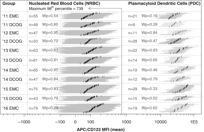
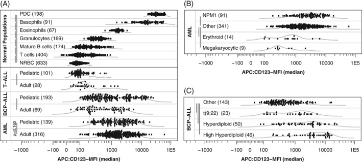
Background: While it is known that CD123 is normally strongly expressed on plasmacytoid dendritic cells and completely absent on nucleated red blood cells, detailed information regarding CD123 expression in acute leukemia is scarce and, if available, hard to compare due to different methodologies.
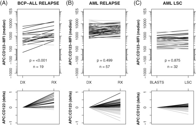
Methods: CD123 expression was evaluated using standardized EuroFlow immunophenotyping in 139 pediatric AML, 316 adult AML, 193 pediatric BCP-ALL, 69 adult BCP-ALL, 101 pediatric T-ALL, and 28 adult T-ALL patients. Paired diagnosis-relapse samples were available for 57 AML and 19 BCP-ALL patients. Leukemic stem cell (LSC) data was available for 32 pediatric AML patients. CD123 expression was evaluated based on mean fluorescence intensity, median fluorescence intensity, and percentage CD123 positive cells.
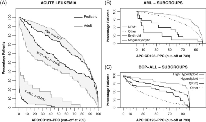
Results: EuroFlow panels were stable over time and between laboratories. CD123 was expressed in the majority of AML and BCP-ALL patients, but absent in most T-ALL patients. Within AML, CD123 expression was lower in erythroid/megakaryocytic leukemia, higher in NPM1 mutated and FLT3-ITD mutated leukemia, and comparable between LSC and leukemic blasts. Within BCP-ALL, CD123 expression was higher in patients with (high) hyperdiploid karyotypes and the BCR-ABL fusion gene. Interestingly, CD123 expression was increased in BCP-ALL relapses while highly variable in AML relapses (compared to CD123 expression at diagnosis).
Conclusions: Authors evaluated CD123 expression in a large cohort of acute leukemia patients, based on standardized and reproducible methodology. Our results may facilitate stratification of patients most likely to respond to CD123 targeted therapies and serve as reference for CD123 expression (in health and disease). © 2018 The Authors. Cytometry Part B: Clinical Cytometry published by Wiley Periodicals, Inc. on behalf of International Clinical Cytometry Society.
Keywords: CD123; acute leukemia; standardized immunophenotyping; targeted therapy.
© 2018 The Authors. Cytometry Part B: Clinical Cytometry published by Wiley Periodicals, Inc. on behalf of International Clinical Cytometry Society.
Conflict of interest statement
None were disclosed by the authors.
Figures
Similar articles
-
Immunoprofiling of leukemic stem cells CD34+/CD38-/CD123+ delineate FLT3/ITD-positive clones.J Hematol Oncol. 2016 Jul 27;9(1):61. doi: 10.1186/s13045-016-0292-z. J Hematol Oncol. 2016. PMID: 27465508 Free PMC article.
-
[Immunophenotyping of leukemic stem cells and chromosome karyotype characteristics in Uyghur leukemia pediatric patients].Zhonghua Zhong Liu Za Zhi. 2013 Jul;35(7):501-4. Zhonghua Zhong Liu Za Zhi. 2013. PMID: 24257300 Chinese.
-
CD123 Expression Is Associated With High-Risk Disease Characteristics in Childhood Acute Myeloid Leukemia: A Report From the Children's Oncology Group.J Clin Oncol. 2022 Jan 20;40(3):252-261. doi: 10.1200/JCO.21.01595. Epub 2021 Dec 2. J Clin Oncol. 2022. PMID: 34855461 Free PMC article.
-
Applications of Flow Cytometric Immunophenotyping in the Diagnosis and Posttreatment Monitoring of B and T Lymphoblastic Leukemia/Lymphoma.Cytometry B Clin Cytom. 2019 Jul;96(4):256-265. doi: 10.1002/cyto.b.21833. Epub 2019 Jun 24. Cytometry B Clin Cytom. 2019. PMID: 31231940 Review.
-
CD123 bi-specific antibodies in development in AML: What do we know so far?Best Pract Res Clin Haematol. 2020 Dec;33(4):101219. doi: 10.1016/j.beha.2020.101219. Epub 2020 Nov 6. Best Pract Res Clin Haematol. 2020. PMID: 33279175 Review.
Cited by
-
Paediatric Strategy Forum for medicinal product development for acute myeloid leukaemia in children and adolescents: ACCELERATE in collaboration with the European Medicines Agency with participation of the Food and Drug Administration.Eur J Cancer. 2020 Sep;136:116-129. doi: 10.1016/j.ejca.2020.04.038. Epub 2020 Jul 17. Eur J Cancer. 2020. PMID: 32688206 Free PMC article.
-
Proteomic Profiling Identifies Specific Leukemic Stem Cell-Associated Protein Expression Patterns in Pediatric AML Patients.Cancers (Basel). 2022 Jul 22;14(15):3567. doi: 10.3390/cancers14153567. Cancers (Basel). 2022. PMID: 35892824 Free PMC article.
-
Bicistronic CAR-T cells targeting CD123 and CLL1 for AML to reduce the risk of antigen escape.Transl Oncol. 2023 Aug;34:101695. doi: 10.1016/j.tranon.2023.101695. Epub 2023 May 22. Transl Oncol. 2023. PMID: 37224766 Free PMC article.
-
CD123 as a Biomarker in Hematolymphoid Malignancies: Principles of Detection and Targeted Therapies.Cancers (Basel). 2020 Oct 23;12(11):3087. doi: 10.3390/cancers12113087. Cancers (Basel). 2020. PMID: 33113953 Free PMC article. Review.
-
IL3RA-Targeting Antibody-Drug Conjugate BAY-943 with a Kinesin Spindle Protein Inhibitor Payload Shows Efficacy in Preclinical Models of Hematologic Malignancies.Cancers (Basel). 2020 Nov 20;12(11):3464. doi: 10.3390/cancers12113464. Cancers (Basel). 2020. PMID: 33233768 Free PMC article.
References
-
- Munoz L, Nomdedeu JF, Lopez O, Carnicer MJ, Bellido M, Aventin A, Brunet S, Sierra J. Interleukin‐3 receptor alpha chain (CD123) is widely expressed in hematologic malignancies. Haematologica. 2001;86:1261–1269. - PubMed
-
- Buckley SA, Walter RB. Update on antigen‐specific immunotherapy of acute myeloid leukemia. Curr Hematol Malig Rep. 2015;10:65–75. - PubMed
-
- Garfin PM, Feldman EJ. Antibody‐based treatment of acute myeloid leukemia. Curr Hematol Malig Rep. 2016;11:545–552. - PubMed
-
- Nijhof IS, Casneuf T, van Velzen J, van Kessel B, Axel AE, Syed K, Groen RW, van Duin M, Sonneveld P, Minnema MC, et al. CD38 expression and complement inhibitors affect response and resistance to daratumumab therapy in myeloma. Blood. 2016;128:959–970. - PubMed
MeSH terms
Substances
LinkOut - more resources
Full Text Sources
Other Literature Sources
Medical
Miscellaneous
