

Increased succinate receptor GPR91 involved in the pathogenesis of Mooren's ulcer
- PMID: 30450301
- PMCID: PMC6232339
- DOI: 10.18240/ijo.2018.11.01
Increased succinate receptor GPR91 involved in the pathogenesis of Mooren's ulcer
Abstract
Aim: To investigate the expression of succinate receptor GPR91 and its pathogenic roles in Mooren's ulcer (MU).
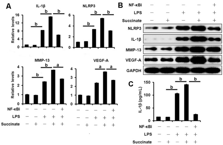
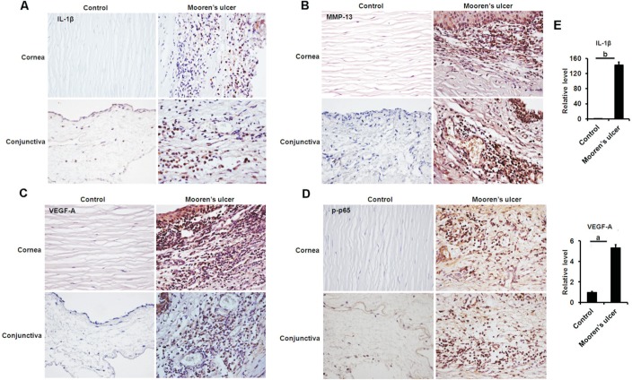
Methods: Biopsy specimens were obtained from 7 patients with MU and 6 healthy donors. The expression of GPR91 in MU tissues was evaluated using quantitative real-time reverse transcription polymerase chain reaction (qRT-PCR) and immunohistochemistry (IHC). Succinate was used to activate GPR91 signaling, and the effect of GPR91 on the expression of interleukin-1β (IL-1β), NLRP3, vascular endothelial growth factor (VEGF) and matrix metalloproteinase-13 (MMP-13) in human peripheral blood mononuclear cells (PBMCs) was determined. The influence of GPR91 on the nuclear factor-κB (NF-κB) signaling in PBMCs was investigated by detecting the phosphorylation of p65. Moreover, the expression of IL-1β, VEGF, MMP-13 and phosphorylated p65 (p-p65) in the tissues of MU was examined by qRT-PCR or IHC.
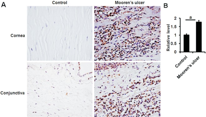
Results: GPR91 mRNA expression showed a higher level in the MU group than in the healthy control group. IHC analysis also revealed that the expression of GPR91 was elevated in patients with MU compared with healthy controls. Moreover, ligation of GPR91 with succinate promoted the lipopolysaccharide-induced production of NLRP3, IL-1β, VEGF and MMP-13 in PBMCs through increased phosphorylation of p65. Pharmacological inhibition of the NF-κB signaling reversed GPR91 induced production of NLRP3, IL-1β, VEGF and MMP-13. These findings, coupled with the elevated amounts of IL-1β, VEGF, MMP-13 and p-p65 observed in the MU biopsies, constituted a rational basis for the involvement of GPR91 in the pathogenesis of MU.
Conclusion: This study indicates the increased succinate receptor GPR91 in conjunctival or corneal tissues is involved in the pathogenesis of MU through elevated NF-κB activity, which may provide a new therapeutic target for MU.
Keywords: Mooren's ulcer; nuclear factor-κB; pathogenesis; succinate receptor.
Figures

Similar articles
-
Increased cGAS/STING signaling components in patients with Mooren's ulcer.Int J Ophthalmol. 2021 Nov 18;14(11):1660-1665. doi: 10.18240/ijo.2021.11.03. eCollection 2021. Int J Ophthalmol. 2021. PMID: 34804854 Free PMC article.
-
Upregulation of NLRP3 inflammasome components in Mooren's ulcer.Graefes Arch Clin Exp Ophthalmol. 2017 Mar;255(3):607-612. doi: 10.1007/s00417-016-3516-6. Epub 2016 Oct 28. Graefes Arch Clin Exp Ophthalmol. 2017. PMID: 27796668
-
Inhibition of high glucose-induced VEGF release in retinal ganglion cells by RNA interference targeting G protein-coupled receptor 91.Exp Eye Res. 2013 Apr;109:31-9. doi: 10.1016/j.exer.2013.01.011. Epub 2013 Feb 1. Exp Eye Res. 2013. PMID: 23379999
-
G protein-coupled receptor 91 signaling in diabetic retinopathy and hypoxic retinal diseases.Vision Res. 2017 Oct;139:59-64. doi: 10.1016/j.visres.2017.05.001. Epub 2017 Jun 23. Vision Res. 2017. PMID: 28539261 Free PMC article. Review.
-
GPR91, a critical signaling mechanism in modulating pathophysiologic processes in chronic illnesses.FASEB J. 2020 Oct;34(10):13091-13105. doi: 10.1096/fj.202001037R. Epub 2020 Aug 19. FASEB J. 2020. PMID: 32812686 Review.
Cited by
-
Increased cGAS/STING signaling components in patients with Mooren's ulcer.Int J Ophthalmol. 2021 Nov 18;14(11):1660-1665. doi: 10.18240/ijo.2021.11.03. eCollection 2021. Int J Ophthalmol. 2021. PMID: 34804854 Free PMC article.
References
-
- Foster CS, Kenyon KR, Greiner J, Greineder DK, Friedland B, Allansmith MR. The immunopathology of Mooren's ulcer. Am J Ophthalmol. 1979;88(2):149–159. - PubMed
-
- Brown SI, Mondino BJ, Rabin BS. Autoimmune phenomenon in Mooren's ulcer. Am J Ophthalmol. 1976;82(6):835–840. - PubMed
-
- Gottsch JD, Liu SH, Minkovitz JB, Goodman DF, Srinivasan M, Stark WJ. Autoimmunity to a cornea-associated stromal antigen in patients with Mooren's ulcer. Invest Ophthalmol Vis Sci. 1995;36(8):1541–1547. - PubMed
-
- Shinomiya K, Ueta M, Sotozono C, Inatomi T, Yokoi N, Koizumi N, Kinoshita S. Immunohistochemical analysis of inflammatory limbal conjunctiva adjacent to Mooren's ulcer. Br J Ophthalmol. 2013;97(3):362–366. - PubMed
LinkOut - more resources
Full Text Sources
Research Materials