. 2018 Nov 14;13(11):e0206355.
doi: 10.1371/journal.pone.0206355. eCollection 2018.

Identification and characterization of protein N-myristoylation occurring on four human mitochondrial proteins, SAMM50, TOMM40, MIC19, and MIC25

Affiliations

Identification and characterization of protein N-myristoylation occurring on four human mitochondrial proteins, SAMM50, TOMM40, MIC19, and MIC25

Toshihiko Utsumi et al. PLoS One. .

Abstract

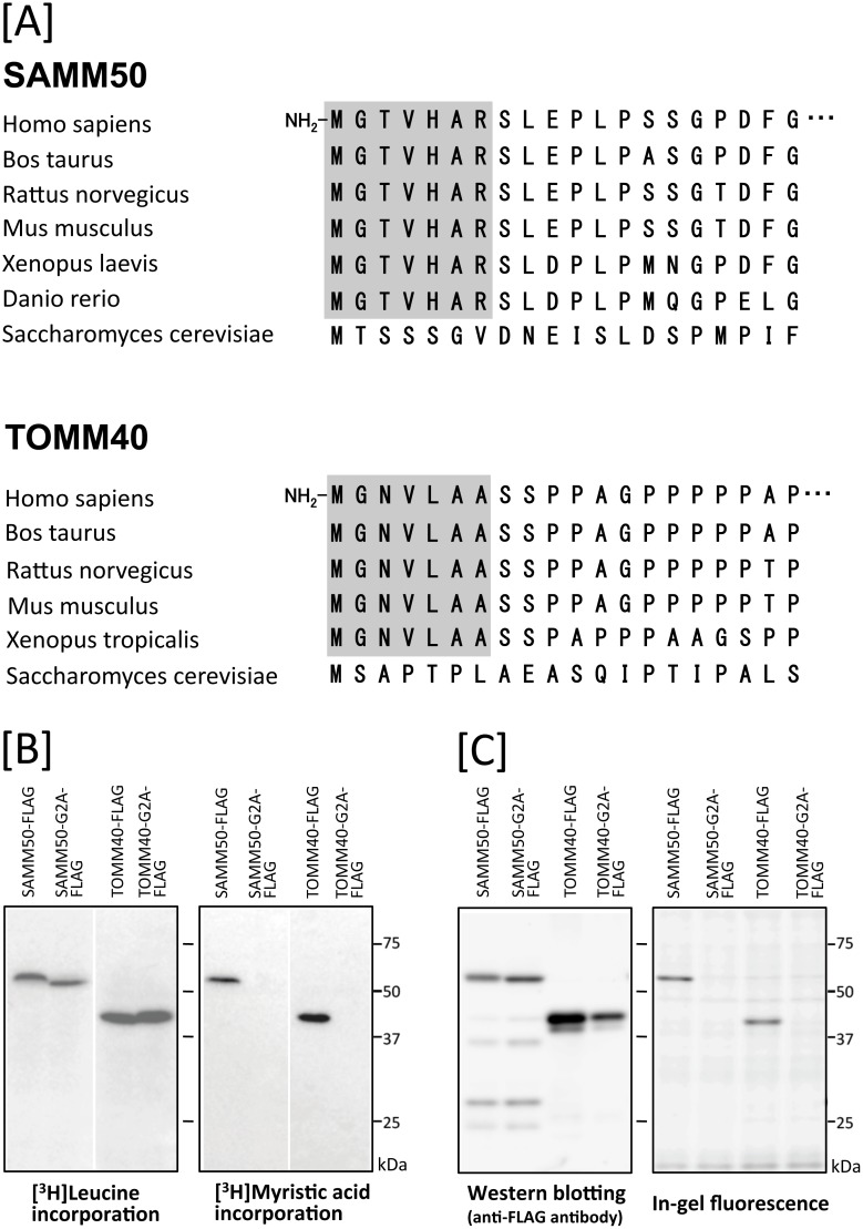
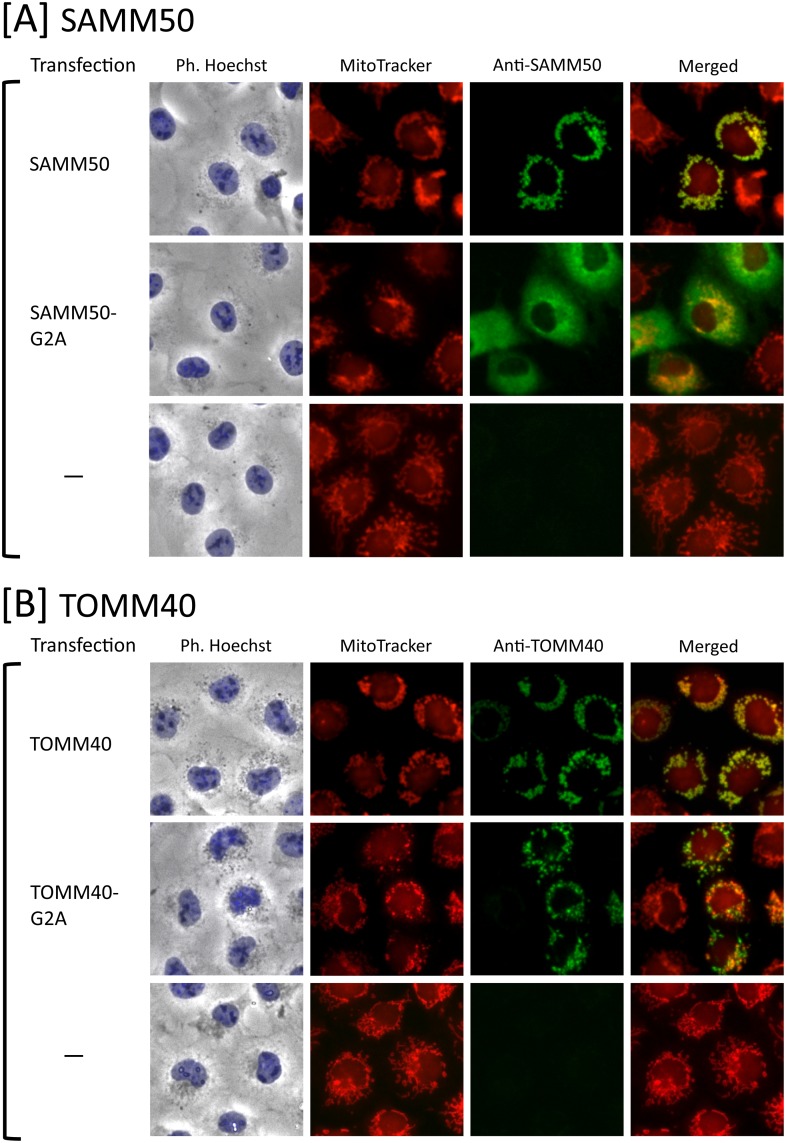
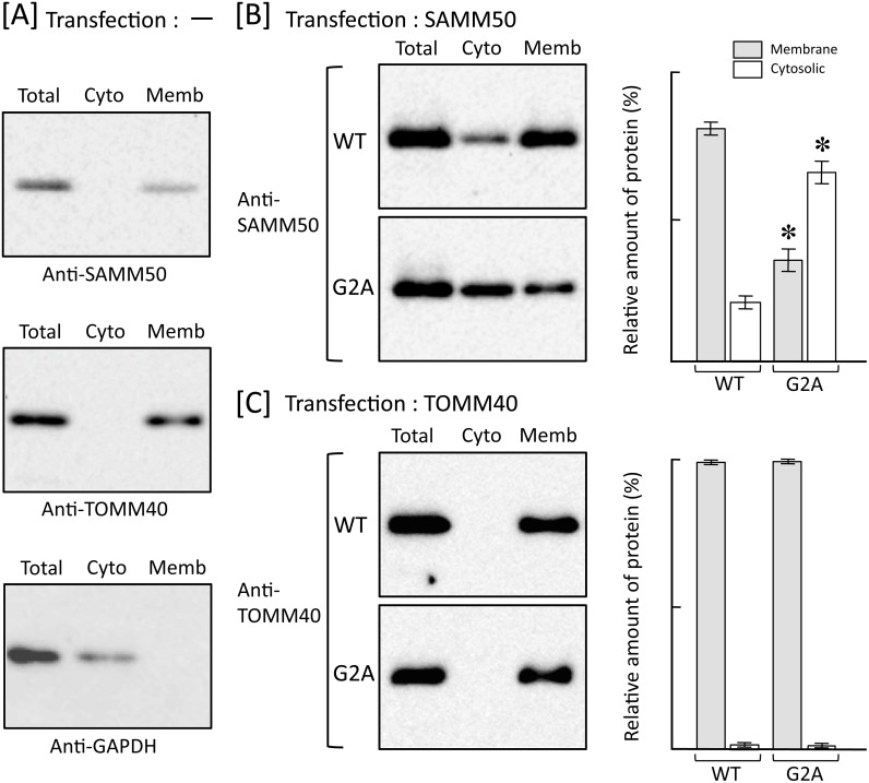
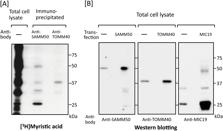
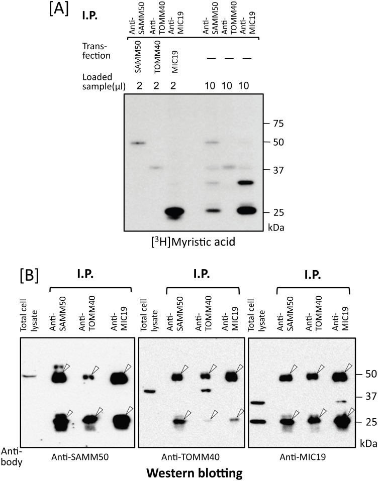
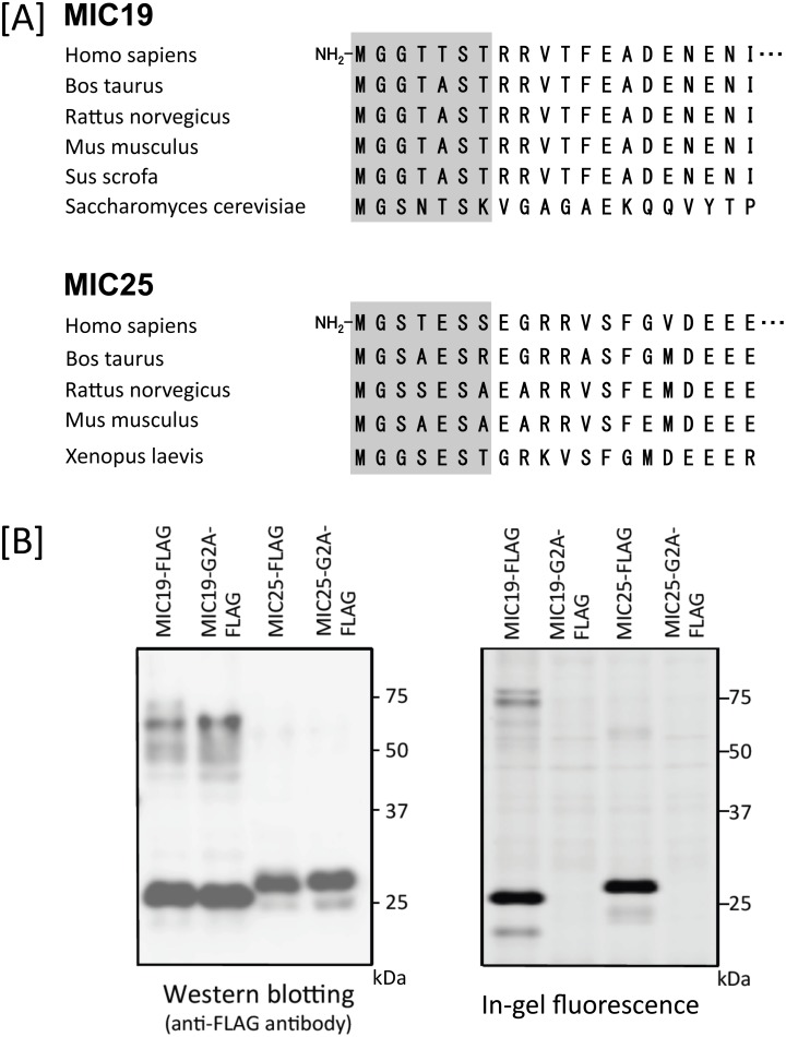
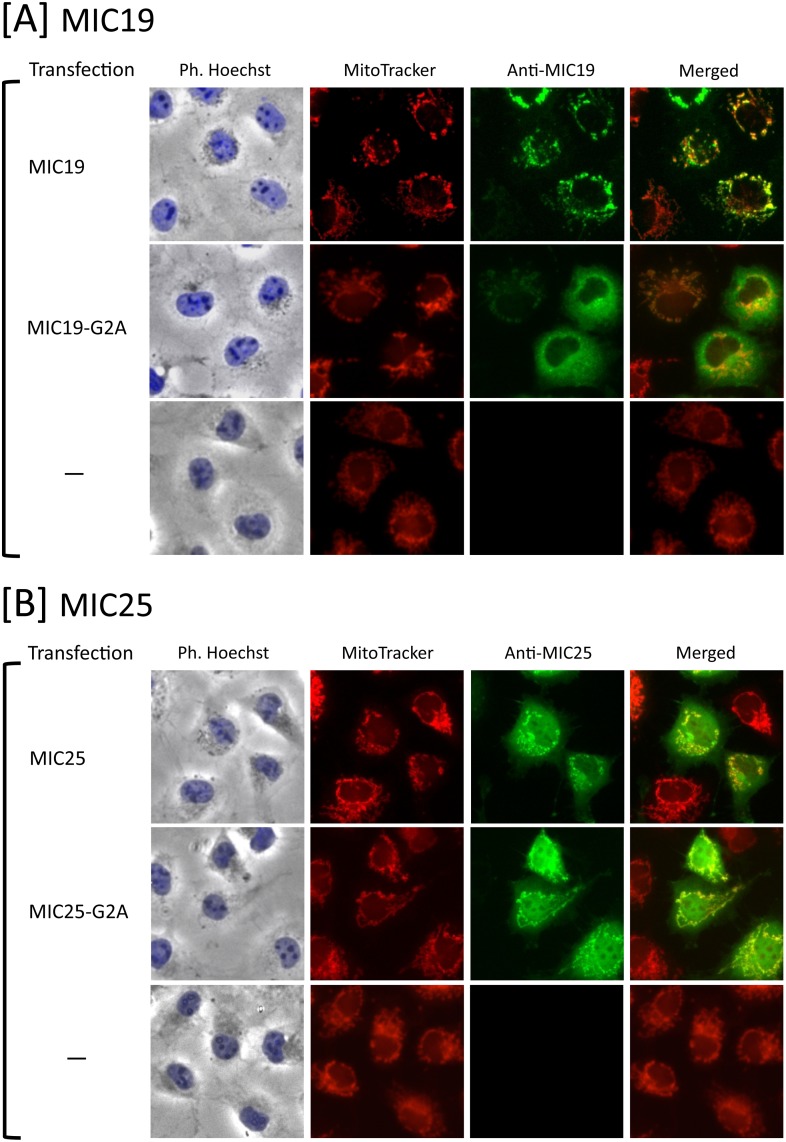
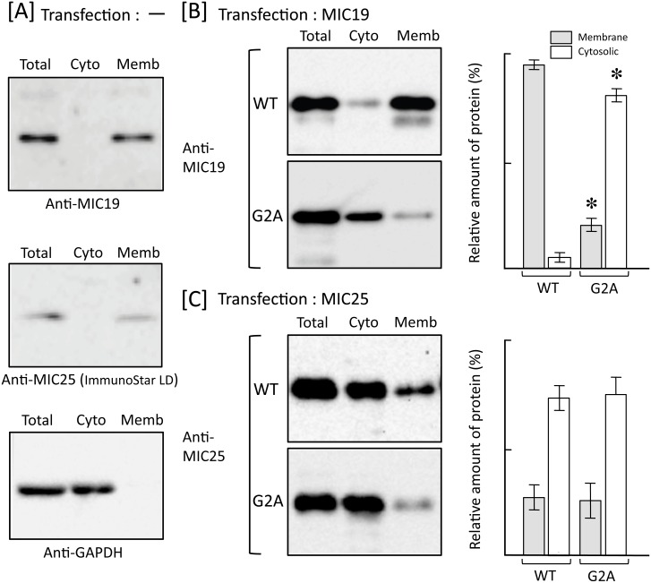
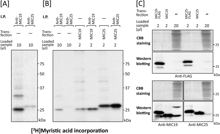
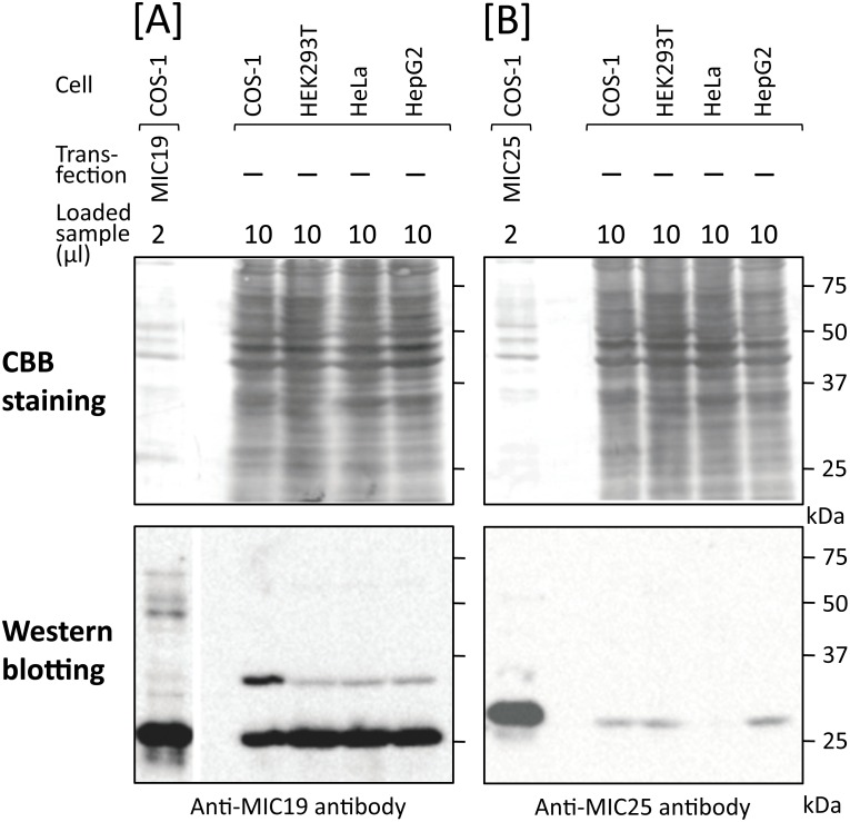
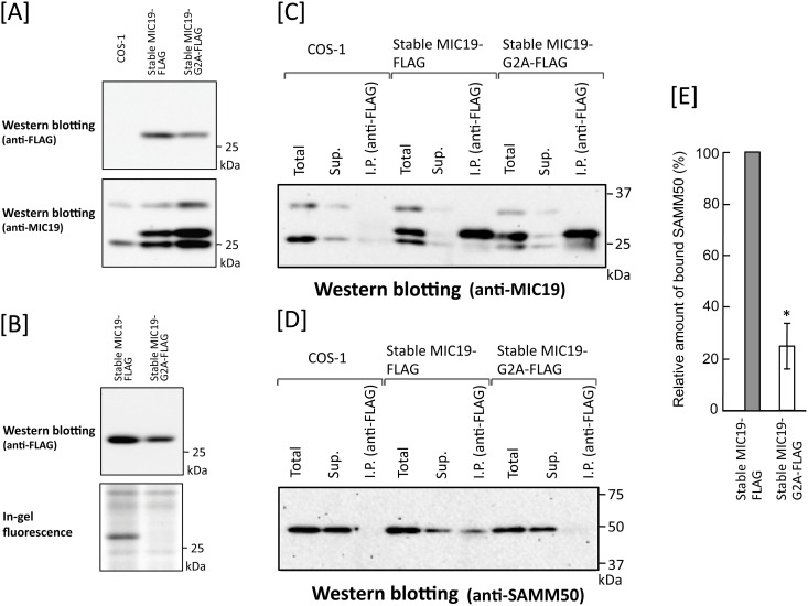
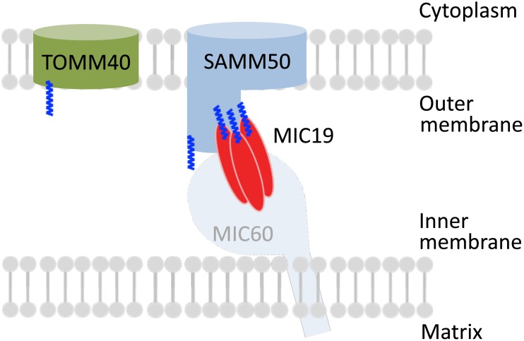
Previously, we showed that SAMM50, a mitochondrial outer membrane protein, is N-myristoylated, and this lipid modification is required for the proper targeting of SAMM50 to mitochondria. In this study, we characterized protein N-myristoylation occurring on four human mitochondrial proteins, SAMM50, TOMM40, MIC19, and MIC25, three of which are components of the mitochondrial intermembrane space bridging (MIB) complex, which plays a critical role in the structure and function of mitochondria. In vitro and in vivo metabolic labeling experiments revealed that all four of these proteins were N-myristoylated. Analysis of intracellular localization of wild-type and non-myristoylated G2A mutants of these proteins by immunofluorescence microscopic analysis and subcellular fractionation analysis indicated that protein N-myristoylation plays a critical role in mitochondrial targeting and membrane binding of two MIB components, SAMM50 and MIC19, but not those of TOMM40 and MIC25. Immunoprecipitation experiments using specific antibodies revealed that MIC19, but not MIC25, was a major N-myristoylated binding partner of SAMM50. Immunoprecipitation experiments using a stable transformant of MIC19 confirmed that protein N-myristoylation of MIC19 is required for the interaction between MIC19 and SAMM50, as reported previously. Thus, protein N-myristoylation occurring on two mitochondrial MIB components, SAMM50 and MIC19, plays a critical role in the mitochondrial targeting and protein-protein interaction between these two MIB components.


Conflict of interest statement

The authors have declared that no competing interests exist.

Figures

Fig 1
Fig 1. Detection of protein N-myristoylation of human SAMM50 and TOMM40 by cell-free and cellular metabolic labeling.
A. Interspecies alignment of the N-terminal sequences of SAMM50 and TOMM40. N-myristoylation motifs are shown in grey in the N-terminal sequences. B. The gene products of cDNAs encoding SAMM50-FLAG, SAMM50-G2A-FLAG, TOMM40-FLAG, and TOMM40-G2A-FLAG were synthesized using an insect cell-free protein synthesis system in the presence of [3H]leucine or [3H]myristic acid. The labeled translation products were analyzed by SDS-PAGE and fluorography, as described in the Materials and methods. C. cDNAs encoding SAMM50-FLAG, SAMM50-G2A-FLAG, TOMM40-FLAG, and TOMM40-G2A-FLAG were transfected into COS-1 cells. The expression of proteins was evaluated by western blotting analysis using an anti-FLAG antibody. Protein N-myristoylation was evaluated by metabolic labeling with a myristic acid analog followed by click chemistry, as described in the Materials and methods.
Fig 2
Fig 2. Protein N-myristoylation of SAMM50 but not of TOMM40 is required for proper targeting to mitochondria.
A. Intracellular localization of SAMM50 and SAMM50-G2A was determined by immunofluorescence staining of COS-1 cells transfected with cDNAs encoding these two proteins using an anti-SAMM50 antibody. Mitochondria were detected using MitoTracker Red. The same experiment was performed using non-transfected COS-1 cells as a control. B. Intracellular localization of TOMM40 and TOMM40-G2A was determined by immunofluorescence staining of COS-1 cells transfected with cDNAs encoding these two proteins using an anti-TOMM40 antibody. Mitochondria were detected using MitoTracker Red. The same experiment was performed using non-transfected COS-1 cells as a control.
Fig 3
Fig 3. Protein N-myristoylation of SAMM50 but not of TOMM40 is required for binding of protein to the membrane.
Total cell extracts of non-transfected COS-1 cells [A] and COS-1 cells transfected with cDNA encoding tag-free SAMM50 [B] or TOMM40 [C] were separated into cytosolic and membrane fractions by using membrane protein extraction kit (Trident). Presence of SAMM50 and TOMM40 in each fraction was determined by western blotting analysis using anti-SAMM50 or anti-TOMM40 antibody, respectively. For this analysis, anti-GAPDH antibody was used to detect GAPDH, a cytoslic marker protein [A]. For the analysis of COS-1 cells transfected with cDNAs coding for SAMM50 [B] and TOMM40 [C], relative amount of protein (%) reside in the cytosolic and total membrane fraction was calculated from densitometric analysis as described in materials and methods. Data are expressed as mean ± SD for three independent experiments. *P < 0.005 vs. wild-type.
Fig 4
Fig 4. Endogenous SAMM50 and TOMM40 expressed in mammalian cells are N-myristoylated.
A. Metabolic labeling of endogenous proteins expressed in COS-1 cells with [3H]myristic acid followed by immunoprecipitation with specific antibodies against SAMM50 and TOMM40 was performed. The labeled proteins were separated by SDS-PAGE and then detected by fluorography. B. Expression of endogenous SAMM50, TOMM40, and MIC19 in COS-1 cells was determined by western blotting analysis using the respective specific antibodies. For this analysis, tag-free-SAMM50, tag-free-TOMM40, and tag-free-MIC19 exogenously expressed in COS-1 cells were used as controls.
Fig 5
Fig 5. Identification of MIC19 as a major N-myristoylated binding partner of SAMM50.
A. Metabolic labeling of N-myristoylated proteins expressed in COS-1 cells with [3H]myristic acid followed by immunoprecipitation with specific antibodies against SAMM50, TOMM40, and MIC19 was performed. The labeled proteins were separated by SDS-PAGE and then detected by fluorography (lanes 4–6). For this analysis, tag-free-SAMM50, -TOMM40, and -MIC19 exogenously expressed in COS-1 cells were used as controls (lanes 1–3). B. Western blotting analysis of immunoprecipitated samples used in A. Presence of endogenous SAMM50, TOMM40, and MIC19 in the immunoprecipitated samples used in A lanes 4–6 was determined by western blotting analysis. For this analysis, total cell lysates of COS-1 cells were used as control (lanes 1, 5, and 9). Arrowheads in B indicate the position of heavy- and light-chains of IgG used for immunoprecipitation.
Fig 6
Fig 6. Detection of protein N-myristoylation occurring on human MIC19 and MIC25 by cellular metabolic labeling.
A. Interspecies alignment of the N-terminal sequences of MIC19 and MIC25. N-myristoylation motifs are shown in grey in the N-terminal sequences. B. cDNAs encoding MIC19-FLAG, MIC25-FLAG, and their G2A mutants were transfected into COS-1 cells. The expression of proteins was evaluated by western blotting analysis using anti-FLAG antibodies. Protein N-myristoylation was evaluated by metabolic labeling with a myristic acid analog followed by click chemistry, as described in the Materials and methods.
Fig 7
Fig 7. Protein N-myristoylation of MIC19 but not of MIC25 is required for proper targeting to mitochondria.
A. Intracellular localization of MIC19 and MIC19-G2A was determined by immunofluorescence staining of COS-1 cells transfected with cDNAs encoding these two proteins using an anti-MIC19 antibody. Mitochondria were detected using MitoTracker Red. The same experiment was performed using non-transfected COS-1 cells as a control. B. Intracellular localization of MIC25 and MIC25-G2A was determined by immunofluorescence staining of COS-1 cells transfected with cDNAs encoding these two proteins using an anti-MIC25 antibody. Mitochondria were detected using MitoTracker Red. The same experiment was performed using non-transfected COS-1 cells as a control.
Fig 8
Fig 8. Protein N-myristoylation of MIC19 but not of MIC25 is required for binding of protein to the membrane.
Total cell extracts of non-transfected COS-1 cells [A] and COS-1 cells transfected with cDNA encoding tag-free MIC19 [B] or MIC25 [C] were separated into cytosolic and membrane fractions by using membrane protein extraction kit (Trident). Presence of MIC19 and MIC25 in each fraction was determined by western blotting analysis using anti-MIC19 or anti-MIC25 antibody, respectively. For this analysis, anti-GAPDH antibody was used to detect GAPDH, a cytoslic marker protein [A]. For the analysis of COS-1 cells transfected with cDNAs coding for MIC19 [B] and MIC25 [C], relative amount of protein (%) reside in the cytosolic and total membrane fraction was calculated from densitometric analysis as described in materials and methods. Data are expressed as mean ± SD for three independent experiments. *P < 0.005 vs. wild-type.
Fig 9
Fig 9. Expression and N-myristoylation of endogenous MIC19 and MIC25 in COS-1 cells.
A. Metabolic labeling of endogenous proteins expressed in COS-1 cells with [3H]myristic acid followed by immunoprecipitation with specific antibodies against MIC19 and MIC25 was performed. The labeled proteins were separated by SDS-PAGE and then detected by fluorography. B. Comparison of the molecular size of endogenous N-myristoylated MIC19 and MIC25 immunoprecipitated from COS-1 cells with that of N-myristoylated tag-free MIC19 and MIC25 exogenously expressed in COS-1 cells. [3H]myristic acid labeling of COS-1 cells transfected with cDNA coding for tag-free MIC19 or MIC25 followed by immunoprecipitation with specific antibodies against these two proteins was performed. The immunoprecipitated samples and the samples obtained in A were separated by SDS-PAGE and then detected by fluorography. C. Comparison of the relative amounts of MIC19 and MIC25 expressed in COS-1 cells. The expression level of endogenous MIC25 in COS-1 cells was compared with that of MIC19 by western blotting analysis using MIC19-FLAG and MIC25-FLAG as standard proteins. For this analysis, total cell lysates derived from non-transfected COS-1 cells or COS-1 cells transfected with cDNAs coding for MIC19-FLAG, tag-free MIC19, MIC25-FLAG, or tag-free MIC25 were subjected to western blotting analysis using anti-FLAG, anti-MIC19, or anti-MIC25 antibodies.
Fig 10
Fig 10. Comparison of the relative amounts of MIC19 and MIC25 expressed in mammalian cells.
The expression levels of endogenous MIC25 in four mammalian cell lines (COS-1, HEK293T, HeLa, HepG2 cells) were compared with those of MIC19 by western blotting analysis using tag-free MIC19 and tag-free MIC25 expressed in COS-1 cells as standard proteins. For this analysis, total cell lysates derived from the four cell lines were subjected to western blotting analysis using anti-MIC19 or anti-MIC25 antibodies. A; Expression of MIC19, B; Expression of MIC25, Upper panels; Coomassie-brilliant blue (CBB) staining, Lower panels; Western blotting analysis.
Fig 11
Fig 11. Protein N-myristoylation of MIC19 is required for the association of MIC19 with SAMM50.
Association of MIC19-FLAG stably expressed in COS-1 cells with endogenously expressed SAMM50 was studied by immunoprecipitation analysis. A. Expression of MIC19-FLAG and MIC19-G2A-FLAG in respective stable transformants. Upper panel; Western blotting analysis using an anti-FLAG antibody, Lower panel; Western blotting analysis using an anti-MIC19 antibody. B. Detection of protein N-myristoylation of MIC19-FLAG expressed in stable transformants of MIC19-FLAG. Upper panel; Western blotting analysis using an anti-FLAG antibody. Lower panel; Detection of protein N-myristoylation by metabolic labeling with myristic acid analog followed by detection with click chemistry. C. Western blotting analysis of MIC19-FLAG or MIC19-G2A-FLAG immunoprecipitated from stable transformants of MIC19-FLAG and MIC19-G2A-FLAG using an anti-MIC19 antibody. D. Analysis of the binding of MIC19-FLAG or MIC19-G2A-FLAG to endogenous SAMM50 by western blotting analysis of immunoprecipitated samples of stable transformants of MIC19-FLAG and MIC19-G2A-FLAG using an anti-SAMM50 antibody. The experiments were performed in triplicate. A typical pattern of western blotting is presented. E. Quantitative analysis of MIC19-FLAG or MIC19-G2A-FLAG bound to SAMM50 was performed using the results obtained in D. Data are expressed as mean ± SD for three independent experiments. *P < 0.01 vs. wild-type.
Fig 12
Fig 12. Schematic representation of the intracellular localization and protein-protein interactions of SAMM50, TOMM40, and MIC19.
SAMM50, TOMM40, MIC19, and MIC25 were cotranslationally N-myristoylated in the cytoplasm. These proteins are transported into the intermembrane space of mitochondria through the TOM complex. For SAMM50 and MIC19, protein N-myristoylation is required for mitochondrial targeting. An oligomeric form of N-myristoylated MIC19 seems to associate with N-myristoylated SAMM50. Protein N-myristoylation of MIC19 is required for the binding of MIC19 to SAMM50.

Similar articles

Cited by

References

    1. Udenwobele DI, Su RC, Good SV, Ball TB, Varma Shrivastav S, Shrivastav A. Myristoylation: an important protein modification in the immune response. Front Immunol. 2017; 8: 751 10.3389/fimmu.2017.00751 - DOI - PMC - PubMed
    1. Peng T, Thinon E, Hang HC. Proteomic analysis of fatty-acylated proteins. Curr Opin Chem Biol. 2016; 30: 77–86. 10.1016/j.cbpa.2015.11.008 - DOI - PMC - PubMed
    1. Giglione C, Fieulaine S, Meinnel T. N-terminal protein modifications: Bringing back into play the ribosome. Biochimie. 2015; 114: 134–146. 10.1016/j.biochi.2014.11.008 - DOI - PubMed
    1. Resh MD. Trafficking and signaling by fatty-acylated and prenylated proteins. Nat Chem Biol. 2006; 2: 584–590. 10.1038/nchembio834 - DOI - PubMed
    1. Farazi TA, Waksman G, Gordon JI. The biology and enzymology of protein N-myristoylation. J Biol Chem. 2001; 276: 39501–39504. 10.1074/jbc.R100042200 - DOI - PubMed

Publication types

MeSH terms

Grants and funding

This work was supported by a Grant-in-Aid for Scientific Research (No. 26450125 and No. 17K07758, https://www.jsps.go.jp/j-grantsinaid/index.html) from the Ministry of Education, Culture, Sports, Science and Technology of Japan (to TU). The funder had no role in study design, data collection and analysis, decision to publish, or preparation of the manuscript. No additional external funding received for this study.

LinkOut - more resources