Characteristics of New Onset Herpes Simplex Keratitis after Keratoplasty
- PMID: 30425853
- PMCID: PMC6217905
- DOI: 10.1155/2018/4351460
Characteristics of New Onset Herpes Simplex Keratitis after Keratoplasty
Abstract
Purpose: To observe clinical characteristics and treatment outcomes of new onset herpes simplex keratitis (HSK) after keratoplasty.
Methods: Among 1,443 patients (1,443 eyes) who underwent keratoplasty (excluding cases of primary HSK) in Shandong Eye Hospital, 17 patients suffered postoperative HSK. The clinical manifestations, treatment regimens, and prognoses of the patients were evaluated.
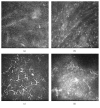
Results: The incidence of new onset HSK after keratoplasty was 1.18%. Epithelial HSK occurred in 10 eyes, with dendritic epithelial infiltration in 6 eyes and map-like epithelial defects in 4 eyes. Nine eyes had lesions at the junction of the graft and recipient. Stromal necrotic and endothelial HSK occurred in 7 eyes, presenting map-shaped ulcers in the entire corneal graft and recipient (two eyes) or at the graft-recipient junction (five eyes). Confocal microscopy revealed infiltration of a large number of dendritic cells at the junction of the lesion and transparent cornea. All 10 eyes with epithelial lesions and two eyes suffering stromal lesions of ≤1/3 corneal thickness healed after systematic and local antiviral treatment. Best-corrected visual acuity and corneal graft transparency were restored. For stromal HSK with an ulcer of >1/3 corneal thickness, amniotic membrane transplantation was performed, and visual acuity and graft transparency decreased significantly.
Conclusion: New onset HSK after keratoplasty primarily resulted in epithelial and stromal lesion, involving both the graft and recipient. Effective treatments included antiviral medications and amniotic membrane transplantation. Delayed treatment may lead to aggravated graft opacification.
Figures


Similar articles
-
Long-term comparison of full-bed deep lamellar keratoplasty with penetrating keratoplasty in treating corneal leucoma caused by herpes simplex keratitis.Am J Ophthalmol. 2012 Feb;153(2):291-299.e2. doi: 10.1016/j.ajo.2011.07.020. Epub 2011 Oct 13. Am J Ophthalmol. 2012. PMID: 21996306
-
[Deep anterior lamellar keratoplasty combined with antiviral therapy in the treatment of severe herpes necrotizing stromal keratitis].Zhonghua Yan Ke Za Zhi. 2018 Feb 11;54(2):97-104. doi: 10.3760/cma.j.issn.0412-4081.2018.02.006. Zhonghua Yan Ke Za Zhi. 2018. PMID: 29429293 Chinese.
-
Short-term results of acellular porcine corneal stroma keratoplasty for herpes simplex keratitis.Xenotransplantation. 2019 Jul;26(4):e12509. doi: 10.1111/xen.12509. Epub 2019 Apr 10. Xenotransplantation. 2019. PMID: 30968461 Clinical Trial.
-
Evidence in the prevention of the recurrence of herpes simplex and herpes zoster keratitis after eye surgery.Arch Soc Esp Oftalmol (Engl Ed). 2022 Mar;97(3):149-160. doi: 10.1016/j.oftale.2022.02.003. Epub 2022 Feb 19. Arch Soc Esp Oftalmol (Engl Ed). 2022. PMID: 35248396 Review.
-
Ocular surgery after herpes simplex and herpes zoster keratitis.Int Ophthalmol. 2020 Dec;40(12):3599-3612. doi: 10.1007/s10792-020-01539-6. Epub 2020 Sep 10. Int Ophthalmol. 2020. PMID: 32910331 Review.
Cited by
-
Herpes Simplex Keratitis Following Corneal Crosslinking for Keratoconus: A One-Year Case Series Follow-Up.Diagnostics (Basel). 2024 Oct 11;14(20):2267. doi: 10.3390/diagnostics14202267. Diagnostics (Basel). 2024. PMID: 39451590 Free PMC article.
-
Role of Innate Interferon Responses at the Ocular Surface in Herpes Simplex Virus-1-Induced Herpetic Stromal Keratitis.Pathogens. 2023 Mar 10;12(3):437. doi: 10.3390/pathogens12030437. Pathogens. 2023. PMID: 36986359 Free PMC article. Review.
-
Regulation of herpes simplex virus type 1 latency-reactivation cycle and ocular disease by cellular signaling pathways.Exp Eye Res. 2022 May;218:109017. doi: 10.1016/j.exer.2022.109017. Epub 2022 Mar 1. Exp Eye Res. 2022. PMID: 35240194 Free PMC article.
-
Cluster of Symptomatic Graft-to-Host Transmission of Herpes Simplex Virus Type 1 in an Endothelial Keratoplasty Setting.Ophthalmol Sci. 2021 Aug 12;1(3):100051. doi: 10.1016/j.xops.2021.100051. eCollection 2021 Sep. Ophthalmol Sci. 2021. PMID: 36247820 Free PMC article.
-
Ocular manifestations of herpes simplex virus.Curr Opin Ophthalmol. 2019 Nov;30(6):525-531. doi: 10.1097/ICU.0000000000000618. Curr Opin Ophthalmol. 2019. PMID: 31567695 Free PMC article. Review.
References
LinkOut - more resources
Full Text Sources

