

Epstein-Barr virus BORF2 inhibits cellular APOBEC3B to preserve viral genome integrity
- PMID: 30420783
- PMCID: PMC6294688
- DOI: 10.1038/s41564-018-0284-6
Epstein-Barr virus BORF2 inhibits cellular APOBEC3B to preserve viral genome integrity
Abstract
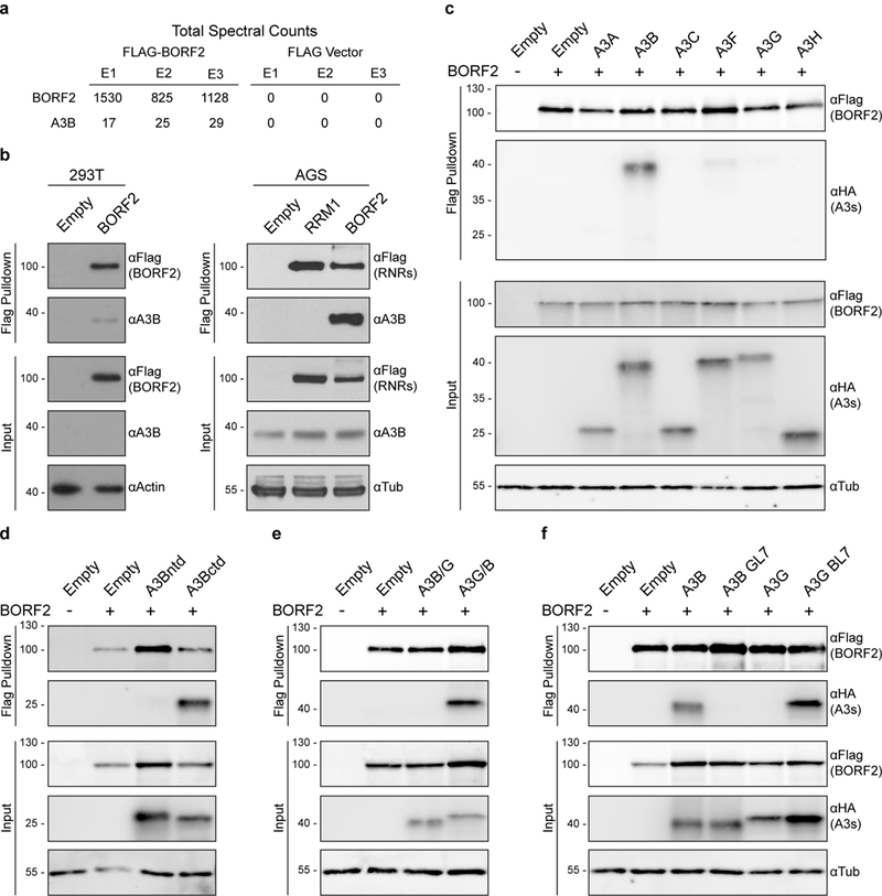
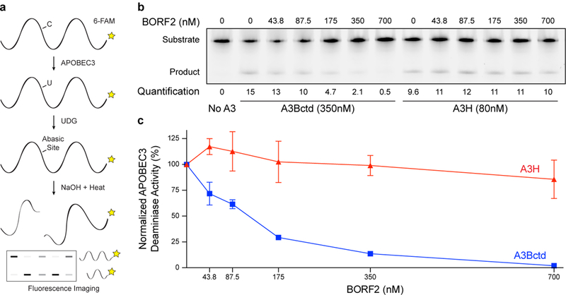
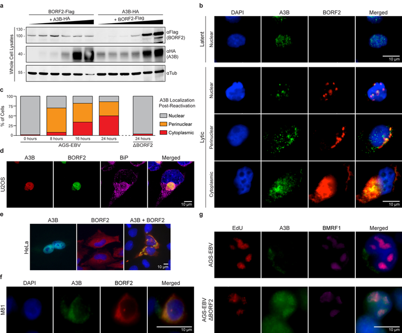
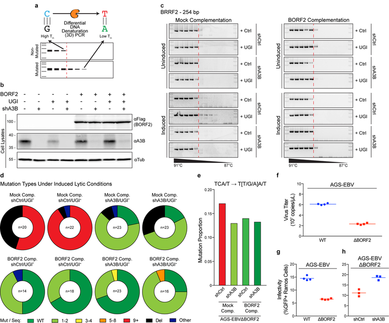
The apolipoprotein B messenger RNA editing enzyme, catalytic polypeptide-like (APOBEC) family of single-stranded DNA (ssDNA) cytosine deaminases provides innate immunity against virus and transposon replication1-4. A well-studied mechanism is APOBEC3G restriction of human immunodeficiency virus type 1, which is counteracted by a virus-encoded degradation mechanism1-4. Accordingly, most work has focused on retroviruses with obligate ssDNA replication intermediates and it is unclear whether large double-stranded DNA (dsDNA) viruses may be similarly susceptible to restriction. Here, we show that the large dsDNA herpesvirus Epstein-Barr virus (EBV), which is the causative agent of infectious mononucleosis and multiple cancers5, utilizes a two-pronged approach to counteract restriction by APOBEC3B. Proteomics studies and immunoprecipitation experiments showed that the ribonucleotide reductase large subunit of EBV, BORF26,7, binds APOBEC3B. Mutagenesis mapped the interaction to the APOBEC3B catalytic domain, and biochemical studies demonstrated that BORF2 stoichiometrically inhibits APOBEC3B DNA cytosine deaminase activity. BORF2 also caused a dramatic relocalization of nuclear APOBEC3B to perinuclear bodies. On lytic reactivation, BORF2-null viruses were susceptible to APOBEC3B-mediated deamination as evidenced by lower viral titres, lower infectivity and hypermutation. The Kaposi's sarcoma-associated herpesvirus homologue, ORF61, also bound APOBEC3B and mediated relocalization. These data support a model where the genomic integrity of human γ-herpesviruses is maintained by active neutralization of the antiviral enzyme APOBEC3B.
Conflict of interest statement
Figures
Comment in
-
APOBEC restriction goes nuclear.Nat Microbiol. 2019 Jan;4(1):6-7. doi: 10.1038/s41564-018-0323-3. Nat Microbiol. 2019. PMID: 30546096 No abstract available.
Similar articles
-
G1/S Cell Cycle Induction by Epstein-Barr Virus BORF2 Is Mediated by P53 and APOBEC3B.J Virol. 2022 Sep 28;96(18):e0066022. doi: 10.1128/jvi.00660-22. Epub 2022 Sep 7. J Virol. 2022. PMID: 36069545 Free PMC article.
-
A Conserved Mechanism of APOBEC3 Relocalization by Herpesviral Ribonucleotide Reductase Large Subunits.J Virol. 2019 Nov 13;93(23):e01539-19. doi: 10.1128/JVI.01539-19. Print 2019 Dec 1. J Virol. 2019. PMID: 31534038 Free PMC article.
-
Human cytomegalovirus mediates APOBEC3B relocalization early during infection through a ribonucleotide reductase-independent mechanism.J Virol. 2023 Aug 31;97(8):e0078123. doi: 10.1128/jvi.00781-23. Epub 2023 Aug 11. J Virol. 2023. PMID: 37565748 Free PMC article.
-
APOBECs and Herpesviruses.Viruses. 2021 Feb 28;13(3):390. doi: 10.3390/v13030390. Viruses. 2021. PMID: 33671095 Free PMC article. Review.
-
Roles of APOBEC3A and APOBEC3B in Human Papillomavirus Infection and Disease Progression.Viruses. 2017 Aug 21;9(8):233. doi: 10.3390/v9080233. Viruses. 2017. PMID: 28825669 Free PMC article. Review.
Cited by
-
G1/S Cell Cycle Induction by Epstein-Barr Virus BORF2 Is Mediated by P53 and APOBEC3B.J Virol. 2022 Sep 28;96(18):e0066022. doi: 10.1128/jvi.00660-22. Epub 2022 Sep 7. J Virol. 2022. PMID: 36069545 Free PMC article.
-
ATM, KAP1 and the Epstein-Barr virus polymerase processivity factor direct traffic at the intersection of transcription and replication.Nucleic Acids Res. 2023 Nov 10;51(20):11104-11122. doi: 10.1093/nar/gkad823. Nucleic Acids Res. 2023. PMID: 37852757 Free PMC article.
-
Insights into the Structures and Multimeric Status of APOBEC Proteins Involved in Viral Restriction and Other Cellular Functions.Viruses. 2021 Mar 17;13(3):497. doi: 10.3390/v13030497. Viruses. 2021. PMID: 33802945 Free PMC article. Review.
-
Epstein-Barr virus and malaria upregulate AID and APOBEC3 enzymes, but only AID seems to play a major mutagenic role in Burkitt lymphoma.Eur J Immunol. 2022 Aug;52(8):1273-1284. doi: 10.1002/eji.202249820. Epub 2022 May 12. Eur J Immunol. 2022. PMID: 35503749 Free PMC article.
-
Single-nucleotide polymorphism of the DNA cytosine deaminase APOBEC3H haplotype I leads to enzyme destabilization and correlates with lung cancer.NAR Cancer. 2020 Sep;2(3):zcaa023. doi: 10.1093/narcan/zcaa023. Epub 2020 Sep 17. NAR Cancer. 2020. PMID: 32984821 Free PMC article.
References
-
- Malim MH & Emerman M HIV-1 accessory proteins--ensuring viral survival in a hostile environment. Cell Host Microbe 3, 388–398 (2008). - PubMed
-
- Yang B, Li X, Lei L & Chen J APOBEC: from mutator to editor. J Genet Genomics 44, 423–437 (2017). - PubMed
-
- Rickinson A & Kieff E in Virology Fields (eds Knipe DM & Howley PM) 2655–2700 (Lippincott Williams & Wilkins, 2007).
Publication types
MeSH terms
Substances
Grants and funding
LinkOut - more resources
Full Text Sources
Research Materials
