. 2018 Nov 5;373(1762):20180170.
doi: 10.1098/rstb.2018.0170.

Role of oligouridylation in normal metabolism and regulated degradation of mammalian histone mRNAs

Affiliations

Role of oligouridylation in normal metabolism and regulated degradation of mammalian histone mRNAs

Stacie A Meaux et al. Philos Trans R Soc Lond B Biol Sci. .

Abstract

Metazoan replication-dependent histone mRNAs are the only known cellular mRNAs that are not polyadenylated. Histone mRNAs are present in large amounts only in S-phase cells, and their levels are coordinately regulated with the rate of DNA replication. In mammals, the stemloop at the 3' end of histone mRNA is bound to stemloop binding protein, a protein required for both synthesis and degradation of histone mRNA, and an exonuclease, 3'hExo (ERI1). Histone mRNAs are rapidly degraded when DNA synthesis is inhibited in S-phase cells and at the end of S-phase. Upf1 is also required for rapid degradation of histone mRNA as is the S-phase checkpoint. We report that Smg1 is required for histone mRNA degradation when DNA replication is inhibited, suggesting it is the PI-like kinase that activates Upf1 for histone mRNA degradation. We also show that some mutant Upf1 proteins are recruited to histone mRNAs when DNA replication is inhibited and act as dominant negative factors in histone mRNA degradation. We report that the pathway of rapid histone mRNA degradation when DNA replication is inhibited in S-phase cells that are activating the S-phase checkpoint is similar to the pathway of rapid degradation of histone mRNA at the end of S-phase.This article is part of the theme issue '5' and 3' modifications controlling RNA degradation'.

Keywords: RNA degradation; cell cycle; histone mRNA; uridylation.


Conflict of interest statement

The authors declare that they have no competing interests.

Figures

Figure 1.
Figure 1.
Cytoplasmic histone mRNAs. (a) A schematic of a mammalian histone mRNA showing the cap on the 5′ end, the short 5′ and 3′ UTRs and the stemloop at the 3′ end, which is bound by SLBP and 3′hExo. (b) Histone mRNA regulation during the cell cycle. Histone mRNAs increase just before entry into S-phase and decrease at the end of S-phase [2]. The levels of SLBP protein are also cell cycle regulated [1], while there is very little change in SLBP mRNA levels. Inhibition of DNA replication during S-phase (dotted line) results in rapid degradation of histone mRNA but not of SLBP. (c) The 3′ ends of the HIST2H2AA3 mRNAs from HCT116 S-phase cells, determined by high-throughput sequencing using the End-Seq approach [7]. The x-axis displays the nts at the 3′ end of the mRNA, with numbers referring to the drawing of the stemloop. The bars indicate the number of RNAs (y-axis) that have their last templated nt at that position. Blue indicates the RNA ends with the last templated nt, green indicates there was a single U added at that site, orange indicates there were two Us added at that site, and red indicates that there were more than 2 nts added at that site.
Figure 2.
Figure 2.
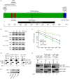
Upf1/Smg1 are required for histone mRNA degradation. (a) Diagram of the C-terminal-tagged SLBP containing a CBP, a streptavidin-binding peptide and an HA tag (i). A stable HeLa cell line expressing the SLBP–SBP was generated, and SLBP and SLBP–SBP were detected by western blotting with the SLBP antibody (ii). (b) Knockdown of Smg1 or Upf1 slows the degradation of histone mRNA after inhibiting DNA replication. Cells treated with the indicated siRNAs were treated with HU for the indicated times, and the levels of histone mRNA were determined by Northern blotting and quantified with a PhosphorImager. The western blots below the graph show the effectiveness of the knockdown. (c) Lysates of synchronized SLBP–SBP-expressing cells were prepared 3 h after release into S-phase, before and 20 min after treating the cells with 5 mM HU. (i) The lysates were bound to streptavidin beads, and bound proteins were resolved by SDS gel electrophoresis and detected with the anti-HA antibody (bottom) or the anti-Upf1 antibody (top). (ii) The lysates were treated the same way, and the bound proteins were detected with anti-HA or with anti-Smg1 antibody. 5% of the input was analysed in the lane next to the antibody precipitate. Smg1 was analysed using a 4% polyacrylamide gel, while Upf1 was detected with a 6% polyacrylamide gel. IN, input. (d) Cells expressing SLBP–SBP were treated with control (C2) siRNA or SMG1 siRNA for 72 h. Lysates were prepared from C2 siRNA cells (lanes 1 and 2) or SMG siRNA-treated cells (lanes 3 and 4) 20 min after treatment with 5 mM HU, and lysates were prepared from the cells. The lysates were immunoprecipitated with anti-HA antibody. 5% of the input and the immunoprecipitate (lanes AP) were resolved by gel electrophoresis. SLBP–SBP and endogenous SLBP were detected by western blotting with anti-SLBP antibody and Upf1 was detected with an anti-Upf1 antibody. Lanes 1 and 3 are 5% of the input compared to the antibody precipitate in lanes 2 and 4. (Online version in colour.)
Figure 3.
Figure 3.
Requirements for Upf1 for histone mRNA degradation. (a) A schematic of the Upf1 protein. (b) Effect of deleting the N- or C-terminal domains of Upf1 on histone mRNA degradation. The SLBP–SBP-expressing HeLa cells (figure 2a) were transfected with the indicated HA-tagged Upf1 proteins. After 72 h, cells were treated with HU, and RNA samples analysed by Northern blotting to detect histone H2a mRNA and 7SK mRNA as a control. (c) The results of three independent biological replicates of the HU treatment are plotted, with s.d. shown. The * indicates that there is a significant retardation of histone mRNA degradation (p < 0.05). (d) Lysates were prepared from SLBP–SBP cells expressing the indicated HA-tagged Upf1 proteins, K498A, R843C or HA-Upf1, 20 min after HU treatment. The proteins bound to SLBP–SBP (lanes 2,4,6,8) were isolated as in figure 2 and detected by western blotting with SLBP antibody and with anti-HA to detect the transfected Upf1. 5% of the input is shown in lanes 1,3,5,7. (e) Lysates were prepared from SLBP–SBP cells expressing the indicated HA-tagged Upf1 deletion proteins, 215–1148 and 1–984, or HA-Upf1, 20 min after HU treatment. The proteins bound to SLBP–SBP (lanes 2,4,6) were isolated as in figure 2 and detected by western blotting with SLBP antibody and with anti-HA to detect the transfected Upf1. 5% of the input is shown in lanes 1,3,5. IN, input. (f) Western blot showing the expression of the HA-tagged Upf1 proteins 72 h after transfection. PTB (polypyrimidine tract binding protein) was used as a control. (Online version in colour.)
Figure 4.
Figure 4.
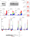
Histone mRNA is degraded by similar pathways after HU treatment and at the end of S-phase. (a) HCT116 cells were synchronized and released into S-phase. At 3 h, cell samples were harvested before treatment with 5 mM HU and 20 or 40 min after treatment with HU. Total RNA was prepared and analysed by Northern blotting for histone H2a mRNAs and 7SK RNA. (b) Cells were harvested at the indicated times after release into S-phase, and total cell RNA was harvested and analysed as in (a). The values for histone mRNA were determined relative to 7SK RNA. Note that the 7SK probe in (a) was of much lower specific activity than the probe used in (b). Below the gel, the amount of histone mRNA present relative to 3 h after release is indicated. (c) Cells from the experiment in (b) were analysed by flow cytometry to determine the percentage of cells in S-phase. The red dots indicate EdU-stained cells (intensity of staining on the y-axis). The cells in the lower left corner are cells that never entered S-phase. The percentage of cells in S-phase and the ratio of S-phase:G2-M cells are indicated under the time. (d,e) Libraries were prepared for analysis of histone mRNA 3′ ends from the cells in mid-phase (3 h), HU treated cells and cells near the end of S-phase (6 h), and the histone mRNAs were sequenced. The RNAs that had some degradation into the stemloop were plotted (ranging from nt 5 to nt 20, which is the end of the stem). The height of the bar indicating the percentage of total reads is indicated on each y-axis. (e) The 3′ ends of intermediates that extended up to 100 nts from the 3′ end are plotted. Note that these start at position 7. The position of the stop codon is indicated by the red octagon. The peak at about nt 75 is 15 nts after the stop codon. Note that there are many fewer degradation intermediates present in the mid-phase (3 h) sample. (f) A schematic of the 3′ end of the histone mRNA with the degradation intermediate after the stop codon indicated.
Figure 5.
Figure 5.
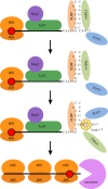
Intermediates in 3′–5′ degradation of histone mRNA. The histone mRNP associated with SLBP, 3′hExo and TUT7 (figure 1a) is the actively translating form of histone mRNA. The proposed initial step in histone mRNA degradation is loss of efficient translation termination (possibly by an unknown modification of SLBP). This results in binding of Upf1 and Smg1, phosphorylation of Upf1 and subsequent weakening of the SLBP/SL interaction, allowing 3′hExo to initiate degradation into the stemloop. The U-tails added in the stem bind Lsm1–7, which can also bind directly to SLBP and 3′hExo through the C-terminal tail of Lsm4. This intermediate accumulates but is resolved by loss of SLBP and 3′hExo, followed by loss of Upf1, resulting in the recruitment of the exosome and rapid degradation until the exosome encounters the ribosome bound to the stop codon, resulting in the formation of a uridylated intermediate. Subsequent degradation can occur either (or both) 5′–3′ as a result of continued exonucleolytic degradation and/or decapping of the mRNA and 5′–3′ degradation.

Similar articles

Cited by

References

    1. Whitfield ML, Zheng L-X, Baldwin A, Ohta T, Hurt MM, Marzluff WF. 2000. Stem-loop binding protein, the protein that binds the 3′ end of histone mRNA, is cell cycle regulated by both translational and posttranslational mechanisms. Mol. Cell. Biol. 20, 4188–4198. (10.1128/MCB.20.12.4188-4198.2000) - DOI - PMC - PubMed
    1. Harris ME, Böhni R, Schneiderman MH, Ramamurthy L, Schümperli D, Marzluff WF. 1991. Regulation of histone mRNA in the unperturbed cell cycle: evidence suggesting control at two posttranscriptional steps. Mol. Cell. Biol. 11, 2416–2424. (10.1128/MCB.11.5.2416) - DOI - PMC - PubMed
    1. Scharl EC, Steitz JA. 1994. The site of 3′ end formation of histone messenger RNA is a fixed distance from the downstream element recognized by the U7 snRNP. EMBO J. 13, 2432–2440. (10.1002/j.1460-2075.1994.tb06528.x) - DOI - PMC - PubMed
    1. Skrajna A, Yang XC, Dadlez M, Marzluff WF, Dominski Z. 2018. Protein composition of catalytically active U7-dependent processing complexes assembled on histone pre-mRNA containing biotin and a photo-cleavable linker. Nucleic Acids Res. 46, 4752–4770. (10.1093/nar/gky133) - DOI - PMC - PubMed
    1. Tan D, Marzluff WF, Dominski Z, Tong L. 2013. Structure of histone mRNA stem-loop, human stem-loop binding protein, and 3'hExo ternary complex. Science 339, 318–321. (10.1126/science.1228705) - DOI - PMC - PubMed

Publication types