. 2018 Dec 28;293(52):20273-20284.
doi: 10.1074/jbc.RA118.004141. Epub 2018 Oct 31.

Conformational flexibility of histone variant CENP-ACse4 is regulated by histone H4: A mechanism to stabilize soluble Cse4

Affiliations

Conformational flexibility of histone variant CENP-ACse4 is regulated by histone H4: A mechanism to stabilize soluble Cse4

Nikita Malik et al. J Biol Chem. .

Abstract

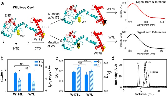
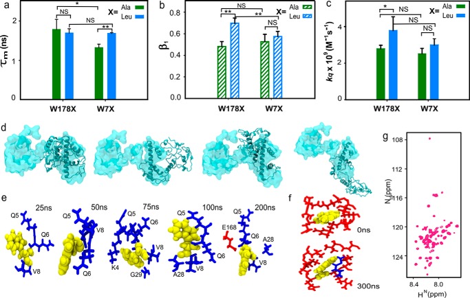
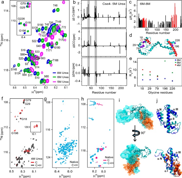
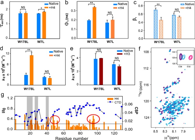
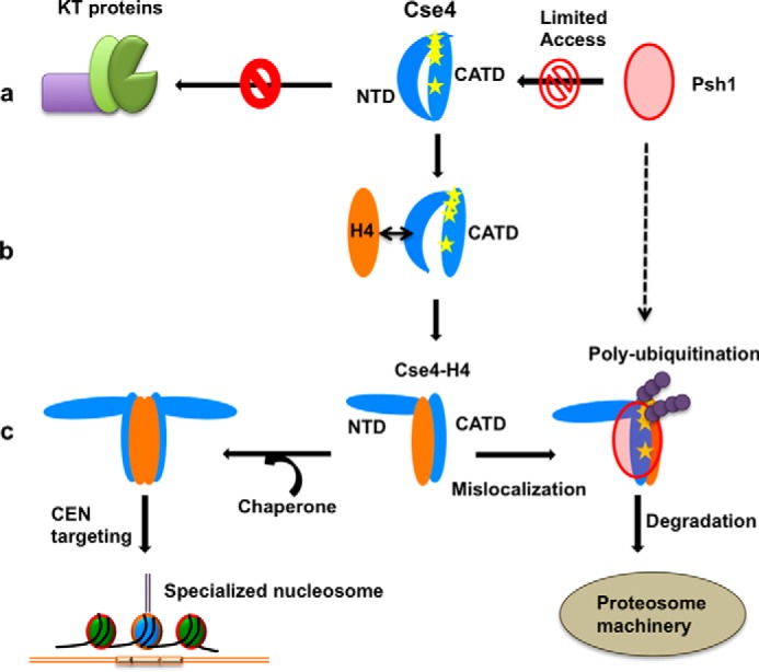
The histone variant CENP-ACse4 is a core component of the specialized nucleosome at the centromere in budding yeast and is required for genomic integrity. Accordingly, the levels of Cse4 in cells are tightly regulated, primarily by ubiquitin-mediated proteolysis. However, structural transitions in Cse4 that regulate its centromeric localization and interaction with regulatory components are poorly understood. Using time-resolved fluorescence, NMR, and molecular dynamics simulations, we show here that soluble Cse4 can exist in a "closed" conformation, inaccessible to various regulatory components. We further determined that binding of its obligate partner, histone H4, alters the interdomain interaction within Cse4, enabling an "open" state that is susceptible to proteolysis. This dynamic model allows kinetochore formation only in the presence of H4, as the Cse4 N terminus, which is required for interaction with other centromeric components, is unavailable in the absence of H4. The specific requirement of H4 binding for the conformational regulation of Cse4 suggests a structure-based regulatory mechanism for Cse4 localization. Our data suggested a novel structural transition-based mechanism where conformational flexibility of the Cse4 N terminus can control Cse4 levels in the yeast cell and prevent Cse4 from interacting with kinetochore components at ectopic locations for formation of premature kinetochore assembly.

Keywords: cell division; centromere; centromeric protein A; chromosome; fluorescence anisotropy; histone; histone tails; histone variants; kinetochore assembly; molecular dynamics; protein-protein interactions; structural biology.


Conflict of interest statement

The authors declare that they have no conflicts of interest with the contents of this article

Figures

Figure 1.
Figure 1.
CENP-ACse4 N-terminal tail is restricted. a, strategy for the fluorescence assay to create single Trp mutants in Cse4 to study the two domains individually. The END and HFD are highlighted in red. NTD and CTD correspond to residues 1–129 and 130–229, respectively. Note that according to a proposed model, Trp-7 is in a disordered region, implying that it is expected to have more conformational freedom than Trp-178. b, comparison of fluorescence lifetimes (filled bars) and solvent accessibility (striped bars) of the two domains. c, conformational flexibility of the two Trp residues in the native state. Filled bars represent φ1, and the striped bars represent β1. d, gel-filtration profile of folded Cse4 protein (solid line). The dotted lines represent the protein markers ovalbumin (O; 44 kDa) and carbonic anhydrase (CA; 29 kDa). The Cse4 structure represented in all figures is the modeled structure by Bloom et al. (Protein Data Bank (PDB) code 2FSC; Ref. 21) and is rendered in PyMOL 1.8. The statistical significance was calculated by one-way analysis of variance: **, p < 0.01; NS (not significant), p > 0.05; error bars represent S.D. A.U., arbitrary units.
Figure 2.
Figure 2.
CENP-ACse4 N-terminal tail interacts with C terminus. a, comparison between the fluorescence lifetime values of the Ala and Leu mutants for the two domains. b, β1 associated with the short correlation time for each mutant. c, solvent accessibility of the two domains in the Ala and Leu mutants. d, overlap of the structure of Cse4 at the start (space-filled) and end (ribbon) of all four simulations. The conformation of the N terminus changes from extended to closed even though the four simulations end in different conformational basins. e, change in the conformation of Trp-7 throughout simulation 1. f, conformation of Trp-178 at the start and end of the simulation. g, 1H-15N HSQC spectrum of folded Cse4. In all graphs, X represents the mutant type with blue and green representing Leu and Ala mutants, respectively. Trp residues are shown in yellow, and N-terminal and C-terminal residues are shown in blue and red sticks, respectively. The statistical significance was calculated by one-way analysis of variance: *, p < 0.05; **, p < 0.01; NS (not significant), p > 0.05; error bars represent S.D.
Figure 3.
Figure 3.
H4 stabilizes CENP-ACse4. a, representative regions of 1H-15N HSQC spectra of Cse4 at different urea concentrations. Inset, peak positions of five Gly residues in similar conditions. b, structural propensities of Cse4 in 5 m urea. Plots of secondary chemical shifts from ΔδCα, ΔδCO, and ΔδHα of residues Asp-15 to Ser-18 (not marked), 30–42, and 75–78 show helical propensity (gray bars). c, difference between the residue-wise R2 rates of Cse4 in 8 and 6 m urea. Residues showing maximum difference for each domain are marked in red. d, modeled Cse4 structure (21) shows the position of residues showing higher R2 difference (red). Note that they are a part of the END and HFD regions. The Gly residues are shown in violet. e, R2 for Gly residues in different denaturant concentrations. f, overlapped regions of 1H-15N HSQC spectra of Cse4 and Cse4–H4 in 4 m urea buffer. Gly residues are marked to show broadening of the C-terminal residues. Inset, Trp side-chain peaks. g, 1H-15N HSQC spectrum of Cse4–H4 complex without denaturant. h, overlap between 1H-15N HSQC spectra of Cse4 and Cse4–H4 in the native state. The arrows indicate reappearance of the NTD Gly residues in the Cse4–H4 spectrum. i, overlap of the structure of Cse4–H4 at the start (space-filled) and end (ribbon) of simulation 1. The N terminus does not fold back on the C terminus. j, arrangement of the C-terminal helices with and without H4 binding at the start (Cse4, blue; H4, orange) and end (Cse4, cyan; H4, brown) of the simulation.
Figure 4.
Figure 4.
H4 binding alters NTD conformation. a, comparison between the fluorescence lifetime values of Cse4 and Cse4–H4 for the two domains. b, change in conformational flexibility of the Trp at the two domains with and without H4. c, β1 associated with the short correlation time for each mutant. d, solvent accessibility of Trp residues at the two domains in Cse4 and Cse4–H4. e, solvent accessibility of Trp residues at the two domains in Cse4 and Cse4–H3. f, overlap of 1H-15N HSQC spectra of Cse4–H4 (light blue) and Cse4(1–129) (purple). Inset, position of Trp side-chain peaks in Cse4, Cse4–H4, and Cse4ΔC. g, comparison of the residue-specific CSPs calculated for the NTD upon addition of H4 (orange histogram) and the change in intensity profile upon titration with CTD (blue scatter plot). Gray bars represent the residues involved in interaction with both the CTD and H4. Some residues show significant CSP but do not interact with the CTD (highlighted by red circles). In the graphs, blue, orange, and brown represent Cse4, Cse4–H4, and Cse4–H3, respectively. Mutants are specified at the x axis. NMR spectra are shown for Cse4–H4 (light blue), Cse4 (pink), and Cse4ΔC (residues 1–129) (purple). The statistical significance was calculated by one-way analysis of variance: *, p < 0.05; **, p < 0.01; NS (not significant), p > 0.05; error bars represent S.D.
Figure 5.
Figure 5.
Proposed role of the NTD conformational change on regulation of Cse4. a, the closed state of the Cse4 NTD prevents interaction with kinetochore (KT) proteins. It also alters the positions of the helices in the C terminus, thereby masking the binding sites (Lys residues highlighted as yellow stars) for Psh1. b, a possible transient state when H4 comes in contact with Cse4. The affinity of NTD residues for H4 can help in dislodging the NTD from the CTD before H4 stably binds to the C terminus. c, upon H4 binding, the NTD adopts an open conformation. The Cse4–H4 dimer/tetramer can be deposited on the centromere by the chaperone Scm3 where the NTD is free to interact with the kinetochore. The C-terminal helices are reoriented in a manner that facilitates ubiquitination and further degradation if Cse4–H4 mislocalizes to an ectopic location.

Similar articles

Cited by

References

    1. Foltz D. R., Jansen L. E., Black B. E., Bailey A. O., Yates J. R. 3rd, and Cleveland D. W. (2006) The human CENP-A centromeric nucleosome-associated complex. Nat. Cell Biol. 8, 458–469 10.1038/ncb1397 - DOI - PubMed
    1. Stoler S., Keith K. C., Curnick K. E., and Fitzgerald-Hayes M. (1995) A mutation in CSE4, an essential gene encoding a novel chromatin-associated protein in yeast, causes chromosome nondisjunction and cell cycle arrest at mitosis. Genes Dev. 9, 573–586 10.1101/gad.9.5.573 - DOI - PubMed
    1. Furuyama S., and Biggins S. (2007) Centromere identity is specified by a single centromeric nucleosome in budding yeast. Proc. Natl. Acad. Sci. U.S.A. 104, 14706–14711 10.1073/pnas.0706985104 - DOI - PMC - PubMed
    1. Meluh P. B., and Koshland D. (1997) Budding yeast centromere composition and assembly as revealed by in vivo cross-linking. Genes Dev. 11, 3401–3412 10.1101/gad.11.24.3401 - DOI - PMC - PubMed
    1. Zhang W., Mellone B. G., and Karpen G. H. (2007) A specialized nucleosome has a “point” to make. Cell 129, 1047–1049 10.1016/j.cell.2007.05.054 - DOI - PMC - PubMed

Publication types

MeSH terms

Associated data

LinkOut - more resources