Review
. 2018 Oct 24;8(4):123.
doi: 10.3390/biom8040123.

Pre-Ribosomal RNA Processing in Human Cells: From Mechanisms to Congenital Diseases

Affiliations
Review

Pre-Ribosomal RNA Processing in Human Cells: From Mechanisms to Congenital Diseases

Maxime Aubert et al. Biomolecules. .

Abstract

Ribosomal RNAs, the most abundant cellular RNA species, have evolved as the structural scaffold and the catalytic center of protein synthesis in every living organism. In eukaryotes, they are produced from a long primary transcript through an intricate sequence of processing steps that include RNA cleavage and folding and nucleotide modification. The mechanisms underlying this process in human cells have long been investigated, but technological advances have accelerated their study in the past decade. In addition, the association of congenital diseases to defects in ribosome synthesis has highlighted the central place of ribosomal RNA maturation in cell physiology regulation and broadened the interest in these mechanisms. Here, we give an overview of the current knowledge of pre-ribosomal RNA processing in human cells in light of recent progress and discuss how dysfunction of this pathway may contribute to the physiopathology of congenital diseases.

Keywords: Diamond–Blackfan anemia; RNA processing; endonucleases; exonucleases; ribosomal RNAs (rRNAs); ribosomal stress; ribosomopathies.


Conflict of interest statement

The authors declare no conflicts of interest.

Figures

Figure 1
Figure 1
Structure of the human ribosome solved by cryo-electron microscopy. The figure shows the ribosomal RNAs in the large subunit (28S: pink, 5.8S: green, 5S: blue) and in the small subunit (18S: purple). (a) Structure with the ribosomal proteins (grey). (b) Ribosomal RNAs only. Adapted from Protein Data Bank (PDB) file 4UG0 [19].
Figure 2
Figure 2
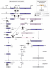
Pre-ribosomal RNA processing in human cells. Three of the four ribosomal RNAs arise from a long primary transcript (47S pre-rRNA) synthesized by RNA polymerase I from loci containing highly-repeated ribosomal DNA (rDNA) genes (also called nucleolar organizer regions or NORs) located in the cell nucleoli. The sequences of 18S (1870 nucleotides (nt)), 5.8SS/L (157 and 162 nt, respectively), and 28S rRNAs (>5000 nt) are flanked by external (5′-ETS, 3656 nt; 3′ ETS, 345 nt) and internal transcribed spacers (ITS1, 1090 nt; ITS2, 1155 nt), which are gradually removed by endo- and exonucleases. Depending on the relative kinetics of endoribonucleolytic cleavage, various rRNA precursors are formed. In the main maturation pathway (pre-rRNAs colored in violet), cleavage at site 2 occurs prior to cleavage at site A0. Less abundant precursors are characteristic of alternative (pale lavender) or minor (pink) routes. The 5S rRNA (121 nt) is transcribed by RNA polymerase III from repeated gene copies located on chromosome 1. As little is known about the maturation of 5S precursors in humans (depicted in orange) [34], data recently obtained in Drosophila were used [35]. When identified, endoribonucleases are quoted in black, and 5′-3′ or 3′-5′ exoribonucleases in grey. Question marks refer to uncertain enzymatic activities (UTP23), or enzymes found in other organisms which could putatively play similar functions in humans (REXO5, ERI1). Site E is also called 2a by some authors.
Figure 3
Figure 3
Folding of the human 18S rRNA. The 18S rRNA folds into 45 helices that distribute into four structural domains distinguished here by different colors. These domains assemble around the pseudoknot (red) formed by nucleotides located at the 5′ end that hybridize with nucleotides located ~1200 residues farther down in the primary sequence, at the junction between the central domain and the 3′ major domain. (a) Secondary structure of the 18S rRNA. (b) Structure of the 18S rRNA within the 40S subunit. (c) Enlarged view of the pseudoknot. The 3D structure in (b,c) was generated from PDB file 4UG0 [19].
Figure 4
Figure 4
Impairment of ribosome production in human diseases. (a) Ribosome production is impaired in a growing number of human diseases through global reduction of rRNA synthesis by RNA polymerase I (yellow ochre), defective pre-rRNA processing steps (purple), decrease of nucleotide chemical modifications (blue), or assembly of 60S ribosomal subunits (pink). The pre-rRNA maturation step that is affected is indicated (see also Table 1). (b) Examples of modification of the pre-rRNA pattern in HeLa cells after knockdown of various RPs with siRNAs. Total RNAs were analyzed by northern blot as previously described [40]. Pre-rRNA precursors to the small and large ribosomal subunits were revealed with probes complementary to ITS1 or ITS2.
Figure 5
Figure 5
The molecular effects of mutations affecting pre-rRNA processing and ribosome assembly. Mutations in genes encoding RPs or RAFs can lead to pre-rRNA processing or ribosome assembly defects. In human cells, different hypotheses may explain how these defects affect cell fate. Ribosome deficit corresponds to reduction in the number/concentration of ribosomes per cell. Depending on their translation initiation rate (Ki), mRNAs translation may be differently affected: (a) Translation of mRNAs that have a modest initiation rate will be mildly affected; (b) mRNAs having a high initiation rate may be more efficiently translated, as reduction in ribosome availability will reduce ribosome crowding and thus improve elongation efficiency. (c) In contrast, reduced concentrations of ribosomes will penalize translation of mRNAs that have low initiation rates [172]. Ribosome alteration refers to the ribosomes that are still produced despite ribosome assembly defects. Quality of these ribosomes (protein composition, RNA processing, and modification) may be suboptimal and change their translational properties, so that translation of specific mRNAs is not properly ensured or regulated. These different mechanisms are not mutually exclusive and may contribute in several ways to the variety of symptoms presented by patients suffering ribosomopathies. Ribosomal stress is induced by impairment of ribosome biogenesis, which promotes accumulation of free RPs that can assume alternative function. The 5S RNP particle, in which the 5S rRNA associates with RPs RPL5 and RPL11, plays a major role in this stress response. As a free complex, it binds and sequesters HDM2, the ubiquitin ligase that constitutively targets the tumor suppressor p53 for degradation. Inactivation of HDM2 promotes stabilization of p53, which regulates progression through the cell cycle. Chronic stimulation of p53 in ribosomopathies may also favor the selection of cells resisting p53 activity and prone to develop a tumorigenic program.

Similar articles

Cited by

References

    1. Palade G.E. A small particulate component of the cytoplasm. J. Biophys. Biochem. Cytol. 1955;1:59–68. doi: 10.1083/jcb.1.1.59. - DOI - PMC - PubMed
    1. Ban N., Nissen P., Hansen J., Capel M., Moore P.B., Steitz T.A. Placement of protein and RNA structures into a 5 Å-resolution map of the 50S ribosomal subunit. Nature. 1999;400:841–847. doi: 10.1038/23641. - DOI - PubMed
    1. Cate J.H., Yusupov M.M., Yusupova G.Z., Earnest T.N., Noller H.F. X-ray crystal structures of 70S ribosome functional complexes. Science. 1999;285:2095–2104. doi: 10.1126/science.285.5436.2095. - DOI - PubMed
    1. Clemons W.M., May J.L., Wimberly B.T., McCutcheon J.P., Capel M.S., Ramakrishnan V. Structure of a bacterial 30S ribosomal subunit at 5.5 Å resolution. Nature. 1999;400:833–840. doi: 10.1038/23631. - DOI - PubMed
    1. Ben-Shem A., Garreau de Loubresse N., Melnikov S., Jenner L., Yusupova G., Yusupov M. The structure of the eukaryotic ribosome at 3.0 Å resolution. Science. 2011;334:1524–1529. doi: 10.1126/science.1212642. - DOI - PubMed

Publication types

LinkOut - more resources