Review
. 2019 Jan;76(1):99-128.
doi: 10.1007/s00018-018-2947-0. Epub 2018 Oct 20.

Molecular pathways of nonalcoholic fatty liver disease development and progression

Affiliations
Review

Molecular pathways of nonalcoholic fatty liver disease development and progression

Fernando Bessone et al. Cell Mol Life Sci. 2019 Jan.

Abstract

Nonalcoholic fatty liver disease (NAFLD) is a main hepatic manifestation of metabolic syndrome. It represents a wide spectrum of histopathological abnormalities ranging from simple steatosis to nonalcoholic steatohepatitis (NASH) with or without fibrosis and, eventually, cirrhosis and hepatocellular carcinoma. While hepatic simple steatosis seems to be a rather benign manifestation of hepatic triglyceride accumulation, the buildup of highly toxic free fatty acids associated with insulin resistance-induced massive free fatty acid mobilization from adipose tissue and the increased de novo hepatic fatty acid synthesis from glucose acts as the "first hit" for NAFLD development. NAFLD progression seems to involve the occurrence of "parallel, multiple-hit" injuries, such as oxidative stress-induced mitochondrial dysfunction, endoplasmic reticulum stress, endotoxin-induced, TLR4-dependent release of inflammatory cytokines, and iron overload, among many others. These deleterious factors are responsible for the triggering of a number of signaling cascades leading to inflammation, cell death, and fibrosis, the hallmarks of NASH. This review is aimed at integrating the overwhelming progress made in the characterization of the physiopathological mechanisms of NAFLD at a molecular level, to better understand the factor influencing the initiation and progression of the disease.

Keywords: Hepatocellular death; Lipotoxicity; Liver fibrosis; Nonalcoholic fatty liver disease; Nonalcoholic steatohepatitis; Oxidative stress.


Figures

Fig. 1
Fig. 1
‘Multiple-hit’ theory of clinical progression of NAFLD. NAFLD comprises a wide spectrum of liver damage, ranging from simple macrovesicular steatosis to steatohepatitis, with combined inflammation, fibrosis and liver injury (steatohepatitis), progressing to cirrhosis and eventually to hepatocellular carcinoma (HCC). The initial hit is insulin resistance, leading to an increased uptake and synthesis of free fatty acids (FFAs) stored as triglycerides, resulting in simple steatosis. Further multiple hits acting as superimposed insults may be involved in the progression to more advanced stages of the disease, including oxidative stress, inflammatory mediators from hepatic origin (e.g., Kupffer cell-derived inflammatory cytokines) and from extrahepatic sources (e.g., adipokines and lipopolysaccharides of gut microbiota), apoptotic insults and the resulting compensatory regeneration, acquisition of a pro-fibrotic phenotype, and prosteatotic genetic factors, among others. NASH may occur even without steatosis, thus supporting the view that steatosis is not a causal factor in NASH development and progression
Fig. 2
Fig. 2
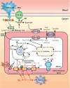
Metabolic basis of NAFLD pathogenesis. Insulin resistance associated with metabolic syndrome leads to unbalance between gain (red arrows) and loss (green arrows) of fat in liver. Insulin resistance in adipocyte tissue leads to massive mobilization of fatty acids (FFAs) from fat-overloaded adipocytes into liver, due to the suppression of the inhibitory effect of insulin on hormone-sensitive lipase (HLS), the rate-limiting enzyme catalyzing adipocyte TG lipolysis. Peripheral insulin resistance also leads to an overload of glucose (not internalized by peripheral tissues) and insulin (due to compensatory hyperinsulinaemia). Delayed insulin resistance in liver as compared with other tissues allows for the exacerbated insulin- and glucose-induced hepatic effects, with overproduction of hepatic FFAs via activation of the transcription factors sterol regulatory binding protein-1c (SREBP-1c) and carbohydrate response element binding protein (ChREBP), respectively. FFAs are stored as TGs in lipid droplets, exported as very low density lipoprotein (VLDL), and oxidized by mitochondrial β-oxidation, as compensatory mechanisms to the increased FFA uptake and synthesis. Lipophagy helps to degrade TGs from lipid droplets and to deliver FFAs for these exportation routes. However, VLDL production and lipophagy are impaired in NAFLD, and enhanced mitochondrial FFA β-oxidation leads to oxidative stress due to exacerbated leakage of electrons from the electron transport chain. Oxidative stress is aggravated by the concomitant FFA-mediated induction of CYP2E1, a cytochrome that performs futile cycles with release of electrons into the cytosol
Fig. 3
Fig. 3
Enzymatic pathways of the methyl group metabolism involved in VLDL formation. The primary methyl donor S-adenosylmethionine (SAMe) provides methyl groups for numerous methyltransferase reactions, and is regenerated by a number of interrelated metabolic pathways. Following SAMe-dependent transmethylations, SAMe is converted into S-adenosylhomocysteine (SAH) by phosphatidylethanolamine N-methyltransferase (PEMT), and SAH is further converted into homocysteine by SAH hydrolase (SAHH). Homocysteine can be remethylated back to methionine using betaine as a methyl donor, with the additional formation of dimethylglycine, in a reaction catalyzed by betaine-homocysteine S-methyltransferase (BHMT). The methionine thus formed, together with that provided by the diet, regenerates SAMe after receiving an adenosyl group from ATP, in a reaction catalyzed by methionine adenosyltransferase (MAT). Betaine is provided by the diet, and it is also produced from diet-derived choline by the enzyme choline dehydrogenase (ChDH), which represents the final step in a catabolic pathway initiated by the formation of phosphatidylcholine (PCh) from phosphatidylethanolamine (PEth), catalyzed by PEMT. Alternatively, PCh can be incorporated with other lipids and apolipoprotein (apo) B into nascent VLDL in the ER, where it plays a critical role in VLDL assembly and secretion. In NAFLD, there is a shortage of SAMe due to the fact that betaine levels are decreased in NASH patients, thus affecting VLDL exportation
Fig. 4
Fig. 4
Mitochondrial dysfunction in NAFLD. Long-chain free fatty acids (FFAs) from different sources, including uptake of FFAs mobilized from adipocyte tissue via CD16, de novo lipogenesis stimulated by insulin via sterol regulatory binding protein-1c (SREBP-1c) activation and triglyceride (TG) hydrolysis, are metabolized by mitochondria to supply energy. Acyl-CoA resulting from FFA activation penetrates the mitochondrial directly (for short and medium chain acyl-CoA) or via the carnitine shuttle (for long chain acyl-CoA), a process consisting of its conversion into carnitine-CoA catalyzed by the mitochondrial outer membrane enzyme carnitine palmitoyltransferase 1 (CPT1) and its further reconversion to acyl-CoA by the inner membrane enzyme CPT2; this makes acyl-CoA available for β-oxidation in the matrix. Acetyl-CoA resulting from β-oxidation enters the tricarboxylic acid (TCA) cycle, which renders NADH and FADH2 molecules. These reduced mediators carry their electrons to the respiratory electron carriers (complexes I-IV) in the mitochondrial inner membrane, which pumps protons (H+) through this membrane to generate the electrochemical H+ gradient that drives mitochondrial ATP production by the ATP synthase (F1F0 complex), a process known as ‘oxidative phosphorylation’. Alternatively, the electrochemical H+ gradient can be dissipated as heat via the uncoupling protein (UCP). Leakage of electrons at complexes I-III results in the formation of reactive oxygen species (ROS), such as superoxide (O2) or H2O2; this may damage the organelle, leading to impaired mitochondrial function and to a decrease in FFA metabolization. Failure in FFA β-oxidation is aggravated further by the inhibition of CPT1 by malonyl-CoA, which is produced from TCA cycle-derived citrate, via conversion to acetyl-CoA by the ATP citrate lyase (ACL) and further carboxylation by the acetyl coenzyme A carboxylase (ACC); the latter enzyme is induced by SREBP-1c
Fig. 5
Fig. 5
Mechanisms of hepatic insulin resistance in NAFLD. This pathology occurs in a context of sub-acute inflammation (e.g., obesity, type 2 mellitus diabetes), which promotes insulin resistance via the influx of pro-inflammatory cytokines, such as TNF-α and IL-6, from both the adipose tissue and Kupffer cells. After interaction with their respective membrane receptors, TNFR and IL-6R, these cytokines activate different signaling pathways that result in ubiquitin-mediated proteosomal degradation of the insulin receptor substrates 1 and 2 (IRS1/2), two signaling molecules required for insulin transduction via the phosphoinositide 3-kinase (PI3K)/Akt pathway. TNF-α and IL-6 promote IRS1/2 degradation by inducing synthesis of suppressor of cytokine signaling (SOCS), via activation of the transcription factors NF-κB and the signal transducer and activator of transcription 3 (STAT3), respectively. NF-κB is activated by TNF-α via inhibitor of κB (I-κB) kinase (IKK)- and c-Jun N-terminal kinase (JNK)-mediated phosphorylation, and further proteosomal degradation of the NF-κB inhibitor I-κB. IL-6 activates STAT3 via phosphorylation of this transcription factor by Janus activated kinase (JAK). Alternatively, proteosomal degradation of IRS1/2 is promoted by lipopolysaccharide (LPS) from intestinal microbiota. LPS interacts with the toll-like receptor (TLR4) to activate the ceramide/PKCθ signaling pathway, which induces, in turn, serine phosphorylation of IRS1/2 and further proteosomal degradation; ceramide also activates phospholipase-A2 (PLA2), which inhibits Akt activation. Lack of Akt signaling impairs Akt-mediated phosphorylation and further ubiquitin-mediated proteosomal degradation of forkhead box protein O1 (FoxO1), a transcription factor whose activation induces upregulation of gluconeogenic enzymes, such as phosphoenolpyruvate carboxykinase (PCK1) and glucokinase-6 phosphatase (G6P). The resulting increase in hepatocellular glucose (Glu) levels stimulates the transcription factor carbohydrate response element-binding protein (ChREBP), which in turn reinforces G6P activation and induces upregulation of enzymes involved in glycogenolysis, such as liver pyruvate kinase (L-PK), thus resulting in a higher hepatocellular glucose increase. Glycogenolysis is exacerbated by lack of Akt-mediated glycogen synthase kinase (GSK), an enzyme that inhibits glycogen synthase (GS). Blue, continuous lines: activation pathways. Red, continuous lines: inhibitory pathways. Red, dashed lines: inactivated pathways
Fig. 6
Fig. 6
Main pro-inflammatory mechanisms involved in NASH pathogenesis. NAFLD is associated with activation of a plethora of inflammatory pathways, with mediators of this process arising from multiple origins. Hypertrophic, dysregulated adipocytes release an increased amount of several pro-inflammatory chemokines and cytokines, and a reduced amount of the insulin-sensitizing and anti-inflammatory adipocytokine, adiponectin. Intestine contributes to the inflammatory process by releasing bacterial products such as lipopolysaccharide (LPS), which targets and activates the TLR4 receptor in Kupffer cells and hepatocytes, with the consequent production of cytokines and chemokines via activation of both the transcription factor nuclear factor-κB (NF-κB) and the nucleotide oligomerization domain (NOD)-like receptor protein 3 (NLRP3) inflammasome. These cyto/chemokines promote inflammation and hepatocellular apoptosis via specific receptors in hepatocytes and immunocompetent cells, thus accelerating NAFLD progression to NASH. In addition, the increase in free fatty acids (FFAs) in hepatocytes produces sustained oxidative damage that also induces apoptosis via redox-sensitive pro-apoptotic pathways. These cyto/chemokines, particularly transforming growth factor β (TGF-β), also promote fibrogenesis by activating hepatic stellate cells (HSCs). Activated HSCs produce collagen, which is deposited in the space of Disse, and release fibrogenic cytokines with autocrine and paracrine effects, including transforming growth factor β (TGF-β), which perpetuate the fibrotic injury
Fig. 7
Fig. 7
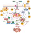
Main mechanisms of free fatty acid (FFA)-induced hepatocellular apoptosis in NASH. FFAs promote apoptosis by virtually all known apoptotic pathways. (1) The intrinsic or mitochondrial pathway of apoptosis, which is activated by FFA-induced cellular stress, and mediated by the release of cytochrome c (Cyt c) from the mitochondrial intermembraneous space to cytosol; Cyt c promotes binding of pro-caspase 9 to apoptosis protease-activating factor-1 (APAF-1), pro-caspase 9 activation by autocatalysis, and finally, recruitment and activation by caspase 9 of the executioner caspases 3, 6, and 7. FFAs activate this pro-apoptotic pathway by a activating the glycogen synthase kinase-3 (GSK-3)/JNK and the protein phosphatase 2A (PP2A) pathways, which causes the transcription factors FoxOa3 and AP-1 to translocate to the nucleus and to induce transcription of genes encoding pro-apoptotic proteins of the Bcl-2 family (PUMA, Bim); Bim is a pore-forming protein whereas PUMA activates the pore-forming protein Bax by binding inhibitory members of the Bcl-2 family, which release Bax from this inhibitory constraint (see mitochondrion, left side), and b by promoting the formation of mitochondrial permeability transition pores (MPTP), a process facilitated by reactive oxygen species (ROS) generated from leakage of electrons from the respiratory chain in mitochondria, from oxidases in peroxisomes and endoplasmic reticulum (ER) involved in FFA β- and ω-oxidations, and from induction of pro-oxidizing enzymes, such as NADPH oxidase 4 (NOX4) and CYP2E1 (see mitochondrion, right side); mitochondria permeabilization also exacerbates the leakage of electrons from the mitochondrial electron transport chain and further cytosolic ROS generation. (2) The endoplasmic reticulum (ER) stress pathway of apoptosis, which is induced by both FFAs and ROS, and mediated by a inositol 1,4,5-triphosphate receptor (Ins3PR)-induced Ca2+ release to cytosol and further Ca2+-dependent activation of the executor caspase 12, b Ca2+-dependent MPTP formation in mitochondria, and c ER stress-induced CCAAT-enhancer-binding protein homologous protein (CHOP) expression, which enhances AP-1 transcriptional activation. (3) The extrinsic pathway of apoptosis, which is induced sequentially by a binding of pro-inflammatory cytokines (e.g., Fas-L and TRAIL) to their respective plasma membrane receptors (Fas and TRAILR), b autophosphorylation of these receptors, c association of the activated receptors with FADD and with pro-caspases 8 and 10 to form the death complex DISC, d proteolytic activation of these pro-caspases, and e caspase-dependent cleavage of Bid to truncated Bid (tBid), which promotes Bcl-2-dependent mitochondrial pore formation. (4) The lysosomal pathway of apoptosis, which involves FFA-induced Bax translocation to lysosomes and further cytosolic release of cathepsin B; this protease activates caspases 12, which would interact directly with mitochondria to induce membrane permeabilization
Fig. 8
Fig. 8
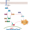
Molecular mechanisms of autophagy inhibition in NAFLD. Downregulation of the protein tyrosine phosphatase receptor type O (PTPRO) in NAFLD leads to impairment of signaling pathways involved in autophagy activation. Since PTPRO is involved in inactivation of the phosphoinositide 3-kinase (PI3K)/Akt signaling pathway, its downregulation leads to activation of this pathway and the further Akt-dependent stabilization of MDM4 via both mammalian target of rapamycin (mTOR)-mediated phosphorylation and ubiquitin-specific cysteine protease 2a (USP2a)-mediated deubiquitination. After stabilization, MDM4 forms the MDM4/MDM2 complex, a regulator of the p53 cytoplasmic levels via p53 poly-ubiquitination; this leads to cytoplasmic accumulation of p53, a well-recognized inhibitor of autophagy function. Akt-mediated mTOR activation also inhibits autophagy by blocking the activation of the pro-autophagic kinase unc-51 like autophagy activating kinase 1 (Ulk1) by AMP-activated protein kinase (AMPK)
Fig. 9
Fig. 9
Pathways of initiation and perpetuation of fibrosis mediated by hepatic stellate cell (HSC) activation. Following liver injury, a number of hormonal, paracrine, and autocrine stimuli induce HSC activation, a process initiated by the transition from quiescent, vitamin A-rich HSCs into fibrogenic, proliferative ones; TGF-β produced by Kupffer cells is the main stimulus that drives this phenotypic change. This ‘initiation phase’ is followed by the ‘perpetuation phase’, in which this phenotype is preserved and potentiated by additional stimuli that promote HSC proliferation (via the platelet-derived growth factor [PDGF]), sustained collagen production and deposition (via TGF-β), matrix degradation (via matrix metalloproteinases 1 and 3 [MMP-1/3]), inhibition of collagen degradation (via the MMP-2 inhibitor, tissue inhibitor of metalloproteinase-1 [TIM-1]), HSC chemotaxis (via PDGF and monocyte chemoattractant protein-1 [MCP-1]), and white cell chemoattraction (via MCP-1)

Similar articles

Cited by

References

    1. Angulo P. Nonalcoholic fatty liver disease. N Engl J Med. 2002;346:1221–1231. doi: 10.1056/NEJMra011775. - DOI - PubMed
    1. Tolman KG, Dalpiaz AS. Treatment of non-alcoholic fatty liver disease. Ther Clin Risk Manag. 2007;3:1153–1163. doi: 10.1136/pgmj.2005.042200. - DOI - PMC - PubMed
    1. Vernon G, Baranova A, Younossi ZM. Systematic review: the epidemiology and natural history of non-alcoholic fatty liver disease and non-alcoholic steatohepatitis in adults. Aliment Pharmacol Ther. 2011;34:274–285. doi: 10.1111/j.1365-2036.2011.04724.x. - DOI - PubMed
    1. Younossi ZM, Stepanova M, Afendy M, Fang Y, Younossi Y, Mir H, Srishord M. Changes in the prevalence of the most common causes of chronic liver diseases in the United States from 1988 to 2008. Clin Gastroenterol Hepatol. 2011;9:524–530. doi: 10.1016/j.cgh.2011.03.020. - DOI - PubMed
    1. Calzadilla Berlot L, Adams LA. The natural course of non-alcoholic fatty liver disease. Int J Mol Sci. 2016;17:E774. doi: 10.3390/ijms17050774. - DOI - PMC - PubMed

Grants and funding