. 2018 Dec 14;46(22):11759-11775.
doi: 10.1093/nar/gky923.

Long non-coding RNA ChRO1 facilitates ATRX/DAXX-dependent H3.3 deposition for transcription-associated heterochromatin reorganization

Affiliations

Long non-coding RNA ChRO1 facilitates ATRX/DAXX-dependent H3.3 deposition for transcription-associated heterochromatin reorganization

Jinyoung Park et al. Nucleic Acids Res. .

Abstract

Constitutive heterochromatin undergoes a dynamic clustering and spatial reorganization during myogenic differentiation. However the detailed mechanisms and its role in cell differentiation remain largely elusive. Here, we report the identification of a muscle-specific long non-coding RNA, ChRO1, involved in constitutive heterochromatin reorganization. ChRO1 is induced during terminal differentiation of myoblasts, and is specifically localized to the chromocenters in myotubes. ChRO1 is required for efficient cell differentiation, with global impacts on gene expression. It influences DNA methylation and chromatin compaction at peri/centromeric regions. Inhibition of ChRO1 leads to defects in the spatial fusion of chromocenters, and mislocalization of H4K20 trimethylation, Suv420H2, HP1, MeCP2 and cohesin. In particular, ChRO1 specifically associates with ATRX/DAXX/H3.3 complex at chromocenters to promote H3.3 incorporation and transcriptional induction of satellite repeats, which is essential for chromocenter clustering. Thus, our results unveil a mechanism involving a lncRNA that plays a role in large-scale heterochromatin reorganization and cell differentiation.


Figures
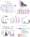
Figure 1.
Figure 1.
ChRO1 is a muscle-specific lncRNA induced during myogenesis (A) Schematic representation of the ChRO1 genomic locus. Occupancies of myogenic transcription factors are shown. MB, myoblasts; MT, myotubes; OB, osteoblasts; Pol II, RNA polymerase II; Pol II-S2, Serine 2 phosphorylated RNA polymerase II. (B) Fold changes of lncRNA levels during myogenesis were calculated from FPKM. (C) ChRO1 RNA was measured by RT-qPCR under growth (myoblasts, MB) or differentiation condition (Day 1, 2, 3) using amplicon primers as shown in (A). (D) Occupancy of pol II (8WG16) on the 0.6K and 1.3K upstream regions from the ChRO1 transcription start site in MB and MT, differentiated for 3 days (D3). (E) RT-qPCR for MyoD, Myogenin and ChRO1 in myogenesis-induced NIH3T3. (F) RT-qPCR for ChRO1 in various mouse tissues. RNA level was normalized by 18S rRNA. n = 4 biological replicates, one-way ANOVA, P < 0.0001, Degree of Freedom = 6, F-value = 15.83. (G) RT-qPCR of ChRO1 and MyHC during muscle regeneration after injection of CTX into TA muscle of mice. RNA level was normalized by 18S rRNA, n = 4. One-way ANOVA, P < 0.0001, Degree of Freedom = 4, F-value = 64.12 for ChRO1 and 107.7 for MyHC. (H) Expression of ChRO1 in human cells. GRCh27/hg19 was used as a human genome reference. (I) Conserved regions identified by sliding-window local alignment are shown at bottom. Identical sequence alignments (conserved domains) between human and mouse ChRO1 are represented in red.
Figure 2.
Figure 2.
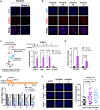
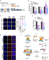
ChRO1 is enriched at chromocenters and associated with heterochromatin reorganization (A) RNA-FISH for ChRO1 in MT (Day 7). RNase A was either treated or not. Scale bars, 50 μm. (B) Representative MT nuclei of RNA-FISH analysis. (C) Experimental scheme of RNA pull-down for analysis of ChRO1-interacting DNA/RNA by using biotinylated ChRO1-AS oligonucleotides (AS-ChRO1) (left). RT-qPCR of ChRO1, MajS, MinS and U2 RNAs after RNA pull-down (right). AS-LacZ (AS oligonucleotides targeting LacZ) was used for a negative control. (D) DNA was analyzed by qPCR with primers for pericentromere (PeriCEN) and centromere (CEN) after ChIRP. (E) RT-qPCR of ChRO1 with amplicons to confirm ChRO1 knockdown using two specific siRNAs. (F) DAPI-staining of ChRO1-depleted MT (left). Number of chromocenters per MT nuclei is shown (right).
Figure 3.
Figure 3.
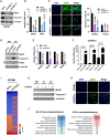
Muscle differentiation is impaired by ChRO1 depletion. (A) Western blot for MyHC and Troponin T in 3-day differentiated MT after siRNA treatment. α-tubulin was used as a loading control. (B) RT-qPCR for MyHC and muscle creatin kinase (MCK) in siRNA-treated C2C12 that was differentiated for 3 days. (C) Representative immunofluorescence for MyHC and DAPI staining. Scale bars, 50 μm (left). MyHC-positive cells and fusion index in MT (right). (D) Western blots for MyHC, Troponin T and Myogenin in either shCtrl or shChRO1 MB and MT (Day 3). Lamin B1 was used as a loading control. *: non-specific band, arrow indicates Myogenin. (E) RT-qPCR for ChRO1, MyHC, MCK and Myogenin in shCtrl or shChRO1 MT (Day 3). (F) RT-qPCR of ChRO1a in empty or ChRO1a over-expressing C2C12 cells in growing (MB) or differentiation (Day 1, Day 2) medium. n = 4 independent experiments. (G) Western blots for MyHC and Troponin T with ChRO1a over-expressing MB and MT (Day 1, Day 2). α-tubulin was used as a loading control. (H) MyHC immunostaining of 1 day-differentiated C2C12 cells over-expressing ChRO1a. Scale bars, 50μm. (I) Differentially expressed genes of shCtrl or shChRO1 C2C12 (Fold Change > 2, P < 0.05). Data are shown as log 10 of fold change. (J) Gene ontology analysis of ChRO1-depleted cells, shown in log P-value.
Figure 4.
Figure 4.
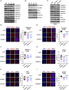
ChRO1 is responsible for heterochromatin compaction via facilitating localization of chromatin factors to chromocenters. (A) Western blots for various histone modifications in MB and MT (Day 1–3). (B) Western blots for Suv420H2, Suv39H1, Ezh2, MyHC and Troponin T in MB and MT (Day 1–3). (C) Western blots for histone modifications and HMTs in ChRO1-depleted MT (Day 3). H4 and Lamin B1 are the loading controls. (DI) Representative MT nuclei of immunostaining (left) and correlation R (right) are shown for H4K20me3, Suv420H2, Smc3, H3K9me3, HP1γ and MeCP2, respectively, in ChRO1-depleted or control MT.
Figure 5.
Figure 5.
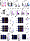
ChRO1 mediates satellite RNA accumulation through ATRX/DAXX/H3.3 localization at peri/centromeric chromocenters. (A) RT-qPCR for MajS and MinS RNA transcripts in MB and MT (Day 1–Day 3). (B) RT-qPCR for MajS and MinS in siRNA-treated C2C12. (C) ChIP-qPCR demonstrating pol II occupancy at peri/centromeres in ChRO1-depleted MTs (Day 3). (D) ChIP-qPCR for H3.3 occupancy at indicated genomic regions in ChRO1-depleted MTs (Day 3). (E) Western blots for IP of Flag/HA tagged H3s using antibodies against Flag or HA (left panel). Detection of ChRO1 by RT-qPCR following H3 IP (right panel). (FI) RT-qPCR for ChRO1 after IP of endogenous H3.3, DAXX, ATRX or HIRA in MTs (Day 3). IP/Input of ChRO1 was normalized by the value of IgG. Pre-rRNA or GAPDH was used as a negative control. (J and K) Representative immunofluorescence and correlation R for DAXX and ATRX in ChRO1-depleted MTs (Day 3). (L) Representative immunofluorescence and correlation R for DAXX in ATRX-depleted MTs (Day 3). (M) RNA-FISH for ChRO1 in ATRX-depleted MTs (Day 3). Correlation R for ChRO1 in ATRX-depleted MTs is shown. (NP) DAPI staining for ATRX-, DAXX- or H3.3-depleted MTs (Day 3). The number of chromocenters per nucleus is demonstrated.
Figure 6.
Figure 6.
Satellite repeat RNAs are involved in chromocenter reorganization during muscle differentiation. (A) Schematic of the structure of the peri/centromeric region that consists of MajS and MinS repeats. The location of antisense oligonucleotides (ASOs) for targeting satellite RNAs is demonstrated. (B) RT-qPCR for MajS, MinS and ChRO1 RNAs in ASO-treated MTs (Day 3). n.s.: not significant. (C) DAPI-staining of nuclei from MTs treated with MajS- or MinS-specific ASOs (left). The number of chromocenters is shown at right. (D) RT-qPCR for MyHC and MCK in MajS- or MinS-depleted MTs (Day 3). (E) RNA-FISH for ChRO1 in MajS- or MinS-ASO treated MTs (Day 3). (F) Schematic of ChRO1-mediated large-scale reorganization of constitutive heterochromatin during myogenesis.

Similar articles

Cited by

References

    1. Politz J.C.R., Scalzo D., Groudine M.. Something silent this way forms: the functional organization of the repressive nuclear compartment. Annu. Rev. Cell Dev. Biol. 2013; 29:241–270. - PMC - PubMed
    1. Solovei I., Wang A.S., Thanisch K., Schmidt C.S., Krebs S., Zwerger M., Cohen T.V., Devys D., Foisner R., Peichl L. et al. . LBR and lamin A/C sequentially tether peripheral heterochromatin and inversely regulate differentiation. Cell. 2013; 152:584–598. - PubMed
    1. Fussner E., Djuric U., Strauss M., Hotta A., Perez-Iratxeta C., Lanner F., Dilworth F.J., Ellis J., Bazett-Jones D.P.. Constitutive heterochromatin reorganization during somatic cell reprogramming. EMBO J. 2011; 30:1778–1789. - PMC - PubMed
    1. Guetg C., Lienemann P., Sirri V., Grummt I., Hernandez-Verdun D., Hottiger M.O., Fussenegger M., Santoro R.. The NoRC complex mediates the heterochromatin formation and stability of silent rRNA genes and centromeric repeats. EMBO J. 2010; 29:2135–2146. - PMC - PubMed
    1. Padeken J., Mendiburo M.J., Chlamydas S., Schwarz H.J., Kremmer E., Heun P.. The nucleoplasmin homolog NLP mediates centromere clustering and anchoring to the nucleolus. Mol. Cell. 2013; 50:236–249. - PubMed

Publication types

MeSH terms