. 2018 Oct 8;14(10):e1007293.
doi: 10.1371/journal.ppat.1007293. eCollection 2018 Oct.

Potent neutralizing antibodies in humans infected with zoonotic simian foamy viruses target conserved epitopes located in the dimorphic domain of the surface envelope protein

Affiliations

Potent neutralizing antibodies in humans infected with zoonotic simian foamy viruses target conserved epitopes located in the dimorphic domain of the surface envelope protein

Caroline Lambert et al. PLoS Pathog. .

Abstract

Human diseases of zoonotic origin are a major public health problem. Simian foamy viruses (SFVs) are complex retroviruses which are currently spilling over to humans. Replication-competent SFVs persist over the lifetime of their human hosts, without spreading to secondary hosts, suggesting the presence of efficient immune control. Accordingly, we aimed to perform an in-depth characterization of neutralizing antibodies raised by humans infected with a zoonotic SFV. We quantified the neutralizing capacity of plasma samples from 58 SFV-infected hunters against primary zoonotic gorilla and chimpanzee SFV strains, and laboratory-adapted chimpanzee SFV. The genotype of the strain infecting each hunter was identified by direct sequencing of the env gene amplified from the buffy coat with genotype-specific primers. Foamy virus vector particles (FVV) enveloped by wild-type and chimeric gorilla SFV were used to map the envelope region targeted by antibodies. Here, we showed high titers of neutralizing antibodies in the plasma of most SFV-infected individuals. Neutralizing antibodies target the dimorphic portion of the envelope protein surface domain. Epitopes recognized by neutralizing antibodies have been conserved during the cospeciation of SFV with their nonhuman primate host. Greater neutralization breadth in plasma samples of SFV-infected humans was statistically associated with smaller SFV-related hematological changes. The neutralization patterns provide evidence for persistent expression of viral proteins and a high prevalence of coinfection. In conclusion, neutralizing antibodies raised against zoonotic SFV target immunodominant and conserved epitopes located in the receptor binding domain. These properties support their potential role in restricting the spread of SFV in the human population.


Conflict of interest statement

The authors have declared that no competing interests exist.

Figures
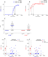
Fig 1
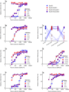
Fig 1. Neutralization of primary gorilla SFV strains by human plasma samples.
Neutralization assays were carried out by infecting the GFAB indicator cells with SFV strains previously incubated with serial dilutions of human plasma samples. GI-D468 (A, blue) and GII-K74 (B, red) strains were incubated with autologous samples collected at two time points. The number of infected units per well (mean, SD) is presented as a function of plasma dilution. Arrows indicate neutralization titers. C. GI-D468 and GII-K74 strains were incubated with serial dilutions of plasma samples from 44 people infected with gorilla SFV (SFVpos) and eight uninfected controls (SFVneg). The neutralization titers against both strains are shown and the bars indicate the median values. D and E: The neutralization titers against GI-D468 (x axis) and GII-K74 (y axis) are presented for the SFV-infected individuals, with symbols colored according to their genotype. Panel D shows the genotypes previously obtained by PCR amplification of the env gene followed by sequencing [33]. Panel E shows genotypes defined by GI and GII-specific PCR described in this study. The dashed lines indicate the neutralization detection thresholds (1:20).
Fig 2
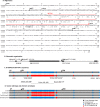
Fig 2. Gorilla SFV envelope proteins: alignment, schematic organization, localization of sequence variations, and design of the chimeric proteins used for epitope mapping.
A. Env sequences from GI-D468 and GII-K74 strains were aligned using CLC Mainworkbench software. Identical residues are indicated with dots. The three subdomains (gp18LP, leader peptide; gp80SU, surface protein; gp48TM, transmembrane protein) are indicated above the sequences. The lines indicate the localization of the variable (SUvar, red) and conserved (SUcon, black) regions, respectively. B. Schematic organization of the SFV Env protein. Amino acid positions are those of the GI-D468 strain. Furin cleavage sites (arrows) and envelope subunits are indicated above the grey line. Major functional or structural domains are shown by the bold line (H, hydrophobic region; RBD, receptor binding domain; FP, fusion peptide; MSD, membrane spanning domain). C. Schematic representations of GI-D468 and GII-K74 Env sequences. Grey symbols represent identical amino acids and blue/red symbols GI/GII-specific amino acids, respectively. Sequences from point mutations outside SUvar are indicated below the diagram, as well as the SUcon and SUvar regions. The color code is not to scale in the SUvar region: amino acid identity is 58% in this region. D. Schematic representation of the Env proteins used to produce the vectors and map the neutralization epitopes. Full-length GI-D468 was used as the wild-type backbone, in which the SU, SUvar, and SUcon coding sequences from GII-K74 were inserted. The same color code is used as in panel B.
Fig 3
Fig 3. Neutralizing antibodies target the SUvar region.
Neutralization assays were performed with plasma samples from gorilla SFV-infected individuals against the four FFV carrying wild-type EnvGI, chimeric EnvGI-SUGII, EnvGI-SUvarGII, and EnvGI-SUconGII. Cells were transduced with untreated FFV to provide the reference value. Relative infectivity was calculated for wells treated with plasma samples and is expressed as the percentage of the reference value. Panels A, B, and D through G present full titration curves for plasma from single neutralizers (A-B), coinfected dual neutralizers (D-E), and dual neutralizers without molecular evidence of coinfection (F-G). Panel C shows the relative infectivity after incubation with plasma samples from single neutralizers diluted 1:80. Fourteen plasma samples were specific for GI-D468 and are presented with dark blue/light pink symbols; six plasma samples were specific for GII-K74 and are presented with light blue/red symbols.
Fig 4
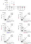
Fig 4. Frequent cross-neutralization of gorilla and chimpanzee SFV belonging to homologous genotypes.
Neutralization assays were performed against four viral strains with plasma samples from individuals infected with three SFV species (chimpanzee, gorilla, and cercopithecus). A. Neutralization titers against four strains (GI-D468, GII-K74, CI-PFV, CII-SFV7) are shown for individuals infected with chimpanzee or cercopithecus SFV; bars indicate the median values. B to G: Neutralization titers against pairs of strains are presented with symbols corresponding to the infecting SFV species (open circles: gorilla SFV; filled squares: chimpanzee SFV). The dashed lines indicate neutralization detection thresholds (1:20). Tables indicate the number of individuals with and without neutralizing antibodies against each of the two strains, with P values from Fisher’s exact test.
Fig 5
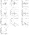
Fig 5. Magnitude and breadth of neutralizing antibodies and parameters of SFV infection Correlations between quantitative measures of neutralization and parameters of infection are presented.
The neutralization titer against the homologous SFV strain was used; for coinfected individuals we considered the highest of the two titers. Breadth was defined as the number of neutralized strains of the four tested. For each viral strain, a score was assigned based on the neutralization titer (neutralization titers < 20 and those ranging from 1:20 to 1:200, 1:200–1:2000, and > 1:2000 correspond to 0, 1, 2 and 3 points, respectively). The sum of these points for the four tested strains defined the neutralization score of a plasma sample. Age (A, B, C), duration of infection (D, E, F), and SFV DNA levels (G, H, I) are presented as a function of the neutralization titers (log10 transformed, A, C, and E), breadth (B, D, and F), and neutralization score (C, F, I). The lines represent the linear regression curves. J: SFV DNA levels are presented as a function of the neutralization breadth for Bantus (triangles) and Pygmies (squares). Results from the Spearman rank test are indicated on the graphs and statistically significant results are shown in bold.
Fig 6
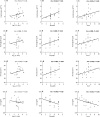
Fig 6. Neutralization magnitude, breadth and score are associated with hematological variables Correlations between quantitative measures of neutralization and hematological parameters are presented.
Erythrocyte counts (A, B, C), hemoglobin (D, E, F), hematocrit (G, H, I), and blood urea (J, K, L) levels are presented as a function of neutralization titers (A, D, G, and J), breadth (B, E, H, and K), and potency (C, F, I and L). The lines represent the linear regression curves. Results from the Spearman tests are indicated on the graphs and statistically significant results are shown in bold.

Similar articles

Cited by

References

    1. Peeters M, Delaporte E. Simian retroviruses in African apes. Clin Microbiol Infect. 2012;18(6):514–20. 10.1111/j.1469-0691.2012.03843.x - DOI - PubMed
    1. Falcone V, Leupold J, Clotten J, Urbanyi E, Herchenroder O, Spatz W, et al. Sites of simian foamy virus persistence in naturally infected African green monkeys: Latent provirus is ubiquitous, whereas viral replication is restricted to the oral mucosa. Virology. 1999;257(1):7–14. 10.1006/viro.1999.9634 - DOI - PubMed
    1. Murray SM, Linial ML. Foamy virus infection in primates. J Med Primatol. 2006;35(4–5):225–35. 10.1111/j.1600-0684.2006.00171.x - DOI - PubMed
    1. Heneine W, Schweizer A, Sandstrom P, Folks T. Human infection with foamy viruses. Curr Top Microbiol Immunol. 2003;277:181–96. - PubMed
    1. Khan AS. Simian foamy virus infection in humans: prevalence and management. Expert Rev Anti-Infect Ther. 2009;7(5):569–80. 10.1586/eri.09.39 - DOI - PubMed

Publication types

MeSH terms

Grants and funding

CL was personally supported by a doctoral grant from the French government program Investissement d'Avenir, Laboratory of Excellence, Integrative Biology of Emerging Infectious Diseases (LabEx IBEID, http://www.agence-nationale-recherche.fr/ProjetIA-10-LABX-0062). LR was personally supported by the Bourse de l’Ecole Normale Supérieure, Faculté Paris Diderot, http://www.ens.fr/. This work was supported by the Institut Pasteur in Paris, France, the Programme Transversal de Recherche from the Institut Pasteur [PTR#437], https://www.pasteur.fr/fr, and the Agence Nationale de la Recherche [grant ANR-10-LABX-62-IBEID; http://www.agence-nationale-recherche.fr/ProjetIA-10-LABX-0062; REEMFOAMY project, ANR 15-CE-15-0008-01; http://www.agence-nationale-recherche.fr/projet-anr/?tx_lwmsuivibilan_pi2%5BCODE%5D=ANR-15-CE15-0008]. The work in the laboratory of DL is supported by the Deutsche Forschungsgemeinschaft [DFG, German Research Foundation grants LI 621/10-1, SPP1923 LI 621/11-1, http://www.dfg.de/]. The funding agencies had no role in the study design, generation of results, or writing of the manuscript.