. 2018 Sep 26;8(1):14418.
doi: 10.1038/s41598-018-32793-5.

NKX2-1-AS1 negatively regulates CD274/PD-L1, cell-cell interaction genes, and limits human lung carcinoma cell migration

Affiliations

NKX2-1-AS1 negatively regulates CD274/PD-L1, cell-cell interaction genes, and limits human lung carcinoma cell migration

Hasmeena Kathuria et al. Sci Rep. .

Abstract

The function of most long noncoding RNAs (lncRNAs) is unknown. However, recent studies reveal important roles of lncRNAs in regulating cancer-related pathways. Human antisense lncRNA-NKX2-1-AS1 partially overlaps the NKX2-1/TTF1 gene within chromosomal region 14q13.3. Amplification of this region and/or differential expression of genes therein are associated with cancer progression. Herein we show higher levels of NKX2-AS1 and NKX2-1 in lung adenocarcinomas relative to non-tumor controls but no correlation between NKX2-1-AS1 and NKX2-1 levels across specimens, or with amplification of the 14q13.3 region, suggesting that NKX2-1-AS1 and NKX2-1 are independently regulated. Loss-and-gain of function experiments showed that NKX2-1-AS1 does not regulate NKX2-1 expression, or nearby genes, but controls genes in trans. Genes up-regulated by NKX2-1-AS1-knockdown belong to cell adhesion and PD-L1/PD-1 checkpoint pathways. NKX2-1-AS1 negatively regulates endogenous CD274/PD-L1, a known target of NKX2-1, and the transcriptional activity of -1kb-CD274 promoter-reporter construct. Furthermore, NKX2-1-AS1 interferes with NKX2-1 protein binding to the CD274-promoter, likely by NKX2-1 protein-NKX2-1-AS1 interactions. Finally, NKX2-1-AS1 negatively regulates cell migration and wound healing, but not proliferation or apoptosis. These findings support potential roles of NKX2-1-AS1 in limiting motility and immune system evasion of lung carcinoma cells, highlighting a novel mechanism that may influence tumorigenic capabilities of lung epithelial cells.


Conflict of interest statement

The authors declare no competing interests.

Figures
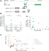
Figure 1
Figure 1
NKX2-1-AS1 expression patterns in human non-small cell lung carcinoma (NSCLC). (A) Schematic representation of the relative chromosomal location of NKX2-1-AS1 lncRNA and adjacent NKX2-1 protein coding-gene in human chromosomal region 14q13.3. Arrows indicate direction of transcription. Boxes indicate exons, dotted lines indicate introns and colored boxes indicate coding regions. (B) Levels of expression of NKX2-1-AS1 and NKX2-1 in NSCLCs (SCC = squamous cell carcinoma; and AC = adenocarcinoma) relative to their corresponding non-tumor control determined by qPCR (n = 8; *p < 0.03). (C) Correlation plot of the expression of NKX2-1-AS1 and NKX2-1 determined by qPCR in the above tumors and their corresponding non-tumor specimens analyzed in this study. (D) Amplification status of the NKX2-1-AS1 locus determined by qPCR of genomic DNA and expressed as copy number of the NKX2-1-AS1 gene per genome plotted relative to NKX2-1-AS1 expression level in each sample (n = 12).
Figure 2
Figure 2
NKX2-1-AS1 follows tissue-specific patterns of expression similar to NKX2-1 in human cells. (A) Expression of NKX2-1-AS1 and NKX2-1 determined by RT-PCR in lung cell lines. The NKX2-1-AS1 PCR fragments were sequenced to confirm the identity of the sequence. (B) Relative expression patterns of NKX2-1-AS1 and NKX2-1 in tissues and cell lines, including normal human adult lung and thyroid and H441 and H661 cell lines, as determined by qPCR (n = 3; *p < 0.05; **p < 0.01). (C) 5′-RACE analysis of NKX2-1-AS1 in human thyroid identified multiple transcription initiation sites within the 500 bp 5′ of the transcription initiation site reported in Ensembl. Thyroid total RNA was used as it has higher levels of NKX2-1-AS1 expression than the lung. The sensitivity of the method did not allow us to analyze lung RNA. Thirty clones, generated in three independent experiments, were sequenced. The black bar indicates the region recently identified in a genome wide analysis of accurate lncRNA transcription initiation sites. (D) Time course analysis of NKX2-1-AS1 and NKX2-1 transcript stability in H441 cells by qPCR after inhibition of transcription by actinomycin D treatment (n = 3; *p < 0.05).
Figure 3
Figure 3
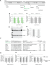
NKX2-1-AS1 does not regulate expression of genes in the 14q13.3 chromosomal region. (A) Scheme of human chr14 within the 14q13.3 cytoband region indicating selected genes neighboring NKX2-1-AS1. (B) qPCR analyses of NKX2-1-AS1 in H441 cells treated with a pool of three siRNAs targeting NKX2-1-AS1 exon 2 show significant down-regulation of NKX2-1-AS1 at 48 h and 72 h post transfection (n = 6; **p = 0.01 and ***p = 0.005 respectively). (C) qPCR analyses of the neighboring protein coding gene NKX2-1 in NKX2-1-AS1 knocked-down cells show no changes in NKX2-1 RNA levels (n = 6). (D) Representative western blots of NKX2-1 protein in non-silencing control [c] and NKX2-1-AS1 siRNA [si] treated H441 cells normalized to β-actin. (E) Densitometry of the western blot signals normalized to β-actin indicates that NKX2-1 protein levels also remained unchanged; n = 3. (F) Expression levels of NKX2-1-AS1 in the knockdown cells at 48 h and of other genes in the 14q13.3 chromosomal region as determined by microarray analysis; n = 6. (G) qPCR analysis in the NKX2-1-AS1 knockdown cells at 24, 48 and 72 h of MBIP; (H) NKX2-8; and (I) PAX9. n = 6, *p = 0.002.
Figure 4
Figure 4
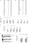
NKX2-1-AS1 knockdown alters gene expression patterns in H441 cells. (A) List of the top 20 genes down- and (B) up-regulated by NKX2-1-AS1 knockdown determined by microarray analysis, 48 h after treatment (n = 6). (C) qPCR validation of down-regulated and up-regulated genes in NKX2-1-AS1 knockdown cells at 48 h after treatment (n = 3, *p < 0.05, **p < 0.01). (D) Real time PCR validation of adherens junction related genes up-regulated by NKX2-1-AS1 knockdown (n = 3; *p < 0.05; **p < 0.01). (E) Representative western blots of PD-L1 and PTPN1 protein in non-silencing control [c] and NKX2-1-AS1 siRNAs [si] treated H441 cells normalized to β-actin. (F) Densitometry of the western blot signals normalized to β-actin indicates that PD-L1 (n = 9; **p = 0.002) and PTPN1 (n = 3; *p = 0.05) protein levels are increased by NKX2-1-AS1 knockdown.
Figure 5
Figure 5
NKX2-1-AS1 overexpression reduces CD274 expression levels in A549 cells in part by impairing NKX2-1 protein binding to the CD274 promoter. ChIP-qPCR analysis of NKX2-1 protein binding to (A) CD274 promoter (n = 4; p = 0.005), (B) CLDN1 promoter (n = 5), and (C) PTPN1 promoter (n = 5) in H441 cells transfected with NKX2-1-AS1 siRNAs or non-silencing control. (D) NKX2-1 co-transfection with the −1kbCD274-Luc vector results in higher luciferase activity (3-fold in the absence of NKX2-1-AS1, 0ug). NKX2-1-AS1 overexpression reduces the activity of the −1kbCD274 promoter in a dose-dependent manner both in the absence (E) (n = 3-4; ANOVA p = 0.003) or presence (F) (n = 3-4; ANOVA p = 0.0001) of NKX2-1 overexpression. NKX2-1-AS1 overexpression reduces the expression of the endogenous CD274 gene in a dose-dependent manner both in the absence (G) (n = 3-4; ANOVA p = 0.001) or presence (H) (n = 3-4; ANOVA p = 0.05) of NKX2-1 overexpression. (I) RIP-qPCR analysis of NKX2-1-AS1 pull down by NKX2-1 antibody compared to IgG control (n = 6; p = 0.05).
Figure 6
Figure 6
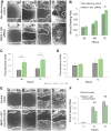
NKX2-1-AS1 inhibits cell motility in H441 cells. (A) Representative wound healing analysis of H441 cells treated with NKX2-1-AS1 siRNAs or non-silencing siRNA control. Cells were treated with the siRNAs for 24 h before the scratch was performed (0 h). Three images per scratch were taken at 0, 24, 48 and 72 h in 3 independent experiments. (B) Average wound area closed determined in the above images (*p < 0.002). (C) Migration of NKX2-1-AS1 knockdown H441 cells was compared to non-silencing control in transwell experiments (n = 3; *p < 0.05). (D) Invasion of H441 cells treated with NKX2-1-AS1 siRNA compared to non-silencing control in collagen-covered transwells (n = 3). (E) Representative wound healing analysis of H441 cells transfected with CMV-NKX2-1-AS1 or with empty vector control. After treatment for 24 h the scratch was performed (0 h). Three images per scratch were taken at 0, 24, and 48 h in three independent experiments. (F) Average wound area closed (**p < 0.00001).
Figure 7
Figure 7
NKX2-1-AS1 knockdown does not affect proliferation or apoptosis of H441 cells. (A) Cell growth was determined by counting cells at 24, 48 and 72 h after treatment with a pool of 3 siRNAs targeting NKX2-1-AS1 (n = 3). (B) Analysis of cell cycle stage by measuring DNA cell content by flow cytometry in NKX2-1-AS1 knockdown cells compared to non-silencing control at 48 h after treatment (n = 3). No significant change in cell number in each cell cycle stage was observed in NKX2-1-AS1 knockdown cells compared to non-silencing control. (C) No significant change in apoptosis was observed in NKX2-1-AS1 knockdown cells compared to non-silencing control as measured by annexin-V binding.

Similar articles

Cited by

References

    1. Campbell JD, et al. Distinct patterns of somatic genome alterations in lung adenocarcinomas and squamous cell carcinomas. Nat. Genet. 2016;48:607–616. doi: 10.1038/ng.3564. - DOI - PMC - PubMed
    1. Weir BA, et al. Characterizing the cancer genome in lung adenocarcinoma. Nature. 2007;450:893–898. doi: 10.1038/nature06358. - DOI - PMC - PubMed
    1. Perner S, et al. TTF1 expression in non-small cell lung carcinoma: association with TTF1 gene amplification and improved survival. J. Pathol. 2009;217:65–72. doi: 10.1002/path.2443. - DOI - PubMed
    1. Kwei KA, et al. Genomic profiling identifies TITF1 as a lineage-specific oncogene amplified in lung cancer. Oncogene. 2008;27:3635–3640. doi: 10.1038/sj.onc.1211012. - DOI - PMC - PubMed
    1. Harris T, et al. Both gene amplification and allelic loss occur at 14q13.3 in lung cancer. Clin. Cancer Res. 2011;17:690–699. doi: 10.1158/1078-0432.CCR-10-1892. - DOI - PMC - PubMed

Publication types