. 2018 Sep 26;3(5):e00399-18.
doi: 10.1128/mSphere.00399-18.

TRAIL (CD253) Sensitizes Human Airway Epithelial Cells to Toxin-Induced Cell Death

Affiliations

TRAIL (CD253) Sensitizes Human Airway Epithelial Cells to Toxin-Induced Cell Death

Yinghui Rong et al. mSphere. .

Abstract

Inhalation of ricin toxin is associated with the onset of acute respiratory distress syndrome (ARDS), characterized by hemorrhage, inflammatory exudates, and tissue edema, as well as the nearly complete destruction of the lung epithelium. Here we report that the Calu-3 human airway epithelial cell line is relatively impervious to the effects of ricin, with little evidence of cell death even upon exposure to microgram amounts of toxin. However, the addition of exogenous soluble tumor necrosis factor (TNF)-related apoptosis-inducing ligand (TRAIL; CD253) dramatically sensitized Calu-3 cells to ricin-induced apoptosis. Calu-3 cell killing in response to ricin and TRAIL exposure was partially inhibited by caspase-8 and caspase-3/7 inhibitors, consistent with involvement of extrinsic apoptotic pathways in cell death. We employed nCounter Technology to define the transcriptional response of Calu-3 cells to ricin, TRAIL, and the combination of ricin plus TRAIL. An array of genes associated with inflammation and cell death were significantly upregulated upon treatment with ricin toxin and were further amplified upon addition of TRAIL. Of particular note was interleukin-6 (IL-6), whose expression in Calu-3 cells increased 300-fold upon ricin treatment and more than 750-fold upon ricin and TRAIL treatment. IL-6 secretion by Calu-3 cells was confirmed by cytometric bead array analysis. On the basis of these finding, we speculate that the severe airway epithelial cell damage observed in animal models following ricin exposure is a result of a positive-feedback loop driven by proinflammatory cytokines such as TRAIL and IL-6.IMPORTANCE Ricin toxin is a biothreat agent that is particularly damaging to lung tissue following inhalation. A hallmark of ricin exposure is widespread inflammation and concomitant destruction of the airway epithelium. In this study, we investigated the possible interaction between ricin and known proinflammatory cytokines associated with lung tissue. Using an established human airway epithelial cell line, we demonstrate that epithelial cell killing by ricin is significantly enhanced in the presence of the proinflammatory cytokine known as TRAIL (CD253). Moreover, epithelial cells that are simultaneously exposed to ricin and TRAIL produced large amounts of secondary proinflammatory signals, including IL-6, which in the context of the lung would be expected to exacerbate toxin-induced tissue damage. Our results suggest that therapies designed to neutralize proinflammatory cytokines such as TRAIL and IL-6 may limit the bystander damage associated with ricin exposure.

Keywords: bioterrorism; cytokines; epithelial cells; inflammation; lung defense; toxins.


Figures

FIG 1
FIG 1
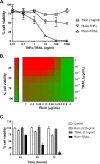
The sensitizing effect of TRAIL on ricin-induced cell death in Calu-3 cells. (A) TNF-ɑ or TRAIL (starting at 1 µg/ml) in a 10-fold serial dilution was mixed with ricin (1 µg/ml) and then administered to the cells seeded in 96-well plates for 24 h. The cells were then washed, and cell viability was measured 72 h later, as described in Materials and Methods. (B) In the dose experiment, cell viability was assessed at 72 h after exposure of the cells to the indicated concentrations of ricin and TRAIL. (C) In the time course experiments, cell viability was assessed 24 h, 48 h, or 72 h after the cells were exposed to ricin (0.25 µg/ml) and TRAIL (0.1 µg/ml). All treatments were performed in triplicate and repeated 3 times. Viability of 100% was defined as the average value obtained from wells in which cells had been treated with medium only.
FIG 2
FIG 2
Protein synthesis in Calu-3 cells following treatments with ricin and/or TRAIL. Calu-3 cells were plated at a density of 5 × 104 cells/well in a 96-well clear-bottom black plate. After confluence was achieved, cells were treated with ricin (0.25 µg/ml) or TRAIL (0.1 μg/ml) or a mixture of ricin and TRAIL with or without anti-ricin MAb PB10 (15 µg/ml) for 24 h. As a control, cells were treated with cycloheximide (250 µg/ml). Cells were then incubated for 2 h in culture medium alone, culture medium containing OPP, or culture media containing OPP and the treatments as described above. The cells were then processed for detection of protein synthesis, as described in Materials and Methods. The results (means ± standard deviations [SD]) represent a single experiment done in triplicate and repeated twice. ***, P  < 0.001; ****, P  < 0.0001 (versus only OPP-treated cells).
FIG 3
FIG 3
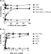
Specificity of ricin and TRAIL in inducing Calu-3 cell death. (A) Anti-TRAIL Abs (starting at 1 µg/ml) or (B) anti-ricin MAbs (starting at 15 µg/ml) in a 2-fold serial dilution were mixed with ricin (0.25 µg/ml) and TRAIL (0.1 µg/ml) and then administered to the cells seeded in 96-well plates for 24 h. The cells were then washed and cell viability was measured 72 h later, as described in Materials and Methods. The results (means ± SD) represent a single experiment done in triplicate and repeated at least three times.
FIG 4
FIG 4
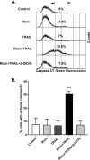
Increased caspase-3/7 activity in ricin- and TRAIL-treated Calu-3 cells. For the quantification of caspase-3/7 activity, Calu-3 cells were treated with ricin (0.25 µg/ml) or TRAIL (0.1 µg/ml) or ricin and TRAIL with or without caspase 3 inhibitor Z-DEVD-FMK(62.5 nM) for 24 h or with medium only (negative control). The caspase-3/7 activity levels were determined by flow cytometry as described in Materials and Methods. Caspase-3/7 activity was expressed as a percentage of total cells. The results are presented as means ± SD of three independent experiments. ***, P  < 0.001 (versus control cells).
FIG 5
FIG 5
Protective effect of caspase inhibitors on cell viability in ricin- and TRAIL-treated Calu-3 cells. Pan-caspase inhibitor Z-VAD-FMK, caspase-3 inhibitor Z-DEVD-FMK, or caspase-8 inhibitor Z-IETD-FMK at 62.5 nM was mixed with ricin (0.25 µg/ml) and TRAIL (0.1 µg/ml) and administered to the cells for 24 h. The cells were then washed, and cell viability was measured 48 h or 72 h later. The results (means ± SD) represent a single experiment done in triplicate and repeated at least three times.
FIG 6
FIG 6
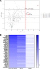
NanoString analysis of genes differentially expressed in ricin- and TRAIL-treated Calu-3 cells. Calu-3 cells were treated with ricin (0.25 µg/ml) or TRAIL (0.1 µg/ml) or a mixture of ricin and TRAIL or medium only (negative control) for 18 h. RNA was extracted and subjected to nCounter analysis using a human immunology array panel. (A) Volcano plot representation of gene expression changes in ricin-plus-TRAIL-treated cells, compared with control cells. Red circles represent transcripts upregulated >32-fold (5 log2-fold). The vertical dashed red line marks the 5 log2-fold change threshold. (B) Heat map showing the relative fold changes in expression of selected genes in each treatment group compared to the control group. Genes were selected based on a minimum of 4-fold (2 log2-fold)-higher expression in comparisons between control and ricin-plus-TRAIL-treated cells. The color scale bar denotes maximum counts in blue and minimal counts in white. The measured fold change values (relative to control cells) are listed in Table S1.
FIG 7
FIG 7
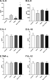
Cytokine secretion by Calu-3 cells following ricin and TRAIL treatment. Calu-3 cells were treated with ricin (0.25 µg/ml) or TRAIL (0.1 µg/ml) or a mixture of ricin and TRAIL or medium only (negative control) for 24 h. Cell supernatants were collected from treated cells. The levels of cytokines IL-6, IL-8, IL-1β, IL-10, TNF-ɑ, and IL-12p70 (panels A to F, respectively) were measured by CBA. The results are presented as the means ± SD of three independent experiments. **, P  < 0.01 (versus untreated cells [negative control]).
FIG 8
FIG 8
Effects of TNF-α- and TRAIL-neutralizing Abs on the viability of Calu-3 cells following ricin and TRAIL treatment. Calu-3 cells were treated with ricin or ricin plus TRAIL in the presence of neutralizing anti-TNF-α Ab or anti-TRAIL Ab or the combination of the two Abs. Cell viability was measured 72 h later. The results (means ± SD) represent a single experiment done in triplicate and repeated at least three times. **, P  < 0.01; *** P  < 0.001 (versus untreated cells [negative control]).

Similar articles

Cited by

References

    1. Cieslak TJ, Kortepeter MG, Wojtyk RJ, Jansen HJ, Reyes RA, Smith JO; NATO Biological Medical Advisory Panel. 2018. Beyond the dirty dozen: a proposed methodology for assessing future bioweapon threats. Mil Med 183:e59–e65. doi:10.1093/milmed/usx004. - DOI - PubMed
    1. Gal Y, Mazor O, Falach R, Sapoznikov A, Kronman C, Sabo T. 2017. Treatments for pulmonary ricin intoxication: current aspects and future prospects. Toxins 9:311. doi:10.3390/toxins9100311. - DOI - PMC - PubMed
    1. Pincus SH, Bhaskaran M, Brey RN III, Didier PJ, Doyle-Meyers LA, Roy CJ. 2015. Clinical and pathological findings associated with aerosol exposure of macaques to ricin toxin. Toxins 7:2121–2133. doi:10.3390/toxins7062121. - DOI - PMC - PubMed
    1. Thompson BT, Chambers RC, Liu KD. 2017. Acute respiratory distress syndrome. N Engl J Med 377:562–572. doi:10.1056/NEJMra1608077. - DOI - PubMed
    1. Brown RF, White DE. 1997. Ultrastructure of rat lung following inhalation of ricin aerosol. Int J Exp Pathol 78:267–276. - PMC - PubMed

Publication types

LinkOut - more resources