

Sperm capacitation is associated with phosphorylation of the testis-specific radial spoke protein Rsph6a†
- PMID: 30239614
- PMCID: PMC6378865
- DOI: 10.1093/biolre/ioy202
Sperm capacitation is associated with phosphorylation of the testis-specific radial spoke protein Rsph6a†
Abstract
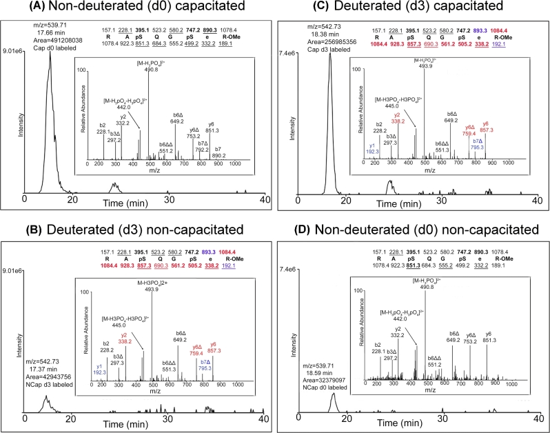
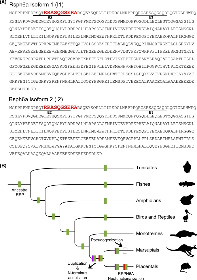
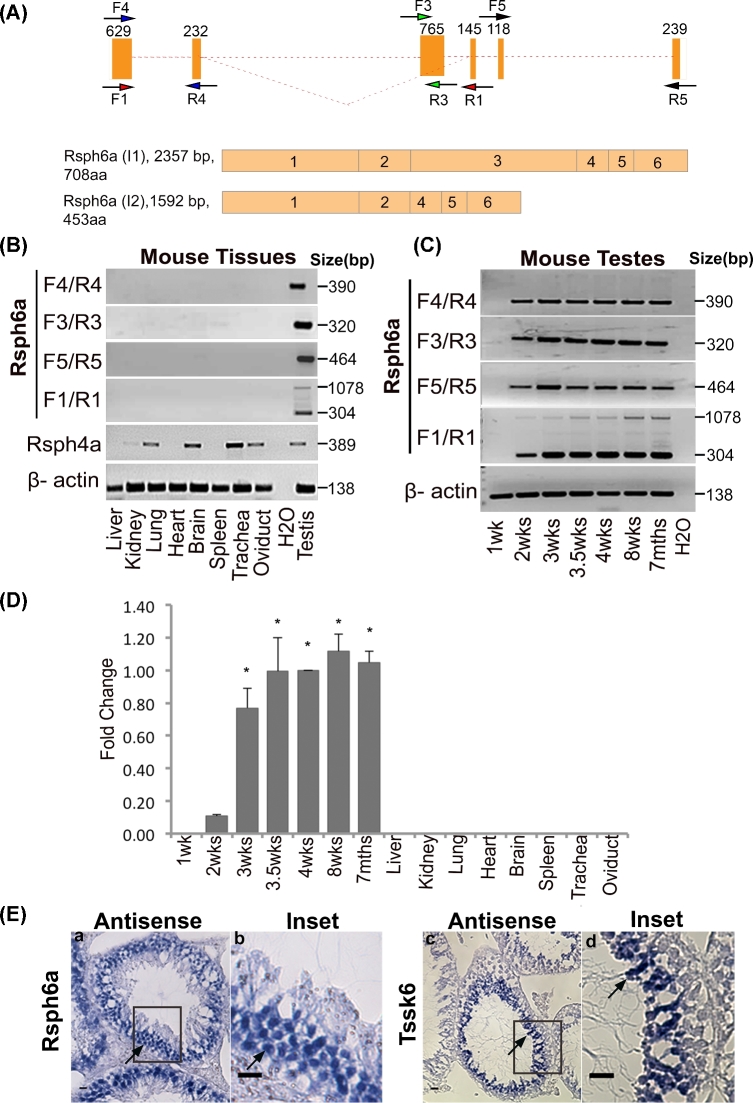
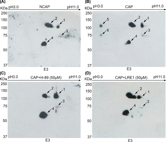
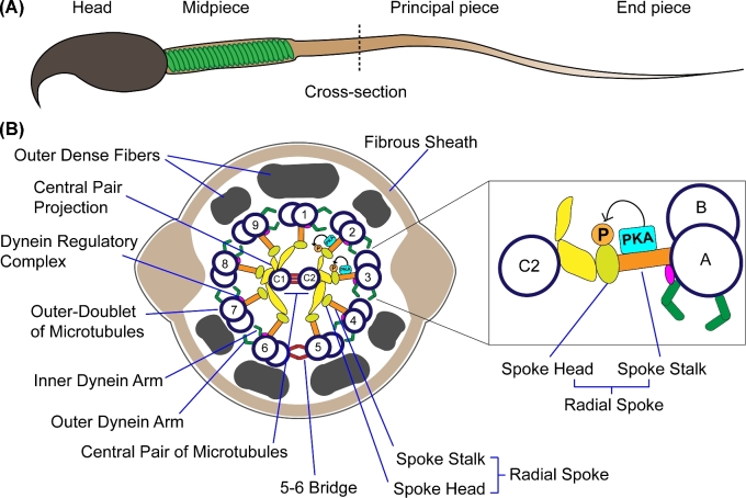
Mammalian sperm undergo a series of biochemical and physiological changes collectively known as capacitation in order to acquire the ability to fertilize. Although the increase in phosphorylation associated with mouse sperm capacitation is well established, the identity of the proteins involved in this signaling cascade remains largely unknown. Tandem mass spectrometry (MS/MS) has been used to identify the exact sites of phosphorylation and to compare the relative extent of phosphorylation at these sites. In the present work, we find that a novel site of phosphorylation on a peptide derived from the radial spoke protein Rsph6a is more phosphorylated in capacitated mouse sperm. The Rsph6a gene has six exons, five of which are conserved during evolution in flagellated cells. The exon containing the capacitation-induced phosphorylation site was found exclusively in eutherian mammals. Transcript analyses revealed at least two different testis-specific splicing variants for Rsph6a.Rsph6a mRNA expression was restricted to spermatocytes. Using antibodies generated against the Rsph6a N-terminal domain, western blotting and immunofluorescence analyses indicated that the protein remains in mature sperm and localizes to the sperm flagellum. Consistent with its role in the axoneme, solubility analyses revealed that Rsph6 is attached to cytoskeletal structures. Based on previous studies in Chlamydomonas reinhardtii, we predict that Rsph6 participates in the interaction between the central pair of microtubules and the surrounding pairs. The findings that Rsph6a is more phosphorylated during capacitation and is predicted to function in axonemal localization make Rsph6a a candidate protein mediating signaling processes in the sperm flagellum.
Keywords: axoneme; capacitation; phosphorylation; radial spoke; sperm.
© The Author(s) 2018. Published by Oxford University Press on behalf of Society for the Study of Reproduction.
Figures

Similar articles
-
RSPH6A is required for sperm flagellum formation and male fertility in mice.J Cell Sci. 2018 Oct 11;131(19):jcs221648. doi: 10.1242/jcs.221648. J Cell Sci. 2018. PMID: 30185526 Free PMC article.
-
FSP95, a testis-specific 95-kilodalton fibrous sheath antigen that undergoes tyrosine phosphorylation in capacitated human spermatozoa.Biol Reprod. 1999 Nov;61(5):1184-97. doi: 10.1095/biolreprod61.5.1184. Biol Reprod. 1999. PMID: 10529264
-
Radial spoke protein 44 (human meichroacidin) is an axonemal alloantigen of sperm and cilia.Gene. 2007 Jul 1;396(1):93-107. doi: 10.1016/j.gene.2007.02.031. Epub 2007 Mar 23. Gene. 2007. PMID: 17451891 Free PMC article.
-
Sperm Capacitation and Acrosome Reaction in Mammalian Sperm.Adv Anat Embryol Cell Biol. 2016;220:93-106. doi: 10.1007/978-3-319-30567-7_5. Adv Anat Embryol Cell Biol. 2016. PMID: 27194351 Review.
-
The protein tyrosine phosphorylation during in vitro capacitation and cryopreservation of mammalian spermatozoa.Cryobiology. 2015 Jun;70(3):211-6. doi: 10.1016/j.cryobiol.2015.03.008. Epub 2015 Mar 28. Cryobiology. 2015. PMID: 25828199 Review.
Cited by
-
Expression of RSPH6A in the first wave of rat spermatogenesis and oxidative stress conditions: Attenuation by melatonin.Reprod Med Biol. 2023 Oct 2;22(1):e12542. doi: 10.1002/rmb2.12542. eCollection 2023 Jan-Dec. Reprod Med Biol. 2023. PMID: 37795044 Free PMC article.
-
RSPH6A is required for sperm flagellum formation and male fertility in mice.J Cell Sci. 2018 Oct 11;131(19):jcs221648. doi: 10.1242/jcs.221648. J Cell Sci. 2018. PMID: 30185526 Free PMC article.
-
CRISPR/CAS9-mediated amino acid substitution reveals phosphorylation residues of RSPH6A are not essential for male fertility in mice†.Biol Reprod. 2020 Oct 29;103(5):912-914. doi: 10.1093/biolre/ioaa161. Biol Reprod. 2020. PMID: 32901808 Free PMC article. No abstract available.
-
Cfap97d1 is important for flagellar axoneme maintenance and male mouse fertility.PLoS Genet. 2020 Aug 12;16(8):e1008954. doi: 10.1371/journal.pgen.1008954. eCollection 2020 Aug. PLoS Genet. 2020. PMID: 32785227 Free PMC article.
-
Overview of the Complex Relationship between Epigenetics Markers, CTG Repeat Instability and Symptoms in Myotonic Dystrophy Type 1.Int J Mol Sci. 2022 Mar 23;23(7):3477. doi: 10.3390/ijms23073477. Int J Mol Sci. 2022. PMID: 35408837 Free PMC article. Review.
References
-
- Gervasi MG, Visconti PE. Chang's meaning of capacitation: A molecular perspective. Mol Reprod Dev 2016; 83:860–874. - PubMed
-
- Jha KN, Salicioni AM, Arcelay E, Chertihin O, Kumari S, Herr JC, Visconti PE. Evidence for the involvement of proline-directed serine/threonine phosphorylation in sperm capacitation. Mol Hum Reprod 2006; 12:781–789. - PubMed
-
- Stival C, La Spina FA, Baro Graf C, Arcelay E, Arranz SE, Ferreira JJ, Le Grand S, Dzikunu VA, Santi CM, Visconti PE, Buffone MG, Krapf D. Src kinase is the connecting player between Protein Kinase A (PKA) activation and hyperpolarization through SLO3 potassium channel regulation in mouse sperm. J Biol Chem 2015; 290:18855–18864. - PMC - PubMed
Publication types
MeSH terms
Substances
Grants and funding
LinkOut - more resources
Full Text Sources
Other Literature Sources
Molecular Biology Databases
