

A Phylogenomic Study of Acanthamoeba polyphaga Draft Genome Sequences Suggests Genetic Exchanges With Giant Viruses
- PMID: 30237791
- PMCID: PMC6135880
- DOI: 10.3389/fmicb.2018.02098
A Phylogenomic Study of Acanthamoeba polyphaga Draft Genome Sequences Suggests Genetic Exchanges With Giant Viruses
Abstract
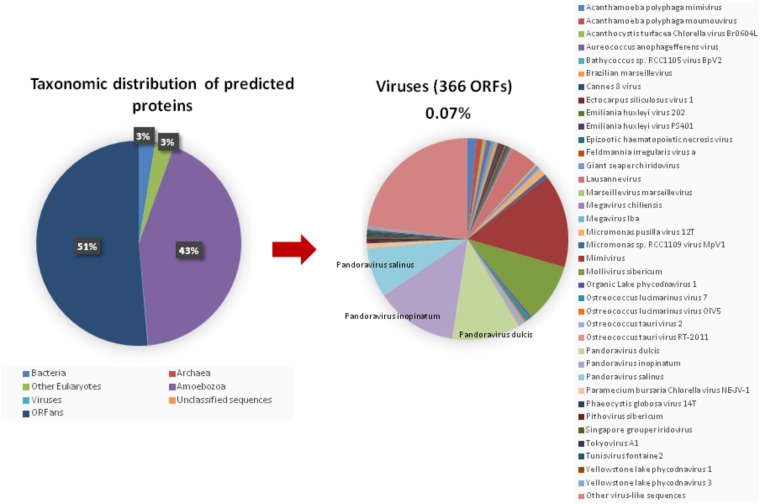
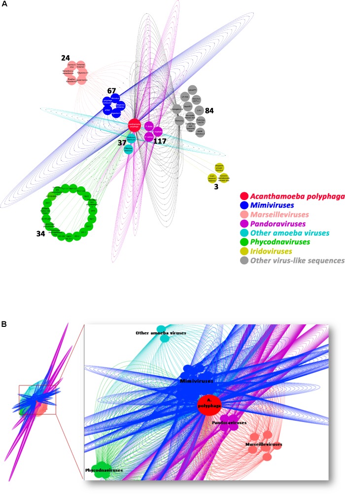
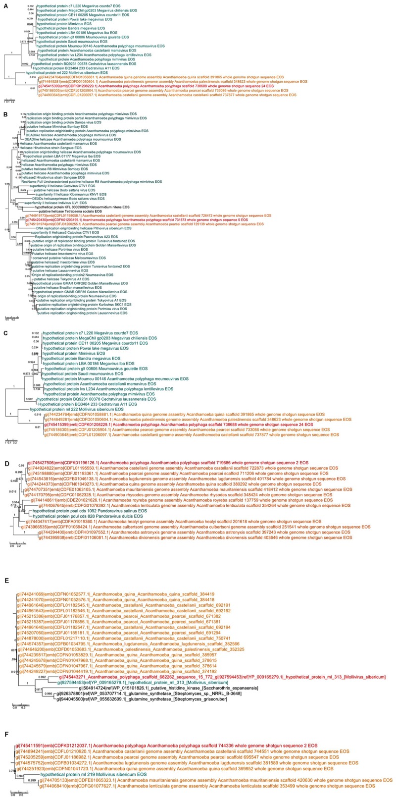
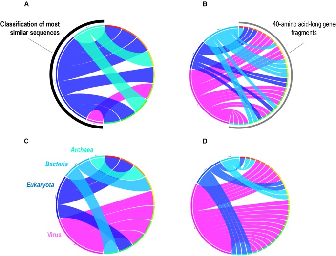
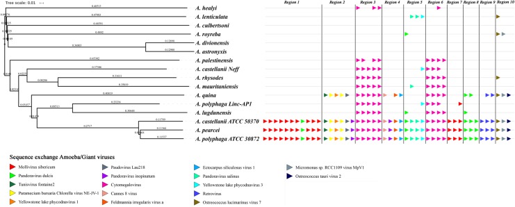
Acanthamoeba are ubiquitous phagocytes predominant in soil and water which can ingest many microbes. Giant viruses of amoebae are listed among the Acanthamoeba-resisting microorganisms. Their sympatric lifestyle within amoebae is suspected to promote lateral nucleotide sequence transfers. Some Acanthamoeba species have shown differences in their susceptibility to giant viruses. Until recently, only the genome of a single Acanthamoeba castellanii Neff was available. We analyzed the draft genome sequences of Acanthamoeba polyphaga through several approaches, including comparative genomics, phylogeny, and sequence networks, with the aim of detecting putative nucleotide sequence exchanges with giant viruses. We identified a putative sequence trafficking between this Acanthamoeba species and giant viruses, with 366 genes best matching with viral genes. Among viruses, Pandoraviruses provided the greatest number of best hits with 117 (32%) for A. polyphaga. Then, genes from mimiviruses, Mollivirus sibericum, marseilleviruses, and Pithovirus sibericum were best hits in 67 (18%), 35 (9%), 24 (7%), and 2 (0.5%) cases, respectively. Phylogenetic reconstructions showed in a few cases that the most parsimonious evolutionary scenarios were a transfer of gene sequences from giant viruses to A. polyphaga. Nevertheless, in most cases, phylogenies were inconclusive regarding the sense of the sequence flow. The number and nature of putative nucleotide sequence transfers between A. polyphaga, and A. castellanii ATCC 50370 on the one hand, and pandoraviruses, mimiviruses and marseilleviruses on the other hand were analyzed. The results showed a lower number of differences within the same giant viral family compared to between different giant virus families. The evolution of 10 scaffolds that were identified among the 14 Acanthamoeba sp. draft genome sequences and that harbored ≥ 3 genes best matching with viruses showed a conservation of these scaffolds and their 46 viral genes in A. polyphaga, A. castellanii ATCC 50370 and A. pearcei. In contrast, the number of conserved genes decreased for other Acanthamoeba species, and none of these 46 genes were present in three of them. Overall, this work opens up several potential avenues for future studies on the interactions between Acanthamoeba species and giant viruses.
Keywords: Acanthamoeba; Acanthamoeba polyphaga; draft genome sequences; giant viruses; horizontal gene transfer; mimivirus; nucleotide sequence transfer.
Figures
Similar articles
-
In-depth study of Mollivirus sibericum, a new 30,000-y-old giant virus infecting Acanthamoeba.Proc Natl Acad Sci U S A. 2015 Sep 22;112(38):E5327-35. doi: 10.1073/pnas.1510795112. Epub 2015 Sep 8. Proc Natl Acad Sci U S A. 2015. PMID: 26351664 Free PMC article.
-
A Comparative Genomic Approach to Determine the Virulence Factors and Horizontal Gene Transfer Events of Clinical Acanthamoeba Isolates.Microbiol Spectr. 2022 Apr 27;10(2):e0002522. doi: 10.1128/spectrum.00025-22. Epub 2022 Apr 13. Microbiol Spectr. 2022. PMID: 35416714 Free PMC article.
-
Cedratvirus, a Double-Cork Structured Giant Virus, is a Distant Relative of Pithoviruses.Viruses. 2016 Nov 3;8(11):300. doi: 10.3390/v8110300. Viruses. 2016. PMID: 27827884 Free PMC article.
-
Mimivirus: leading the way in the discovery of giant viruses of amoebae.Nat Rev Microbiol. 2017 Apr;15(4):243-254. doi: 10.1038/nrmicro.2016.197. Epub 2017 Feb 27. Nat Rev Microbiol. 2017. PMID: 28239153 Free PMC article. Review.
-
Giant Viruses of Amoebas: An Update.Front Microbiol. 2016 Mar 22;7:349. doi: 10.3389/fmicb.2016.00349. eCollection 2016. Front Microbiol. 2016. PMID: 27047465 Free PMC article. Review.
Cited by
-
Assessing the biogeography of marine giant viruses in four oceanic transects.bioRxiv [Preprint]. 2023 Feb 1:2023.01.30.526306. doi: 10.1101/2023.01.30.526306. bioRxiv. 2023. Update in: ISME Commun. 2023 Apr 29;3(1):43. doi: 10.1038/s43705-023-00252-6. PMID: 36778472 Free PMC article. Updated. Preprint.
-
Application of the omics sciences to the study of Naegleria fowleri, Acanthamoeba spp., and Balamuthia mandrillaris: current status and future projections.Parasite. 2021;28:36. doi: 10.1051/parasite/2021033. Epub 2021 Apr 12. Parasite. 2021. PMID: 33843581 Free PMC article. Review.
-
Discovery and Further Studies on Giant Viruses at the IHU Mediterranee Infection That Modified the Perception of the Virosphere.Viruses. 2019 Mar 30;11(4):312. doi: 10.3390/v11040312. Viruses. 2019. PMID: 30935049 Free PMC article. Review.
-
Medusavirus, a Novel Large DNA Virus Discovered from Hot Spring Water.J Virol. 2019 Apr 3;93(8):e02130-18. doi: 10.1128/JVI.02130-18. Print 2019 Apr 15. J Virol. 2019. PMID: 30728258 Free PMC article.
-
Characterization of Mollivirus kamchatka, the First Modern Representative of the Proposed Molliviridae Family of Giant Viruses.J Virol. 2020 Mar 31;94(8):e01997-19. doi: 10.1128/JVI.01997-19. Print 2020 Mar 31. J Virol. 2020. PMID: 31996429 Free PMC article.
References
LinkOut - more resources
Full Text Sources
Other Literature Sources