. 2018 Oct 24;38(43):9228-9239.
doi: 10.1523/JNEUROSCI.0585-18.2018. Epub 2018 Sep 18.

A Subpopulation of Foxj1-Expressing, Nonmyelinating Schwann Cells of the Peripheral Nervous System Contribute to Schwann Cell Remyelination in the Central Nervous System

Affiliations

A Subpopulation of Foxj1-Expressing, Nonmyelinating Schwann Cells of the Peripheral Nervous System Contribute to Schwann Cell Remyelination in the Central Nervous System

Dan Ma et al. J Neurosci. .

Abstract

New myelin sheaths can be restored to demyelinated axons in a spontaneous regenerative process called remyelination. In general, new myelin sheaths are made by oligodendrocytes newly generated from a widespread population of adult CNS progenitors called oligodendrocyte progenitor cells (OPCs). New myelin in CNS remyelination in both experimental models and clinical diseases can also be generated by Schwann cells (SCs), the myelin-forming cells of the PNS. Fate-mapping studies have shown that SCs contributing to remyelination in the CNS are often derived from OPCs and appear not to be derived from myelinating SCs from the PNS. In this study, we address whether CNS remyelinating SCs can also be generated from PNS-derived cells other than myelinating SCs. Using a genetic fate-mapping approach, we have found that a subpopulation of nonmyelinating SCs identified by the expression of the transcription factor Foxj1 also contribute to CNS SC remyelination, as well as to remyelination in the PNS. We also find that the ependymal cells lining the central canal of the spinal cord, which also express Foxj1, do not generate cells that contribute to CNS remyelination. These findings therefore identify a previously unrecognized population of PNS glia that can participate in the regeneration of new myelin sheaths following CNS demyelination.SIGNIFICANCE STATEMENT Remyelination failure in chronic demyelinating diseases such as multiple sclerosis drives the current quest for developing means by which remyelination in CNS can be enhanced therapeutically. Critical to this endeavor is the need to understand the mechanisms of remyelination, including the nature and identity of the cells capable of generating new myelin sheath-forming cells. Here, we report a previously unrecognized subpopulation of nonmyelinating Schwann cells (SCs) in the PNS, identified by the expression of the transcription factor Foxj1, which can give rise to SCs that are capable of remyelinating both PNS and CNS axons. These cells therefore represent a new cellular target for myelin regenerative strategies for the treatment of CNS disorders characterized by persistent demyelination.

Keywords: CNS remyelination; Foxj1; Schwann cells; peripheral nerve.


Figures

Figure 1.
Figure 1.
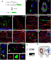
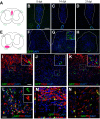
Foxj1 controlled GFP expression labels cells in both the CNS and PNS. A, Diagram illustrating transgene design of Foxj1 promoter-controlled, tamoxifen-induced activation of fGFP reporter gene expression. BK are images from multiple immunostaining for GFP and different cell markers. GFP-expressing cells are detected in ependymal cells lining lateral ventricles (LV; B); ependymal cells in the central canal of spinal cord (CC; C), which coexpress Sox2; and Bergmann glia in cerebellum cortex (CB; D). Foxj1-controlled GFP is also expressed in the PNS. GFP+ cells are scattered between neurofilament-positive axons in spinal dorsal roots (DR) (E, transverse section), sciatic nerves (SN, F, longitudinal section), and trigeminal nerves (TN) (G, longitudinal sections). GFP-expressing cells do not colocalize with myelinating SC markers (H, inset shows transverse view), P0 (I), and S100β (J, transverse section). Image in K is from a dorsal root ganglion (DRG) showing GFP-expressing cells among nerve fibers but few among neuronal cell bodies (asterisk). Occasionally, Foxj1-GFP cells surround a DRG neuron at axonal entry zone (inset in K). Image L illustrates immunoreactive Foxj1+ cells in small number of ependymal cells in CC, which also expressed GFP (solid arrowhead). However, not all GFP+ are detected with Foxj1+ (open arrowhead). Nucleus-localized Foxj1 is detectable in the transverse section of ventral root (VR) of spinal cord in GFP+ or GFP cells (M). The blue color in all images is the DAPI-stained nucleus. Foxj1 protein and mRNA can be detected in peripheral nerves as well as spinal cords (SC) by Western blot and RT-PCR (N). Foxj1 transcript can be visualized by the sensitive RNAScope technique in sciatic nerve and spinal cord central canal (O). Scale bars in the images indicate 20 μm except for E, which is 50 μm, and K, which is 100 μm.
Figure 2.
Figure 2.
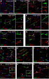
Foxj1-GFP-labeled cells express fibroblast markers but are of a distinct population from PDGFRa-GFP labeled cells in peripheral nerves. Immunofluorescence characterization of normal sciatic nerves (SN) from Foxj1-GFP mice treated with tamoxifen. Double immunostaining for GFP and various markers was performed on longitudinal sections. Colocalized staining is marked with arrowheads. Overlaid images show that Foxj1 labeled cells in SN are not associated with endothelial cells labeled by CD31 (A), nor do they express the macrophage marker IBA1 (B). A proportion of GFP+ cells are labeled with NG2 (C) and proteins usually expressed by fibroblasts such as fibronectin (FN, D), P4HB (E), SMA (F), and HSP47 (G). In DG, the split channel views of the boxed area by dotted lines are shown in separate images. Foxj1-GFP-labeled cells also expressed P75NTRs (H and confocal orthogonal view in H′). GFP-labeled cells do not coexpress PDGFRa (CD140a, I). PDGFRa fate-mapping GFP reporter mice were characterized by double immunostaining. PDGFRa-GFP labeled cells are not associated with neurofilament-labeled axons (J). The GFP+ cells are confirmed expressing CD140a (inset in J) and are double labeled with P4HB and SMA (K and inset in K). PDGFRa-GFP-labeled cells are also detected for P75NTR (L). Scale bar indicates 50 μm for all images.
Figure 3.
Figure 3.
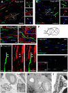
Foxj1 labels nonmyelinating SCs. Images in AF illustrate merged double immunofluorescence staining on longitudinal sections of sciatic nerve (SN) from adult Foxj1-GFP mice treated with tamoxifen. Most GFP-expressing cells coexpress the L1 cell adhesion molecule, a marker of nonmyelinating SCs in peripheral nerves (A). GFP-expressing cells can also express Sox10, a neural crest-related transcription factor (B). The single channel images of the same area in A and B marked by dotted line are shown in image sets on the right side of the main images. PDGFRa-labeled cells (PDGFRa-GFP) in sciatic nerves do not coexpress L1 (C). Low levels of GFAP are detected in Foxj1-GFP-expressing cells (D, inset). Costaining for GFP and neurofilament reveals a close association between GFP and small-diameter axons (arrowheads) (E). Dorsal and ventral roots (DR and VR, respectively) from lumbar spinal cord were stained for GFP, showing greater numbers of Foxj1-GFP+ cells in dorsal root (F) and this was correlated with the proportion of L1 immunoreactivity in corresponding locations (inset in F). GI are images from pre-embedding immunogold electron microscopy against GFP on SN from Foxj1 and Sox10 fate-mapping mice. Silver-enhanced gold particles were mainly detected on “Remak” bundles in Foxj1-GFP samples (G), whereas the majority of gold particles in Sox10 fate-mapping mice are deposited in SC cytoplasm around the myelinated axons but excluded from compact myelin sheaths (H). PDGFRa labeling identifies endoneurial fibroblast like cells (I). Scale bars: AE, 25 μm; F, 50 μm; G, 2 μm; H, I, 1 μm.
Figure 4.
Figure 4.
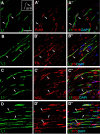
Nonmyelinating SCs express fibroblast markers. Image sets AC illustrate immunostaining of longitudinal sections from unlesioned sciatic nerves for L1Cam (L1, green), a marker labeling nonmyelinating SCs, with markers labeling peripheral nerve fibroblasts (red). The merged images show that a proportion of L1 immunoreactive cells colocalize with P4HB (AA″), fibronectin (FN, BB″), and NG2 (CC″). Image set D shows that the majority of L1-labeled cells are also positive for P75NTR (P75) (D′ and merged image D″) and a proportion of P75+ cells are not labeled with L1. The open arrowheads highlight a P75+ cell that is not labeled with L1 (D′ and D″). The solid arrowheads in all images indicate examples of colocalization. Scale bar indicates 50 μm, applicable to all images.
Figure 5.
Figure 5.
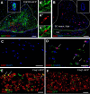
Foxj1-labeled cells give rise to remyelinating SCs in demyelinating lesions in spinal cord white matter. Demyelination lesions were induced in mice Foxj1-GFP-expressing mice by direct injection of lysolecithin in either dorsal or ventral funiculi as shown in A and E, with corresponding images from GFP immunostaining for 5, 14, and 21 dpl. Few GFP-expressing cells have been detected in dorsal lesions (BD), whereas a large number of GFP+ cells are found in ventral lesions from 14 dpl (FH). Double immunostaining indicates that Foxj1-GFP cells do not express the astrocyte marker GFAP (I), the oligodendrocyte lineage marker Olig2 (J), nor the microglial marker IBA1 (K). L shows that considerable numbers of GFP-expressing cells coexpress the myelinating SC marker PRX (arrows). The box area is shown as split channels (L1 and L2) and merged in (L3). M, Orthogonal view of confocal images of double immunostaining confirming the colocalization of GFP and PRX in ventral lesions at 14 dpl. The SC identity of GFP+ cells in lesion is verified by colocalization with myelin P0, as illustrated by orthogonal confocal view in N. Insets in the images show magnified area in lesions in respective images marked with dotted outlines, for either double staining (IK) or single colors (G, L). Scale bars in B indicate 100 μm and apply to C, D, and FH. Scale bar in I indicates 50 μm and applies to J and K. In LN, the scale bars indicate 25 μm.
Figure 6.
Figure 6.
SC remyelination from Foxj1-GFP-labeled cells in spinal cord lesions are not derived from ependymal cells, but likely originate from peripheral nerves. A, Demyelinated lesion at 21 dpl in ventral spinal cord white matter of a FGFR3-GFP reporter mouse treated with tamoxifen. Image shows overlaid double immunostaining for GFP and PRX, with the dotted line marking the boundaries of the lesion. GFP is expressed by ependymal cells lining the central canal of the spinal cord (inset). A magnified area with in the red box in the lesion in A showing separate and overlaid channels indicating nonoverlapping expression of GFP and PRX is shown in A′, A″, and A‴). B illustrates a ventral spinal cord lesion (white dotted line) at 5 dpl immunostained for the proliferation marker Ki67. Ki67+ cells are found in the adjacent ventral roots (VR, green dotted line), but not in ependymal cells in central canal (inset). There are no Ki67+ cells in the contralateral VR (C). The Ki67+ nucleus are found in L1+ cells in the VR of ventral spinal cord lesion side (D). E and F illustrate Foxj1-GFP cells colabeled with PRX in the VR of spinal cord lesion side at 14 dpl, but none in the VR of contralateral side. Scale bars indicate 100 μm in A, 50 μm in B, and 20 μm in CF.
Figure 7.
Figure 7.
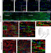
Foxj1-GFP-labeled cells become repair SCs following sciatic nerve injury. Sciatic nerve crush lesions were created in adult mice expressing reporter genes and samples were analyzed by immunofluorescence staining. AD show an area of sciatic nerve in longitudinal section immunostained to reveal Foxj1-GFP-labeled cells in normal (A) and crushed nerve at 5, 14, and 21 dpi (BD, respectively). Inset in B illustrates GFP+ cells expressing Ki67. E is a montage combined from a series of overlapping images spanning the proximal and crushed site at 28 dpi. Doubling immunostaining shows that, at 28 dpl, GFP colocalizes with the mature myelinating SC marker PRX (F). The dotted outlined area is shown in single channels as in insets in F, with examples of colocalization marked with arrows. GFP labels the myelinated axons marked by myelin P0, which appears only localized in the cytoplasm rather than compact myelin (G). GFP-labeled cells enclose the axons (arrows) marked by neurofilament (NF) staining (H). Approximately 30% of remyelinated axons in the crush area are labeled with GFP, but >70% of GFP immunoreactivity colocalizes with PRX (mean ± SE, n = 3; I). J and K show GFP and PRX double immunostaining in control (uninjured nerve) and crushed sciatic nerve from PDGFRa-GFP animals at 28 dpi. The magnified boxed area in K is shown in K′ which shows that there is no colocalization between GFP and PRX. Scale bars indicate 50 μm for all images.
Figure 8.
Figure 8.
Early-appearing SC lineage cells following nerve transection are mainly NMSCs. Immunohistochemistry for GFP and PRX was performed on longitudinal sections of sciatic nerves from mice that received transection injury. Shown is an area of proximal stump (A), rejoining bridge (B), and distal stamp (C) following sciatic cut 7 dpi from a tamoxifen-treated Foxj1-GFP mouse. Staining of GFP-expressing cells at the bridge at the same time point following transection from a Sox10-GFP mouse is shown in D. E shows an area of intact sciatic nerve from Sox10-GFP mice indicating that both myelinating (arrows) and nonmyelinating cells (arrowheads) have been labeled by GFP. The GFP-labeled cells in Sox10-GFP sciatic nerve coexpress L1, as shown in confocal image in F, confirming their identity as nonmyelinating SCs. Scale bars indicate 100 μm in AD and 50 μm in E and F.

Similar articles

Cited by

References

    1. Arthur-Farraj PJ, Latouche M, Wilton DK, Quintes S, Chabrol E, Banerjee A, Woodhoo A, Jenkins B, Rahman M, Turmaine M, Wicher GK, Mitter R, Greensmith L, Behrens A, Raivich G, Mirsky R, Jessen KR (2012) c-jun reprograms Schwann cells of injured nerves to generate a repair cell essential for regeneration. Neuron 75:633–647. 10.1016/j.neuron.2012.06.021 - DOI - PMC - PubMed
    1. Assinck P, Duncan GJ, Plemel JR, Lee MJ, Stratton JA, Manesh SB, Liu J, Ramer LM, Kang SH, Bergles DE, Biernaskie J, Tetzlaff W (2017) Myelinogenic plasticity of oligodendrocyte precursor cells following spinal cord contusion injury. J Neurosci 37:8635–8654. 10.1523/JNEUROSCI.2409-16.2017 - DOI - PMC - PubMed
    1. Barnabé-Heider F, Göritz C, Sabelström H, Takebayashi H, Pfrieger FW, Meletis K, Frisén J (2010) Origin of new glial cells in intact and injured adult spinal cord. Cell Stem Cell 7:470–482. 10.1016/j.stem.2010.07.014 - DOI - PubMed
    1. Blakemore WF. (1975) Remyelination by Schwann cells of axons demyelinated by intraspinal injection of 6-aminonicotinamide in the rat. J Neurocytol 4:745–757. 10.1007/BF01181634 - DOI - PubMed
    1. Carlén M, Meletis K, Göritz C, Darsalia V, Evergren E, Tanigaki K, Amendola M, Barnabé-Heider F, Yeung MS, Naldini L, Honjo T, Kokaia Z, Shupliakov O, Cassidy RM, Lindvall O, **Frisén J (2009) Forebrain ependymal cells are notch-dependent and generate neuroblasts and astrocytes after stroke. Nat Neurosci 12:259–267. 10.1038/nn.2268 - DOI - PubMed

Publication types

MeSH terms

Substances

LinkOut - more resources