. 2018 Nov;53(5):1997-2009.
doi: 10.3892/ijo.2018.4553. Epub 2018 Sep 5.

Distinct phenotypes and 'bystander' effects of senescent tumour cells induced by docetaxel or immunomodulatory cytokines

Affiliations

Distinct phenotypes and 'bystander' effects of senescent tumour cells induced by docetaxel or immunomodulatory cytokines

Olena Sapega et al. Int J Oncol. 2018 Nov.

Abstract

Cellular senescence is the process of the permanent proliferative arrest of cells in response to various inducers. It is accompanied by typical morphological changes, in addition to the secretion of bioactive molecules, including proinflammatory cytokines and chemokines [known as the senescence-associated secretory phenotype (SASP)]. Thus, senescent cells may affect their local environment and induce a so-called 'bystander' senescence through the state of SASP. The phenotypes of senescent cells are determined by the type of agent inducing cellular stress and the cell lineages. To characterise the phenotypes of senescent cancer cells, two murine cell lines were employed in the present study: TC-1 and B16F10 (B16) cells. Two distinct senescence inductors were used: Chemotherapeutic agent docetaxel (DTX) and a combination of immunomodulatory cytokines, including interferon γ (IFNγ) and tumour necrosis factor α (TNFα). It was demonstrated that DTX induced senescence in TC-1 and B16 tumour cell lines, which was demonstrated by growth arrest, positive β-galactosidase staining, increased p21Waf1 (p21) expression and the typical SASP capable of inducing a 'bystander' senescence. By contrast, treatment with a combination of T helper cell 1 cytokines, IFNγ and TNFα, induced proliferation arrest only in B16 cells. Despite the presence of certain characteristic features resembling senescent cells (proliferation arrest, morphological changes and increased p21 expression), these cells were able to form tumours in vivo and started to proliferate upon cytokine withdrawal. In addition, B16 cells were not able to induce a 'bystander' senescence. In summary, the present study described cell line- and treatment-associated differences in the phenotypes of senescent cells that may be relevant in optimization of cancer chemo- and immunotherapy.


Figures
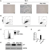
Figure 1
Figure 1
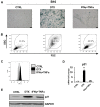
DTX induces senescence in TC-1 cells. (A) Senescence-associated β-galactosidase activity in TC-1 cells treated with DTX or IFNγ + TNFα for 4 days. (B) The size and granularity of control or IFNγ + TNFα-treated senescent TC-1 cells was determined by forward and side scatter flow cytometry analysis. (C) Autofluorescence of the TC-1 control cells is presented in light grey, DTX-treated in black and IFNγ + TNFα-treated in grey. (D) Reverse transcription-quantitative polymerase chain reaction quantification of p21 in control, DTX- and IFNγ + TNFα-treated TC-1 cells. (E) Immunoblotting detection of mouse p21 in control, DTX- and IFNγ + TNFα-treated TC-1 cells harvested on day 4. GAPDH was used as a loading control. Representative results from at least three independent experiments are presented. Data are presented as the mean ± standard deviation. CTRL, control cells; DTX, docetaxel; IFNγ, interferon γ; TNFα, tumour necrosis factor α; FSC, forward scattering; SSC, side scattering; p21, p21Waf1.
Figure 2
Figure 2
DTX induces senescence in the B16 cell line. (A) Senescence-associated β-galactosidase activity in B16 cells treated with DTX or IFNγ + TNFα for 4 days. (B) The size and granularity of control or IFNγ + TNFα-treated, senescent B16 cells was determined by forward and side scatter flow cytometry analysis. (C) Autofluorescence of the B16 control cells is presented in light grey, DTX-treated in black and IFNγ + TNFα-treated in grey. (D) Reverse transcription-quantitative polymerase chain reaction quantification of p21 in control, DTX- and IFNγ + TNFα-treated B16 cells. (E) Immunoblotting detection of mouse p21 in control, DTX- and IFNγ + TNFα-treated B16 cells harvested on day 4. GAPDH was used as a loading control. Representative results from at least three independent experiments are presented. Data are presented as the mean ± standard deviation. **P<0.01 vs. CTRL. CTRL, control cells; DTX, docetaxel; IFNγ, interferon γ; TNFα, tumour necrosis factor α; FSC, forward scattering; SSC, side scattering; p21, p21Waf1; B16, B16F10 cell line.
Figure 3
Figure 3
Analysis of TC-1 and B16 cell proliferation during ‘primary’ induction. (A) TC-1 and (B) B16 cells were treated with DTX and IFNγ + TNFα. Cell proliferation was determined by counting the cell number on days 4 (TC-1 and B16) and 7 (B16 only). Control cells were passaged on day 4. Data are presented as the mean ± standard deviation. ***P<0.001 vs. day 0. For EdU incorporation: Cells were driven to senescence by 4-day treatments with DTX or IFNγ + TNFα, and then incubated with 10 µM EdU for 24 h. Click-iT reaction was performed on fixed cells, and FACS analysis was performed to determine the fraction of proliferating (C) TC-1 and (D) B16 cells in the treated and control samples. Representative results from three independent experiments are presented. Mice (8 per group) were transplanted subcutaneously on day 0 with (E) TC-1 and (F) B16 cells (3×10⁴), with IFNγ + TNFα-treated cells (3×10⁴) or with senescent DTX-treated cells at the doses 3×10⁴ [B16 or TC-1/DTX(a)] or 3×10⁵ [B16 or TC-1/DTX(b)] tumour cells and the tumour growth was monitored. The experiment was repeated two times with similar results. *P<0.05 [TC-1 or TC-1/IFNγ + TNFα vs. TC-1/DTX (a or b)]; [B16 or B16/IFNγ + TNFα vs. B16/ DTX (a or b); analysis of variance]. CTRL, control cells; DTX, docetaxel; IFNγ, interferon γ; TNFα, tumour necrosis factor α; B16, B16F10 cell line.
Figure 4
Figure 4
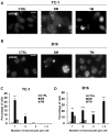
DNA damage detection in TC-1 and B16 tumour cell lines. To detect DNA damage, control, DTX- or IFNγ + TNFα-treated (A) TC-1 and (B) B16 cells were stained with phosphoSer139 H2A histone family, member X antibody and mounted with Mowiol containing 4’,6-diamidine-2-phenylindole. Scale bar, 20 µm. Percentage of cells with 1, 2, 3 or more micronuclei in (C) TC-1 and (D) B16 cells treated with DTX or IFNγ + TNFα was quantified. Data are presented as the mean ± standard deviation. *P<0.05, **P<0.01 and ***P<0.001 vs. CTRL. A total of 100 cells were analysed in each experimental group. CTRL, control cells; DTX, docetaxel; IFNγ, interferon γ; TNFα, tumour necrosis factor α; B16, B16F10 cell line.
Figure 5
Figure 5
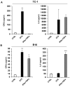
Secretion of IL-6 and GROα by murine TC-1 and B16 tumour cell lines. Enzyme-linked immunosorbent assay of IL-6 and GROα in supernatants of (A) TC-1 and (B) B16 cells treated with DTX and IFNγ + TNFα. Supernatants were tested in triplicate and the results from three independent experiments are presented as the mean ± standard deviation. ***P<0.001, **P<0.01 and *P<0.05 vs. CTRL. Asterisks correspond to comparisons between control and treated groups. CTRL, control cells; DTX, docetaxel; IFNγ, interferon γ; TNFα, tumour necrosis factor α; B16, B16F10 cell line; GROα, growth-regulated oncogene α; IL-6, interleukin 6.
Figure 6
Figure 6
Induction of ‘bystander’ senescence in TC-1 tumour cells. (A) Senescence-associated β-galactosidase activity in TC-1 cells cultured for 4 days in medium (SM), or proliferating cell medium (TM). (B) The size and granularity of control and senescent TC-1 cells was determined by forward and side scatter flow cytometry analysis. (C) Expression of p21 in TC-1 cells cultured for 4 days in different media (reverse transcription-quantitative polymerase chain reaction). (D) Immunoblotting detection of mouse p21 in TC-1 cells harvested on day 4 following cultivation in different media. GAPDH was used as a loading control. Data are presented as the mean ± standard deviation. *P<0.05 vs. CTRL. CTRL, control cells; SM, senescence medium; TM, tumour medium; FSC, forward scattering; SSC, side scattering; p21, p21Waf1.
Figure 7
Figure 7
Induction of ‘bystander’ senescence in B16 tumour cells. (A) Senescence-associated β-galactosidase activity in B16 cells cultured for 4 days in the medium from DTX-treated cells (SM), IFNγ + TNFα-treated cells or proliferating cell medium (TM). (B) The size and granularity of control and senescent B16 cells was determined by forward and side scatter flow cytometry analysis. (C) Expression of p21 in B16 cells cultured for 4 days in different media (reverse transcription-quantitative polymerase chain reaction). (D) Immunoblotting detection of mouse p21 in B16 cells harvested on day 4 after cultivation in different media. GAPDH was used as a loading control. Data are presented as the mean ± standard deviation. **P<0.01. CTRL, control cells; SM, senescence medium; TM, tumour medium; FSC, forward scattering; SSC, side scattering; p21, p21Waf1; B16, B16F10 cell line; IFNγ, interferon γ; TNFα, tumour necrosis factor α.
Figure 8
Figure 8
Analysis of TC-1 and B16 cell proliferation during ‘secondary’ induction. (A) TC-1 and (B) B16 cells were seeded in 25 cm2 cell culture flasks in triplicate and treated with SM and TM. Cell proliferation was determined by counting the cell number on day 4. Data are presented as the mean ± standard deviation. ***P<0.001 vs. day 0. For EdU incorporation: Cells were driven to senescence by cultivation for 4 days in the medium from DTX-treated cells (SM), proliferating cell medium (TM) and in fresh RPMI medium (CTRL), and then incubated with 10 µM EdU for 2 h. Click-iT reaction was performed on fixed cells and FACS analysis was performed to determine the fraction of proliferating (C) TC-1 and (D) B16 cells in the treated and control samples. Mice (8 per group) were transplanted subcutaneously on day 0 with (E) TC-1 and (F) B16 cells (3×10⁴), with SM (3×10⁴) [B16, TC-1/DTX(a)] or with SM at a density of 3×10⁵ [B16, TC-1/DTX(b)] of tumour cells and the tumour growth was monitored. The experiment was repeated two times with similar results. *P<0.05 [TC-1 vs. TC-1/SM (a or b)]; [B16 or B16 vs. B16/SM (a or b); analysis of variance]. CTRL, control cells; SM, senescence medium; TM, tumour medium; B16, B16F10 cell line; DTX, docetaxel.
Figure 9
Figure 9
DNA damage response in ‘bystander’ cells treated with conditioned medium from senescent TC-1 and B16 cell lines. Immunofluorescence detection of phosphoSer139 H2A histone family, member X in (A) TC-1 and (B) B16 cells treated with relevant SM and TM medium for 4 days. Scale bar, 20 µm. Percentage of cells with 1, 2, 3 or more micronuclei in (C) TC-1 and (D) B16 cells treated with SM or TM was quantified. Data are presented as the mean ± standard deviation. **P<0.01 and ***P<0.001 vs. CTRL. A total of 100 cells were analysed in each experimental group. CTRL, control cells; SM, senescence medium; TM, tumour medium; B16, B16F10 cell line.

Similar articles

Cited by

References

    1. Coppé JP, Desprez PY, Krtolica A, Campisi J. The senescence-associated secretory phenotype: The dark side of tumor suppression. Annu Rev Pathol. 2010;5:99–118. doi: 10.1146/annurev-pathol-121808-102144. - DOI - PMC - PubMed
    1. Coppé JP, Patil CK, Rodier F, Sun Y, Muñoz DP, Goldstein J, Nelson PS, Desprez PY, Campisi J. Senescence-asso ciated secretory phenotypes reveal cell-nonautonomous functions of oncogenic RAS and the p53 tumor suppressor. PLoS Biol. 2008;6:2853–2868. doi: 10.1371/journal.pbio.0060301. - DOI - PMC - PubMed
    1. Benvenuti S, Cramer R, Bruce J, Waterfield MD, Jat PS. Identification of novel candidates for replicative senescence by functional proteomics. Oncogene. 2002;21:4403–4413. doi: 10.1038/sj.onc.1205525. - DOI - PubMed
    1. Bringold F, Serrano M. Tumor suppressors and oncogenes in cellular senescence. Exp Gerontol. 2000;35:317–329. doi: 10.1016/S0531-5565(00)00083-8. - DOI - PubMed
    1. Cristofalo VJ, Lorenzini A, Allen RG, Torres C, Tresini M. Replicative senescence: A critical review. Mech Ageing Dev. 2004;125:827–848. doi: 10.1016/j.mad.2004.07.010. - DOI - PubMed

MeSH terms