. 2018 Jul 27;5(Pt 5):574-584.
doi: 10.1107/S2052252518008369. eCollection 2018 Sep 1.

Rapid sample delivery for megahertz serial crystallography at X-ray FELs

Affiliations

Rapid sample delivery for megahertz serial crystallography at X-ray FELs

Max O Wiedorn et al. IUCrJ. .

Abstract

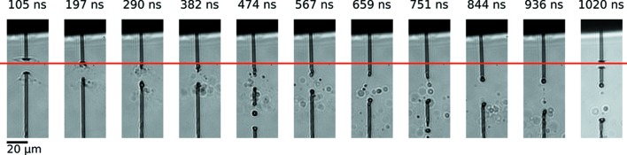
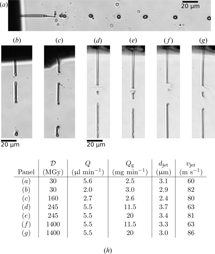
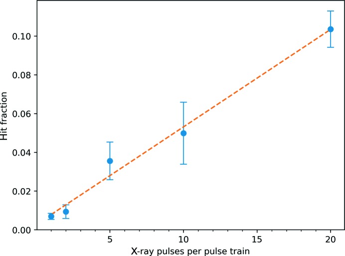
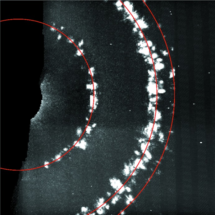
Liquid microjets are a common means of delivering protein crystals to the focus of X-ray free-electron lasers (FELs) for serial femtosecond crystallography measurements. The high X-ray intensity in the focus initiates an explosion of the microjet and sample. With the advent of X-ray FELs with megahertz rates, the typical velocities of these jets must be increased significantly in order to replenish the damaged material in time for the subsequent measurement with the next X-ray pulse. This work reports the results of a megahertz serial diffraction experiment at the FLASH FEL facility using 4.3 nm radiation. The operation of gas-dynamic nozzles that produce liquid microjets with velocities greater than 80 m s-1 was demonstrated. Furthermore, this article provides optical images of X-ray-induced explosions together with Bragg diffraction from protein microcrystals exposed to trains of X-ray pulses repeating at rates of up to 4.5 MHz. The results indicate the feasibility for megahertz serial crystallography measurements with hard X-rays and give guidance for the design of such experiments.

Keywords: FELs; X-ray FEL pulse trains; X-ray free-electron lasers; megahertz repetition rates.


Figures

Figure 1
Figure 1
A slice through an X-ray tomogram of the nozzle used in the FLASH experiments. The nozzle is almost cylindrical/symmetric and consists of a sharpened glass capillary with an inner bore diameter of 30 µm surrounded by an injection-molded ceramic conical piece. The capillary transports the sample liquid to the nozzle exit where a free-standing liquid jet is formed by gas flowing through the interstitial space and out through the orifice. Tomography was performed with 25 keV photon energy at the P05 endstation at the PETRA III synchrotron facility.
Figure 2
Figure 2
Schematic of the experimental setup in the Bauhaus chamber at FLASH. The X-ray-pulse trains pass through the in-line microscope into the interaction region. The nozzle is positioned so that the emerging jet intercepts the X-rays. The diffraction signal generated by the interaction between the jet and the X-ray beam is recorded by the Princeton PI-MTE detector and the effects of this interaction on the jet are monitored with a bright field microscope setup. Here, pulses from a fiber-coupled diode laser (DILAS) illuminate the jet and the image is formed with an in-vacuum microscope objective and a fast camera (Photron SA-4) located outside the vacuum chamber.
Figure 3
Figure 3
Example diffraction patterns recorded with one pulse (a) and (b), and 20 pulses (c) and (d), each at a fluence of 0.5 ± 0.3 µJ µm−2. Panels (a) and (c) display the raw images on a logarithmic gray scale and (b) and (d) show the corrected images on a linear gray scale after background subtraction, masking of jet streak, and identification of the peaks. The peak locations are indicated by red circles. Panels (a) and (b) show a typical pattern with one very weak peak whereas (c) and (d) show a strong pattern with multiple peaks from different crystals recorded in the course of the pulse train. One peak from the 100 class of Bragg reflections and multiple peaks from the 110 class were found. Based on the angle between some of the 110-peaks and the beam center, these cannot originate from the same crystal lattice.
Figure 4
Figure 4
(a) Sideview images of a water jet at various delays after interception by a FLASH FEL pulse. (b) Plot of the evolution of the gap size in the first 50 ns after the FLASH FEL pulse hit the jet (solid circles) and the fit of a logarithmic function to the data (dashed line). The jet was flowing at a rate of 6.7 µl min−1 (helium mass-flow rate Q g = 2.6 mg min−1) with a diameter d jet = 3.1 µm and velocity v jet = 60 m s−1. The dose deposited into the jet was approximately 30 MGy. Note that the position of the gap in the jet varies as a result of nozzle vibrations; the frames shown here are among those with the largest jet gaps recorded. The scale bar in the first panel of (a) is 20 µm and X-rays are incident from the left.
Figure 5
Figure 5
Sideview images showing the arrival of the second X-ray pulse in a train of pulses. The water jet was operated under the same conditions described in Fig. 4 ▸. The horizontal red line indicates that the jet has recovered and is stable in the X-ray interaction region after <290 ns. The last image in the sequence shows the jet after being hit by the second pulse in the pulse train. The scale bar is 20 µm. X-rays are incident from the left.
Figure 6
Figure 6
Comparison of the effects of X-ray-pulse structure and dose on different jets. (a) A water jet similar to the one shown in Figs. 4 ▸ and 5 ▸, exposed to a full train of 250 X-ray pulses at a repetition rate of 1 MHz, measured 90.1 s; after the initial pulse. (b) A thinner and faster water jet exposed to the same pulse train structure, measured 3.14 s after the initial pulse. (c) An ethanol jet also exposed to the same pulse train structure and measured at the same delay. (d) A slow water jet exposed to two FLASH pulses with a spacing of 221.5 ns. (e) A faster water jet exposed to the double-pulse, measured 50 ns after the second pulse. (f) A slow ethanol jet that barely recovers before the second X-ray pulse hits it 221.5 ns later, as measured 50 ns after the second pulse. (g) A fast ethanol jet under the same conditions. (h) A table with the experimental conditions of the jets shown in panels (a)–(g). The jets shown in (a)–(c) were formed with one particular nozzle and (d)–(g) were formed with another. A slice through an X-ray tomogram of the latter nozzle is shown in Fig. 1 ▸. The ethanol jets, especially (f) and (g), exhibit a non-symmetric gap formation where the jet explosion is directed towards the right side. The X-ray beam is incident from the left and is strongly absorbed at the surface of the jet. The energy deposition into the jet is consequently non-uniform.
Figure 7
Figure 7
Hit fraction for different numbers of X-ray pulses in each pulse train. The error bars are derived from the standard deviation of hit fractions from many data collection runs under similar conditions. The dashed line is a linear fit.
Figure 8
Figure 8
Virtual powder pattern from one run during the experiment. Photosystem I crystals have a hexagonal unit cell with a = b = 281 and c = 165.2 Å, space group P63. The innermost ring is comprised of 100-type reflections at a resolution of ∼41 µm−1, the intermediate ring contains nearly overlapping 110- and 101-type reflections at resolutions ≃ 72 µm−1 and the outermost ring consists of 200-type reflections at ∼82 µm−1.

Similar articles

  • Megahertz pulse trains enable multi-hit serial femtosecond crystallography experiments at X-ray free electron lasers.
    Holmes S, Kirkwood HJ, Bean R, Giewekemeyer K, Martin AV, Hadian-Jazi M, Wiedorn MO, Oberthür D, Marman H, Adriano L, Al-Qudami N, Bajt S, Barák I, Bari S, Bielecki J, Brockhauser S, Coleman MA, Cruz-Mazo F, Danilevski C, Dörner K, Gañán-Calvo AM, Graceffa R, Fanghor H, Heymann M, Frank M, Kaukher A, Kim Y, Kobe B, Knoška J, Laurus T, Letrun R, Maia L, Messerschmidt M, Metz M, Michelat T, Mills G, Molodtsov S, Monteiro DCF, Morgan AJ, Münnich A, Peña Murillo GE, Previtali G, Round A, Sato T, Schubert R, Schulz J, Shelby M, Seuring C, Sellberg JA, Sikorski M, Silenzi A, Stern S, Sztuk-Dambietz J, Szuba J, Trebbin M, Vagovic P, Ve T, Weinhausen B, Wrona K, Xavier PL, Xu C, Yefanov O, Nugent KA, Chapman HN, Mancuso AP, Barty A, Abbey B, Darmanin C. Holmes S, et al. Nat Commun. 2022 Aug 11;13(1):4708. doi: 10.1038/s41467-022-32434-6. Nat Commun. 2022. PMID: 35953469 Free PMC article.
  • Liquid sample delivery techniques for serial femtosecond crystallography.
    Weierstall U. Weierstall U. Philos Trans R Soc Lond B Biol Sci. 2014 Jul 17;369(1647):20130337. doi: 10.1098/rstb.2013.0337. Philos Trans R Soc Lond B Biol Sci. 2014. PMID: 24914163 Free PMC article. Review.
  • Megahertz data collection from protein microcrystals at an X-ray free-electron laser.
    Grünbein ML, Bielecki J, Gorel A, Stricker M, Bean R, Cammarata M, Dörner K, Fröhlich L, Hartmann E, Hauf S, Hilpert M, Kim Y, Kloos M, Letrun R, Messerschmidt M, Mills G, Nass Kovacs G, Ramilli M, Roome CM, Sato T, Scholz M, Sliwa M, Sztuk-Dambietz J, Weik M, Weinhausen B, Al-Qudami N, Boukhelef D, Brockhauser S, Ehsan W, Emons M, Esenov S, Fangohr H, Kaukher A, Kluyver T, Lederer M, Maia L, Manetti M, Michelat T, Münnich A, Pallas F, Palmer G, Previtali G, Raab N, Silenzi A, Szuba J, Venkatesan S, Wrona K, Zhu J, Doak RB, Shoeman RL, Foucar L, Colletier JP, Mancuso AP, Barends TRM, Stan CA, Schlichting I. Grünbein ML, et al. Nat Commun. 2018 Aug 28;9(1):3487. doi: 10.1038/s41467-018-05953-4. Nat Commun. 2018. PMID: 30154468 Free PMC article.
  • Serial femtosecond crystallography: the first five years.
    Schlichting I. Schlichting I. IUCrJ. 2015 Feb 3;2(Pt 2):246-55. doi: 10.1107/S205225251402702X. eCollection 2015 Mar 1. IUCrJ. 2015. PMID: 25866661 Free PMC article. Review.
  • Emerging opportunities in structural biology with X-ray free-electron lasers.
    Schlichting I, Miao J. Schlichting I, et al. Curr Opin Struct Biol. 2012 Oct;22(5):613-26. doi: 10.1016/j.sbi.2012.07.015. Epub 2012 Aug 22. Curr Opin Struct Biol. 2012. PMID: 22922042 Free PMC article. Review.

Cited by

  • Recent trends in crystallography - a current IUCr journals perspective.
    Allen AJ. Allen AJ. IUCrJ. 2019 Nov 1;6(Pt 6):984-987. doi: 10.1107/S2052252519014507. eCollection 2019 Nov 1. IUCrJ. 2019. PMID: 31709052 Free PMC article.
  • Membrane protein megahertz crystallography at the European XFEL.
    Gisriel C, Coe J, Letrun R, Yefanov OM, Luna-Chavez C, Stander NE, Lisova S, Mariani V, Kuhn M, Aplin S, Grant TD, Dörner K, Sato T, Echelmeier A, Cruz Villarreal J, Hunter MS, Wiedorn MO, Knoska J, Mazalova V, Roy-Chowdhury S, Yang JH, Jones A, Bean R, Bielecki J, Kim Y, Mills G, Weinhausen B, Meza JD, Al-Qudami N, Bajt S, Brehm G, Botha S, Boukhelef D, Brockhauser S, Bruce BD, Coleman MA, Danilevski C, Discianno E, Dobson Z, Fangohr H, Martin-Garcia JM, Gevorkov Y, Hauf S, Hosseinizadeh A, Januschek F, Ketawala GK, Kupitz C, Maia L, Manetti M, Messerschmidt M, Michelat T, Mondal J, Ourmazd A, Previtali G, Sarrou I, Schön S, Schwander P, Shelby ML, Silenzi A, Sztuk-Dambietz J, Szuba J, Turcato M, White TA, Wrona K, Xu C, Abdellatif MH, Zook JD, Spence JCH, Chapman HN, Barty A, Kirian RA, Frank M, Ros A, Schmidt M, Fromme R, Mancuso AP, Fromme P, Zatsepin NA. Gisriel C, et al. Nat Commun. 2019 Nov 4;10(1):5021. doi: 10.1038/s41467-019-12955-3. Nat Commun. 2019. PMID: 31685819 Free PMC article.
  • Kilohertz droplet-on-demand serial femtosecond crystallography at the European XFEL station FXE.
    Perrett S, Fadini A, Hutchison CDM, Bhattacharya S, Morrison C, Turkot O, Jakobsen MB, Größler M, Licón-Saláiz J, Griese F, Flewett S, Valerio J, Schulz J, Biednov M, Jiang Y, Han H, Yousef H, Khakhulin D, Milne C, Barty A, van Thor JJ. Perrett S, et al. Struct Dyn. 2024 Apr 17;11(2):024310. doi: 10.1063/4.0000248. eCollection 2024 Mar. Struct Dyn. 2024. PMID: 38638699 Free PMC article.
  • Megahertz serial crystallography.
    Wiedorn MO, Oberthür D, Bean R, Schubert R, Werner N, Abbey B, Aepfelbacher M, Adriano L, Allahgholi A, Al-Qudami N, Andreasson J, Aplin S, Awel S, Ayyer K, Bajt S, Barák I, Bari S, Bielecki J, Botha S, Boukhelef D, Brehm W, Brockhauser S, Cheviakov I, Coleman MA, Cruz-Mazo F, Danilevski C, Darmanin C, Doak RB, Domaracky M, Dörner K, Du Y, Fangohr H, Fleckenstein H, Frank M, Fromme P, Gañán-Calvo AM, Gevorkov Y, Giewekemeyer K, Ginn HM, Graafsma H, Graceffa R, Greiffenberg D, Gumprecht L, Göttlicher P, Hajdu J, Hauf S, Heymann M, Holmes S, Horke DA, Hunter MS, Imlau S, Kaukher A, Kim Y, Klyuev A, Knoška J, Kobe B, Kuhn M, Kupitz C, Küpper J, Lahey-Rudolph JM, Laurus T, Le Cong K, Letrun R, Xavier PL, Maia L, Maia FRNC, Mariani V, Messerschmidt M, Metz M, Mezza D, Michelat T, Mills G, Monteiro DCF, Morgan A, Mühlig K, Munke A, Münnich A, Nette J, Nugent KA, Nuguid T, Orville AM, Pandey S, Pena G, Villanueva-Perez P, Poehlsen J, Previtali G, Redecke L, Riekehr WM, Rohde H, Round A, Safenreiter T, Sarrou I, Sato T, Schmidt M, Schmitt B, Schönherr R, Schulz J, Sellberg JA, Seibert MM, Seuring C, Shelby ML, Shoeman RL, Sikorski M, Silenzi A, Stan CA, Shi X, Stern S, Sztuk-Dambietz J, S… See abstract for full author list ➔ Wiedorn MO, et al. Nat Commun. 2018 Oct 2;9(1):4025. doi: 10.1038/s41467-018-06156-7. Nat Commun. 2018. PMID: 30279492 Free PMC article.
  • High-throughput in situ experimental phasing.
    Lawrence JM, Orlans J, Evans G, Orville AM, Foadi J, Aller P. Lawrence JM, et al. Acta Crystallogr D Struct Biol. 2020 Aug 1;76(Pt 8):790-801. doi: 10.1107/S2059798320009109. Epub 2020 Jul 28. Acta Crystallogr D Struct Biol. 2020. PMID: 32744261 Free PMC article.

References

    1. Acero, A. J., Ferrera, C., Montanero, J. M. & Gañán-Calvo, A. M. (2012). J. Micromech. Microeng. 22, 065011.
    1. Barends, T. R. M., Foucar, L., Ardevol, A., Nass, K., Aquila, A., Botha, S., Doak, R. B., Falahati, K., Hartmann, E., Hilpert, M., Heinz, M., Hoffmann, M. C., Köfinger, J., Koglin, J. E., Kovacsova, G., Liang, M., Milathianaki, D., Lemke, H. T., Reinstein, J., Roome, C. M., Shoeman, R. L., Williams, G. J., Burghardt, I., Hummer, G., Boutet, S. & Schlichting, I. (2015). Science, 350, 445–450. - PubMed
    1. Beyerlein, K. R., Adriano, L., Heymann, M., Kirian, R., Knoška, J., Wilde, F., Chapman, H. N. & Bajt, S. (2015). Rev. Sci. Instrum. 86, 125104. - PubMed
    1. Beyerlein, K. R., Dierksmeyer, D., Mariani, V., Kuhn, M., Sarrou, I., Ottaviano, A., Awel, S., Knoska, J., Fuglerud, S., Jönsson, O., Stern, S., Wiedorn, M. O., Yefanov, O., Adriano, L., Bean, R., Burkhardt, A., Fischer, P., Heymann, M., Horke, D. A., Jungnickel, K. E. J., Kovaleva, E., Lorbeer, O., Metz, M., Meyer, J., Morgan, A., Pande, K., Panneerselvam, S., Seuring, C., Tolstikova, A., Lieske, J., Aplin, S., Roessle, M., White, T. A., Chapman, H. N., Meents, A. & Oberthuer, D. (2017). IUCrJ, 4, 769–777. - PMC - PubMed
    1. Beyerlein, K. R., White, T. A., Yefanov, O., Gati, C., Kazantsev, I. G., Nielsen, N. F.-G., Larsen, P. M., Chapman, H. N. & Schmidt, S. (2017). J. Appl. Cryst. 50, 1075–1083. - PMC - PubMed