Regulation of mPGES-1 composition and cell growth via the MAPK signaling pathway in jurkat cells
- PMID: 30214544
- PMCID: PMC6125822
- DOI: 10.3892/etm.2018.6538
Regulation of mPGES-1 composition and cell growth via the MAPK signaling pathway in jurkat cells
Abstract
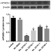
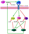
Previous studies have suggested that microsomal prostaglandin E synthase-1 (mPGES-1) is highly expressed and closely associated with mitogen-activated protein kinase (MAPK) signaling pathways in various types of malignant cells. However, their expression patterns and function with respect to T-cell acute lymphoblastic leukemia (T-ALL) remain largely unknown. The present study investigated whether mPGES-1 served a crucial role in T-ALL and aimed to identify interactions between mPGES-1 and the MAPK signaling pathway in T-ALL. The results indicated that mPGES-1 overexpression in T-ALL jurkat cells was significantly decreased by RNA silencing. Decreasing mPGES-1 on a consistent basis may inhibit cell proliferation, induce apoptosis and arrest the cell cycle in T-ALL jurkat cells. Microarray and western blot analyses revealed that c-Jun N-terminal kinase served a role in the mPGES-1/prostaglandin E2/EP4/MAPK positive feedback loops. In addition, P38 and extracellular signal-regulated kinase 1/2 exhibited negative feedback effects on mPGES-1. In conclusion, the results suggested that cross-talk between mPGES-1 and the MAPK signaling pathway was very complex. Therefore, the combined regulation of mPGES-1 and the MAPK signaling pathway may be developed into a new candidate therapy for T-ALL in the future.
Keywords: T-cell acute lymphoblastic leukemia; feedback; jurkat cell; microsomal prostaglandin-E synthase; mitogen activated protein Kinase Signaling System.
Figures


Similar articles
-
Poly(I:C) increases the expression of mPGES-1 and COX-2 in rat primary microglia.J Neuroinflammation. 2016 Jan 18;13:11. doi: 10.1186/s12974-015-0473-7. J Neuroinflammation. 2016. PMID: 26780827 Free PMC article.
-
Up-regulation of microsomal prostaglandin E synthase 1 in osteoarthritic human cartilage: critical roles of the ERK-1/2 and p38 signaling pathways.Arthritis Rheum. 2004 Sep;50(9):2829-38. doi: 10.1002/art.20437. Arthritis Rheum. 2004. PMID: 15457451
-
Signal pathways involved in the regulation of prostaglandin E synthase-1 in human gingival fibroblasts.Cell Signal. 2006 Dec;18(12):2131-42. doi: 10.1016/j.cellsig.2006.04.003. Cell Signal. 2006. PMID: 16766159
-
Andrographolide inhibits growth of human T-cell acute lymphoblastic leukemia Jurkat cells by downregulation of PI3K/AKT and upregulation of p38 MAPK pathways.Drug Des Devel Ther. 2016 Apr 11;10:1389-97. doi: 10.2147/DDDT.S94983. eCollection 2016. Drug Des Devel Ther. 2016. PMID: 27114702 Free PMC article.
-
Inhibition of microsomal prostaglandin E synthase-1 facilitates liver repair after hepatic injury in mice.J Hepatol. 2018 Jul;69(1):110-120. doi: 10.1016/j.jhep.2018.02.009. Epub 2018 Feb 16. J Hepatol. 2018. PMID: 29458169
Cited by
-
Latest progress in the development of cyclooxygenase-2 pathway inhibitors targeting microsomal prostaglandin E2 synthase-1.Future Med Chem. 2022 Mar;14(6):385-388. doi: 10.4155/fmc-2021-0317. Epub 2022 Jan 5. Future Med Chem. 2022. PMID: 34985304 Free PMC article. No abstract available.
-
Licochalcone A Inhibits Prostaglandin E2 by Targeting the MAPK Pathway in LPS Activated Primary Microglia.Molecules. 2023 Feb 17;28(4):1927. doi: 10.3390/molecules28041927. Molecules. 2023. PMID: 36838914 Free PMC article.
-
Growth of T-cell lymphoma cells is inhibited by mPGES-1/PGE2 suppression via JAK/STAT, TGF-β/Smad3 and PI3K/AKT signal pathways.Transl Cancer Res. 2022 Jul;11(7):2175-2184. doi: 10.21037/tcr-21-2834. Transl Cancer Res. 2022. PMID: 35966330 Free PMC article.
-
mPGES-1/PGE2 promotes the growth of T-ALL cells in vitro and in vivo by regulating the expression of MTDH via the EP3/cAMP/PKA/CREB pathway.Cell Death Dis. 2020 Apr 6;11(4):221. doi: 10.1038/s41419-020-2380-9. Cell Death Dis. 2020. PMID: 32251289 Free PMC article.
-
Novel 1,2,4-triazoles derived from Ibuprofen: synthesis and in vitro evaluation of their mPGES-1 inhibitory and antiproliferative activity.Mol Divers. 2023 Oct;27(5):2185-2215. doi: 10.1007/s11030-022-10551-0. Epub 2022 Nov 4. Mol Divers. 2023. PMID: 36331786
References
-
- Larsson K, Kock A, Idborg H, Henriksson Arsenian M, Martinsson T, Johnsen JI, Korotkova M, Kogner P, Jakobsson PJ. COX/mPGES-1/PGE2 pathway depicts an inflammatory-dependent high-risk neuroblastoma subset. Proc Natl Acad Sci U S A. 2015;112:8070–8075. doi: 10.1073/pnas.1424355112. - DOI - PMC - PubMed
LinkOut - more resources
Full Text Sources
Other Literature Sources
Research Materials
Miscellaneous
