. 2019 Jan 1;25(1):369-377.
doi: 10.1158/1078-0432.CCR-18-1776. Epub 2018 Sep 11.

Dual PAK4-NAMPT Inhibition Impacts Growth and Survival, and Increases Sensitivity to DNA-Damaging Agents in Waldenström Macroglobulinemia

Affiliations

Dual PAK4-NAMPT Inhibition Impacts Growth and Survival, and Increases Sensitivity to DNA-Damaging Agents in Waldenström Macroglobulinemia

Na Li et al. Clin Cancer Res. .

Abstract

Purpose: p21-activated kinase 4 (PAK4) plays a significant biological and functional role in a number of malignancies, including multiple myeloma (MM). On the basis of our promising findings in MM, we here characterize PAK4 expression and role in WM cells, as well effect of dual PAK4-NAMPT inhibitor (KPT-9274) against WM cell growth and viability.

Experimental design: We have analyzed mRNA and protein expression levels of PAK4 in WM cells, and used loss-of-function approach to investigate its contribution to WM cell viability. We have further tested the in vitro and in vivo effect of KPT-9274 against WM cell growth and viability.

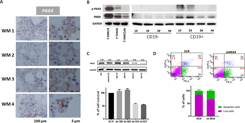
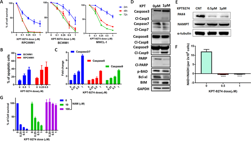
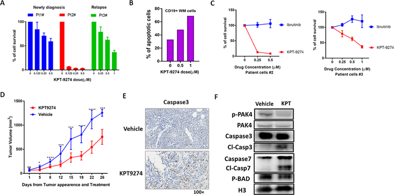
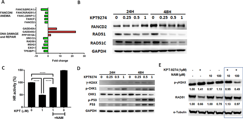
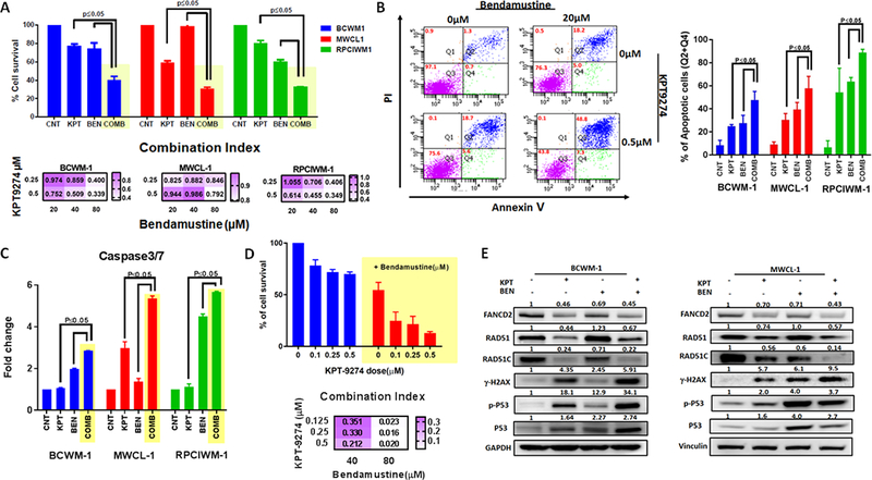
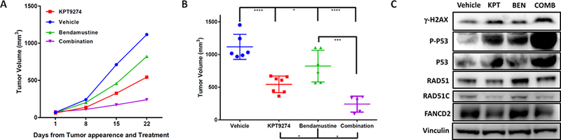
Results: We report here high-level expression and functional role of PAK4 in WM, as demonstrated by shRNA-mediated knockdown; and significant impact of KPT-9274 on WM cell growth and viability. The growth inhibitory effect of KPT-9274 was associated with decreased PAK4 expression and NAMPT activity, as well as induction of apoptosis. Interestingly, in WM cell lines treated with KPT-9274, we detected a significant impact on DNA damage and repair genes. Moreover, we observed that apart from inducing DNA damage, KPT-9274 specifically decreased RAD51 and the double-strand break repair by the homologous recombination pathway. As a result, when combined with a DNA alkylating agents bendamustine and melphalan, KPT-9274 provided a synergistic inhibition of cell viability in WM cell lines and primary patient WM cells in vitro and in vivo.

Conclusions: These results support the clinical investigation of KPT-9274 in combination with DNA-damaging agent for treatment of WM.


Conflict of interest statement

Conflicts of Interest: William Senapedis and Erkan Baloglu are employees of Karyopharm Therapeutics Inc. All other authors declare no potential conflicts of interest.

Figures

Figure 1.
Figure 1.. PAK4 is expressed and functional in WM cells.
A) Representative images of PAK4 immunocytochemistry stain in BM from 4 different WM patients. Positive staining appears in a dark brown color. Scale bars: 100 μm and 5 μm. B) Protein lysates from 3 WM cell lines, CD19+ and CD19- cells isolated from healthy individuals were analyzed for p-PAK4 and PAK4 expression by WB. GAPDH was used as loading control. C) Genetic depletion of PAK4 was achieved using 4 different pGIPZ lentiviral vectors (Dharmacon) containing the target sequence or scrambled control. Cell were collected 3 days after puromycin selection. Protein lysates were analyzed by WB, confirming decreased PAK4 protein levels in cells expressing PAK4 shRNAs compared with scrambled cells. Cell viability was evaluated by CellTiter-Glo Luminescent Cell Assay on puromycin-selected shRNA-expressing cells in culture for 48 hours. D) Apoptotic cell death was assessed by flow cytometric analysis following DAPI and PE-AnnexinV staining in cells expressing scramble or PAK4-targeting shRNA (construct #934).
Figure 2.
Figure 2.. KPT-9274 inhibits WM cell viability and triggers apoptotic cell death.
A) WM cell lines were treated with different concentrations of KPT-9274. Cells viability was evaluated with CellTiter-Glo at indicated time (24h-48–72 hours) and expressed as % change from untreated cells. Data represents mean ±SD of 5 experiments performed in triplicates. B) BCWM-1 and RPCIWM-1 were cultured in the absence or presence of several concentrations of KPT-9274 for 48 hours. Apoptotic cell death was assessed by flow cytometry analysis following Annexin-V and propidium iodide (PI) staining. The % of AnnexinV+ cells is shown in the graphs. C) Indicated caspase activities were evaluated in BCWM1 cells after KPT-9274 treatment for 48 hours using luminescence assay. D) Whole cell lysates from BCWM1 cells treated with control vehicle or KPT-9274 for 48 hours were subjected to Western blot analysis and probed with antibodies against indicated proteins, with GAPDH as loading control. E) BCWM1 cell were treated with different doses of KPT-9274 for 48 hours. Protein lysates were assessed by WB analysis for expression of PAK4 and NAMPT using specific Abs. α-Tubulin was used as loading control. F) Cellular NAD levels in BCWM1 treated with KPT-9274 were measured using an enzyme cyclic assay and normalized to total cell number. G) BCWM-1 cells were treated with KPT9274 (1 μM) in absence or presence of NAM (10 and 100 μM) for 48 hours. Cell viability was tested with CellTiter-Glo Luminescent Cell Assay and expressed as % change from untreated cells.
Figure 3.
Figure 3.. KPT-9274 impairs cell viability and promotes apoptosis in primary WM cells and displays significant activity in vivo in a xenograft murine model of WM.
A) CD19+ WM cells purified from three WM patients, were cultured in the presence of different concentrations of KPT-9274 for 48 hours. Cell viability was assessed by CTG and expressed as percent change from untreated cells. B) Apoptosis was evaluated in primary WM cells after 48h treatment with KPT-9274 by flow cytometry analysis following Annexin-V and propidium iodide (PI) staining. The % of AnnexinV+ cells is shown in the graphs. C) CD19+ primary WM cells were treated with different concentration of either KPT-9274 or Ibrutinib. Cell viability was assessed by CTG after 48 hours of treatment. D) Sublethally irradiated SCID mice were injected subcutaneously with BCWM-1 cells. Following detection of tumor, mice were randomized and treated with either KPT-9274 or vehicle orally, for 5 consecutive days/week for 3 weeks. Tumor volume was evaluated by caliper measurement. Differences between the 2 groups were evaluated using standard T-test. (* p ≤ 0.05, ** p ≤ 0.01, *** p ≤ 0.001, **** P ≤ 0.0001). E) Tumors were isolated from KPT-9274 treated and control mice and sections were evaluated by histological examinations following caspase3 staining. One representative experiment is shown. F) Tumor cells retrieved from mice were lysed in RIPA buffer and whole cell lysate subjected to WB analysis and probed with antibodies against p-PAK4, PAK4, caspase-7 and p-BAD. Histone 3 was used as loading control.
Figure 4.
Figure 4.. KPT-9274 impacts DDR pathways in WM cells.
A) Total RNA from control or KPT-9274-treated BCWM1 cells was evaluated by Human DNA Damage Signaling Pathway RT2 Profiler™ PCR Array to profile the expression of 84 genes involved in DNA damage signaling pathways. The relative expression level compared to control cells for each gene is plotted relative in the graph. B-D) Whole cell lysates from BCWM-1 cells treated with several concentrations of KPT-9274 for 24 and 48 hours were subjected to WB analysis and probed with indicated antibodies. GAPDH was used as loading control. C) Relative HR activity of BCWM-1 cells treated with KPT-9274 in absence or presence of nicotinamide (NAM, 100 μM). E) Whole cell lysates from BCWM-1 treated with KPT9274 (1 μM) in absence and presence nicotinamide (10 and 100 μM) for 48 hours, were subjected to WB analysis and probed with indicated antibodies.
Figure 5.
Figure 5.. KPT-9274 induces synergistic growth inhibitory effects in combination with bendamustine in WM cells in vitro.
A) WM cell lines were simultaneously treated with KPT-9274 and/or bendamustine for 48 hours. Cells viability was tested with CellTiter-Glo and expressed as % of cell viability from untreated cells. Data represents mean ±SD of 3 experiments performed in triplicates (upper panel). The interaction between KPT-9274 and bendamustine was analyzed using the CalcuSyn software program (Biosoft, Ferguson, MO) based on the Chou-Talalay method. The Combination index (CI) is represented as heatmaps (lower panel). CI = 1 indicates additive effects; CI < 1 indicates synergism and CI > 1 indicates antagonism. B) Apoptotic cell death was assessed by flow cytometric analysis following AnnexinV and PI staining in single treated with single agents or combination. One representative experiment is shown on the left panel, while the % of AnnexinV+ in different WM cell lines following treatment is shown on the right panel. Data represent mean ±SD of two experiments. C) Caspase 3/7 activation was evaluated after combination treatment using luminescence assay. D) Primary CD19+ cells were cultured in the presence of several concentrations of KPT-9274 in combination with bendamustine for 48 hours. Cell viability was assessed by CTG uptake and presented as % compared to control cells (upper panel). Drug interactions were assessed by CalcuSyn 2.0 software (Biosoft) and CIs are represented in the heatmap (lower panel). E) Whole cell lysates from BCWM-1 (left panel) and MWCL1 (right panel) cell lines treated with control, or single agents or combination were subjected to WB analysis and probed with indicated antibodies.
Figure 6.
Figure 6.. Combination therapy results in tumor shrinkage in vivo compared to single agent therapy.
A) Sub-lethally irradiated SCID mice were subcutaneously inoculated with BCWM-1 WM cell line. Treatment started following detection of tumor (approximately 2 weeks from cell injection). Mice were treated either with vehicle (100mg/kg once/daily, 5days/week), KPT-9274 (100mg/kg once/daily, 5days/week), bendamustine (25mg/kg, once/weekly, for 2 weeks) and a combination of KPT-9274 and bendamustine. Tumors were measured in two perpendicular dimensions weekly. B) Comparison of tumor volume in control and treated mice 3 weeks after initial assessment of tumor appearance and start of treatment. C) Whole cell lysate from tumor cells retrieved from mice was subjected to WB analysis and probed with antibodies against with indicated antibodies. Vinculin was used as loading control.

Similar articles

Cited by

References

    1. Hunter ZR, Xu L, Yang G, Zhou Y, Liu X, Cao Y, et al. The genomic landscape of Waldenstrom macroglobulinemia is characterized by highly recurring MYD88 and WHIM-like CXCR4 mutations, and small somatic deletions associated with B-cell lymphomagenesis. Blood 2014;123(11):1637–46 doi 10.1182/blood-2013-09-525808. - DOI - PubMed
    1. Hunter ZR, Xu L, Yang G, Tsakmaklis N, Vos JM, Liu X, et al. Transcriptome sequencing reveals a profile that corresponds to genomic variants in Waldenstrom macroglobulinemia. Blood 2016;128(6):827–38 doi 10.1182/blood-2016-03-708263. - DOI - PMC - PubMed
    1. Yang G, Zhou Y, Liu X, Xu L, Cao Y, Manning RJ, et al. A mutation in MYD88 (L265P) supports the survival of lymphoplasmacytic cells by activation of Bruton tyrosine kinase in Waldenstrom macroglobulinemia. Blood 2013;122(7):1222–32 doi 10.1182/blood-2012-12-475111. - DOI - PubMed
    1. Dimopoulos MA, Kastritis E, Owen RG, Kyle RA, Landgren O, Morra E, et al. Treatment recommendations for patients with Waldenstrom macroglobulinemia (WM) and related disorders: IWWM-7 consensus. Blood 2014;124(9):1404–11 doi 10.1182/blood-2014-03-565135. - DOI - PMC - PubMed
    1. Hunter ZR, Yang G, Xu L, Liu X, Castillo JJ, Treon SP. Genomics, Signaling, and Treatment of Waldenstrom Macroglobulinemia. Journal of clinical oncology : official journal of the American Society of Clinical Oncology 2017;35(9):994–1001 doi 10.1200/jco.2016.71.0814. - DOI - PubMed

Publication types

MeSH terms