TRPM2 Exacerbates Central Nervous System Inflammation in Experimental Autoimmune Encephalomyelitis by Increasing Production of CXCL2 Chemokines
- PMID: 30201769
- PMCID: PMC6596171
- DOI: 10.1523/JNEUROSCI.2203-17.2018
TRPM2 Exacerbates Central Nervous System Inflammation in Experimental Autoimmune Encephalomyelitis by Increasing Production of CXCL2 Chemokines
Abstract
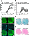
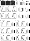
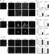
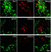
Multiple sclerosis (MS) is a chronic inflammatory disorder of the CNS characterized by demyelination and axonal injury. Current therapies that mainly target lymphocytes do not fully meet clinical need due to the risk of severe side effects and lack of efficacy against progressive MS. Evidence suggests that MS is associated with CNS inflammation, although the underlying molecular mechanism is poorly understood. Transient receptor potential melastatin 2 (TRPM2), a Ca2+-permeable nonselective cation channel, is expressed at high levels in the brain and by immune cells, including monocyte lineage cells. Here, we show that TRPM2 plays a pathological role in experimental autoimmune encephalomyelitis (EAE), an animal model of MS. Knockout (KO) or pharmacological inhibition of TRPM2 inhibited progression of EAE and TRPM2-KO mice showed lower activation of Iba1-immunopositive monocyte lineage cells and neutrophil infiltration of the CNS than WT mice. Moreover, CXCL2 production in TRPM2-KO mice was significantly reduced at day 14, although the severity of EAE was the same as that in WT mice at that time point. In addition, we used BM chimeric mice to show that TRPM2 expressed by CNS-infiltrating macrophages contributes to progression of EAE. Because CXCL2 induces migration of neutrophils, these results indicate that reduced expression of CXCL2 in the CNS suppresses neutrophil infiltration and slows progression of EAE in TRPM2-KO mice. Together, the results suggest that TRPM2 plays an important role in progression of EAE pathology and shed light on its putative role as a therapeutic target for MS.SIGNIFICANCE STATEMENT Current therapies for multiple sclerosis (MS), which mainly target lymphocytes, carry the risk of severe side effects and lack efficacy against the progressive form of the disease. Here, we found that the transient receptor potential melastatin 2 (TRPM2) channel, which is abundantly expressed in CNS-infiltrating macrophages, plays a crucial role in development of experimental autoimmune encephalomyelitis (EAE), an animal model of MS. EAE progression was suppressed by Knockout (KO) or pharmacological inhibition of TRPM2; this was attributed to a reduction in CXCL2 chemokine production by CNS-infiltrating macrophages in TRPM2-KO mice, resulting in suppression of neutrophil infiltration into the CNS. These results reveal an important role of TRPM2 in the pathogenesis of EAE and shed light on its potential as a therapeutic target.
Keywords: TRP channel; TRPM2; cxcl2; macrophage; multiple sclerosis; neutrophil.
Copyright © 2018 the authors 0270-6474/18/388484-12$15.00/0.
Figures


Comment in
-
Intricate Interplay between Innate Immune Cells and TRMP2 in a Mouse Model of Multiple Sclerosis.J Neurosci. 2019 Mar 27;39(13):2366-2368. doi: 10.1523/JNEUROSCI.2982-18.2019. J Neurosci. 2019. PMID: 30918048 Free PMC article. No abstract available.
Similar articles
-
An IFNγ/CXCL2 regulatory pathway determines lesion localization during EAE.J Neuroinflammation. 2018 Jul 16;15(1):208. doi: 10.1186/s12974-018-1237-y. J Neuroinflammation. 2018. PMID: 30012158 Free PMC article.
-
Involvement of TRPM2 in peripheral nerve injury-induced infiltration of peripheral immune cells into the spinal cord in mouse neuropathic pain model.PLoS One. 2013 Jul 30;8(7):e66410. doi: 10.1371/journal.pone.0066410. Print 2013. PLoS One. 2013. PMID: 23935822 Free PMC article.
-
TRPM2 contributes to neuroinflammation and cognitive deficits in a cuprizone-induced multiple sclerosis model via NLRP3 inflammasome.Neurobiol Dis. 2021 Dec;160:105534. doi: 10.1016/j.nbd.2021.105534. Epub 2021 Oct 19. Neurobiol Dis. 2021. PMID: 34673151
-
Role of Th17 cells in the pathogenesis of CNS inflammatory demyelination.J Neurol Sci. 2013 Oct 15;333(1-2):76-87. doi: 10.1016/j.jns.2013.03.002. Epub 2013 Apr 8. J Neurol Sci. 2013. PMID: 23578791 Free PMC article. Review.
-
The contribution of neutrophils to CNS autoimmunity.Clin Immunol. 2018 Apr;189:23-28. doi: 10.1016/j.clim.2016.06.017. Epub 2016 Jul 1. Clin Immunol. 2018. PMID: 27377536 Free PMC article. Review.
Cited by
-
Myelin Oligodendrocyte Glycoprotein 35-55 (MOG 35-55)-induced Experimental Autoimmune Encephalomyelitis: A Model of Chronic Multiple Sclerosis.Bio Protoc. 2019 Dec 20;9(24):e3453. doi: 10.21769/BioProtoc.3453. eCollection 2019 Dec 20. Bio Protoc. 2019. PMID: 33654948 Free PMC article.
-
Intricate Interplay between Innate Immune Cells and TRMP2 in a Mouse Model of Multiple Sclerosis.J Neurosci. 2019 Mar 27;39(13):2366-2368. doi: 10.1523/JNEUROSCI.2982-18.2019. J Neurosci. 2019. PMID: 30918048 Free PMC article. No abstract available.
-
A comprehensive review on the role of chemokines in the pathogenesis of multiple sclerosis.Metab Brain Dis. 2021 Mar;36(3):375-406. doi: 10.1007/s11011-020-00648-6. Epub 2021 Jan 6. Metab Brain Dis. 2021. PMID: 33404937 Review.
-
Altered Expression of Ion Channels in White Matter Lesions of Progressive Multiple Sclerosis: What Do We Know About Their Function?Front Cell Neurosci. 2021 Jun 25;15:685703. doi: 10.3389/fncel.2021.685703. eCollection 2021. Front Cell Neurosci. 2021. PMID: 34276310 Free PMC article. Review.
-
The astrocytic TRPA1 channel mediates an intrinsic protective response to vascular cognitive impairment via LIF production.Sci Adv. 2023 Jul 21;9(29):eadh0102. doi: 10.1126/sciadv.adh0102. Epub 2023 Jul 21. Sci Adv. 2023. PMID: 37478173 Free PMC article.
References
-
- Berghoff SA, Gerndt N, Winchenbach J, Stumpf SK, Hosang L, Odoardi F, Ruhwedel T, Böhler C, Barrette B, Stassart R, Liebetanz D, Dibaj P, Möbius W, Edgar JM, Saher G (2017) Dietary cholesterol promotes repair of demyelinated lesions in the adult brain. Nat Commun 8:14241. 10.1038/ncomms14241 - DOI - PMC - PubMed
Publication types
MeSH terms
Substances
LinkOut - more resources
Full Text Sources
Other Literature Sources
Medical
Molecular Biology Databases
Research Materials
Miscellaneous
