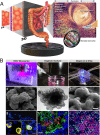
Modeling Host-Pathogen Interactions in the Context of the Microenvironment: Three-Dimensional Cell Culture Comes of Age
- PMID: 30181350
- PMCID: PMC6204695
- DOI: 10.1128/IAI.00282-18
Modeling Host-Pathogen Interactions in the Context of the Microenvironment: Three-Dimensional Cell Culture Comes of Age
Abstract
Tissues and organs provide the structural and biochemical landscapes upon which microbial pathogens and commensals function to regulate health and disease. While flat two-dimensional (2-D) monolayers composed of a single cell type have provided important insight into understanding host-pathogen interactions and infectious disease mechanisms, these reductionist models lack many essential features present in the native host microenvironment that are known to regulate infection, including three-dimensional (3-D) architecture, multicellular complexity, commensal microbiota, gas exchange and nutrient gradients, and physiologically relevant biomechanical forces (e.g., fluid shear, stretch, compression). A major challenge in tissue engineering for infectious disease research is recreating this dynamic 3-D microenvironment (biological, chemical, and physical/mechanical) to more accurately model the initiation and progression of host-pathogen interactions in the laboratory. Here we review selected 3-D models of human intestinal mucosa, which represent a major portal of entry for infectious pathogens and an important niche for commensal microbiota. We highlight seminal studies that have used these models to interrogate host-pathogen interactions and infectious disease mechanisms, and we present this literature in the appropriate historical context. Models discussed include 3-D organotypic cultures engineered in the rotating wall vessel (RWV) bioreactor, extracellular matrix (ECM)-embedded/organoid models, and organ-on-a-chip (OAC) models. Collectively, these technologies provide a more physiologically relevant and predictive framework for investigating infectious disease mechanisms and antimicrobial therapies at the intersection of the host, microbe, and their local microenvironments.
Keywords: 3-D; 3D; RWV; gut-on-a-chip; host-microbe interaction; host-pathogen interactions; mechanotransduction; organ-on-a-chip; organoid; rotating wall vessel.
Figures

Similar articles
-
Three-Dimensional Rotating Wall Vessel-Derived Cell Culture Models for Studying Virus-Host Interactions.Viruses. 2016 Nov 9;8(11):304. doi: 10.3390/v8110304. Viruses. 2016. PMID: 27834891 Free PMC article. Review.
-
Modeling infectious diseases and host-microbe interactions in gastrointestinal organoids.Dev Biol. 2016 Dec 15;420(2):262-270. doi: 10.1016/j.ydbio.2016.09.014. Epub 2016 Sep 14. Dev Biol. 2016. PMID: 27640087 Review.
-
Culturing and applications of rotating wall vessel bioreactor derived 3D epithelial cell models.J Vis Exp. 2012 Apr 3;(62):3868. doi: 10.3791/3868. J Vis Exp. 2012. PMID: 22491366 Free PMC article.
-
Development of a Multicellular Three-dimensional Organotypic Model of the Human Intestinal Mucosa Grown Under Microgravity.J Vis Exp. 2016 Jul 25;(113):54148. doi: 10.3791/54148. J Vis Exp. 2016. PMID: 27500889 Free PMC article.
-
From Single Cells to Engineered and Explanted Tissues: New Perspectives in Bacterial Infection Biology.Int Rev Cell Mol Biol. 2015;319:1-44. doi: 10.1016/bs.ircmb.2015.06.003. Epub 2015 Jul 21. Int Rev Cell Mol Biol. 2015. PMID: 26404465 Review.
Cited by
-
Recent Updates on Research Models and Tools to Study Virus-Host Interactions at the Placenta.Viruses. 2019 Dec 18;12(1):5. doi: 10.3390/v12010005. Viruses. 2019. PMID: 31861492 Free PMC article. Review.
-
Systems vaccinology and big data in the vaccine development chain.Immunology. 2019 Jan;156(1):33-46. doi: 10.1111/imm.13012. Epub 2018 Nov 13. Immunology. 2019. PMID: 30317555 Free PMC article. Review.
-
Role of RpoS in Regulating Stationary Phase Salmonella Typhimurium Pathogenesis-Related Stress Responses under Physiological Low Fluid Shear Force Conditions.mSphere. 2022 Aug 31;7(4):e0021022. doi: 10.1128/msphere.00210-22. Epub 2022 Aug 1. mSphere. 2022. PMID: 35913142 Free PMC article.
-
Antibiofilm activity of marine microbial natural products: potential peptide- and polyketide-derived molecules from marine microbes toward targeting biofilm-forming pathogens.J Nat Med. 2024 Jan;78(1):1-20. doi: 10.1007/s11418-023-01754-2. Epub 2023 Nov 6. J Nat Med. 2024. PMID: 37930514 Review.
-
An Advanced Human Intestinal Coculture Model Reveals Compartmentalized Host and Pathogen Strategies during Salmonella Infection.mBio. 2020 Feb 18;11(1):e03348-19. doi: 10.1128/mBio.03348-19. mBio. 2020. PMID: 32071273 Free PMC article.
References
Publication types
MeSH terms
Grants and funding
LinkOut - more resources
Full Text Sources
Other Literature Sources

