. 2018 Sep 3;10(9):472.
doi: 10.3390/v10090472.

Identification of the Potential Virulence Factors and RNA Silencing Suppressors of Mulberry Mosaic Dwarf-Associated Geminivirus

Affiliations

Identification of the Potential Virulence Factors and RNA Silencing Suppressors of Mulberry Mosaic Dwarf-Associated Geminivirus

Xiuling Yang et al. Viruses. .

Abstract

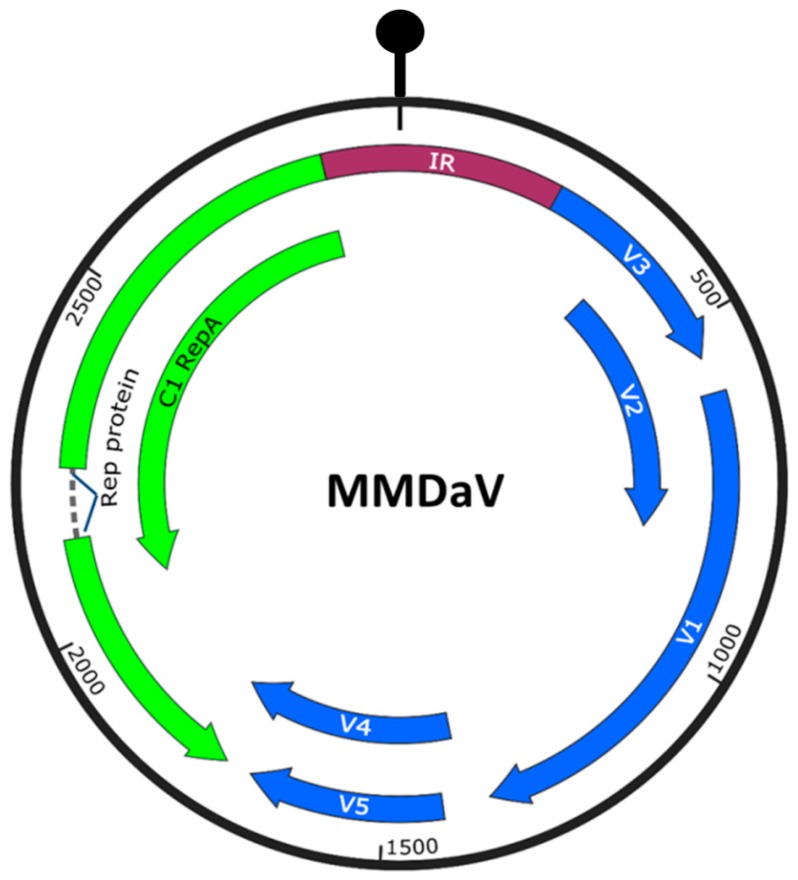
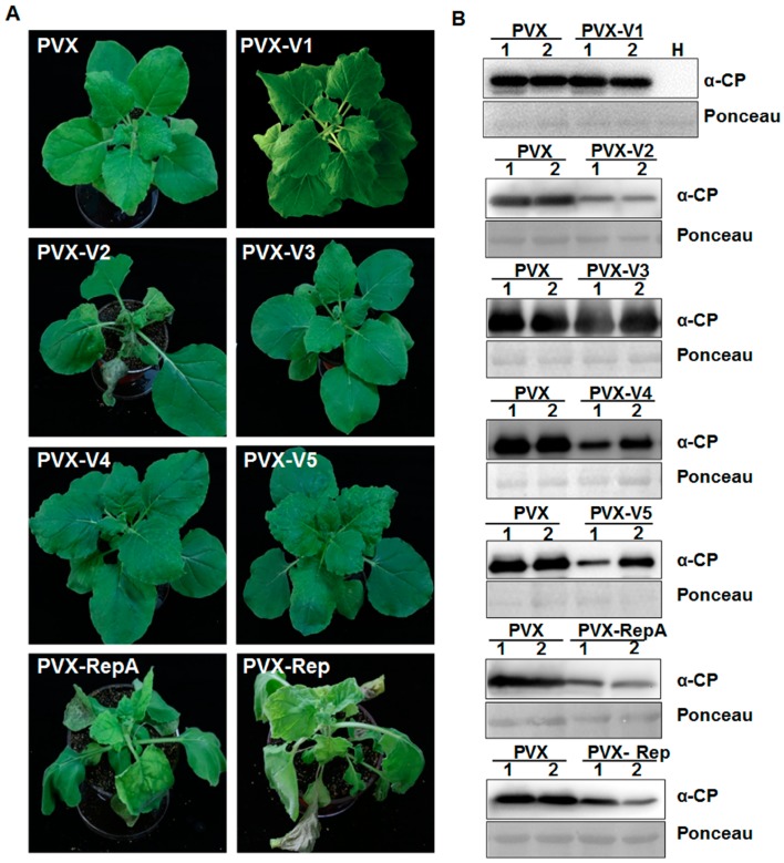
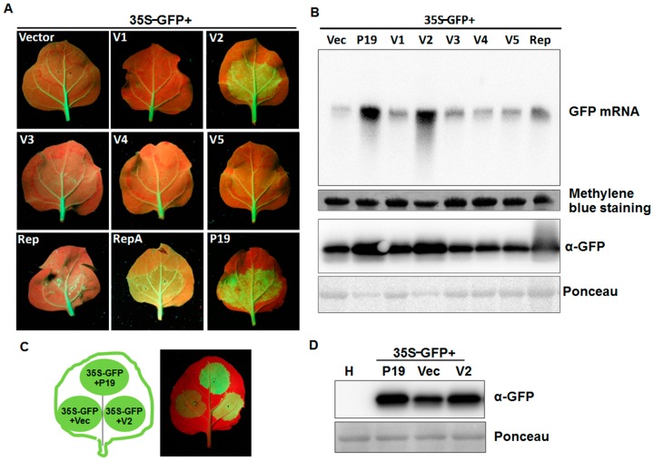
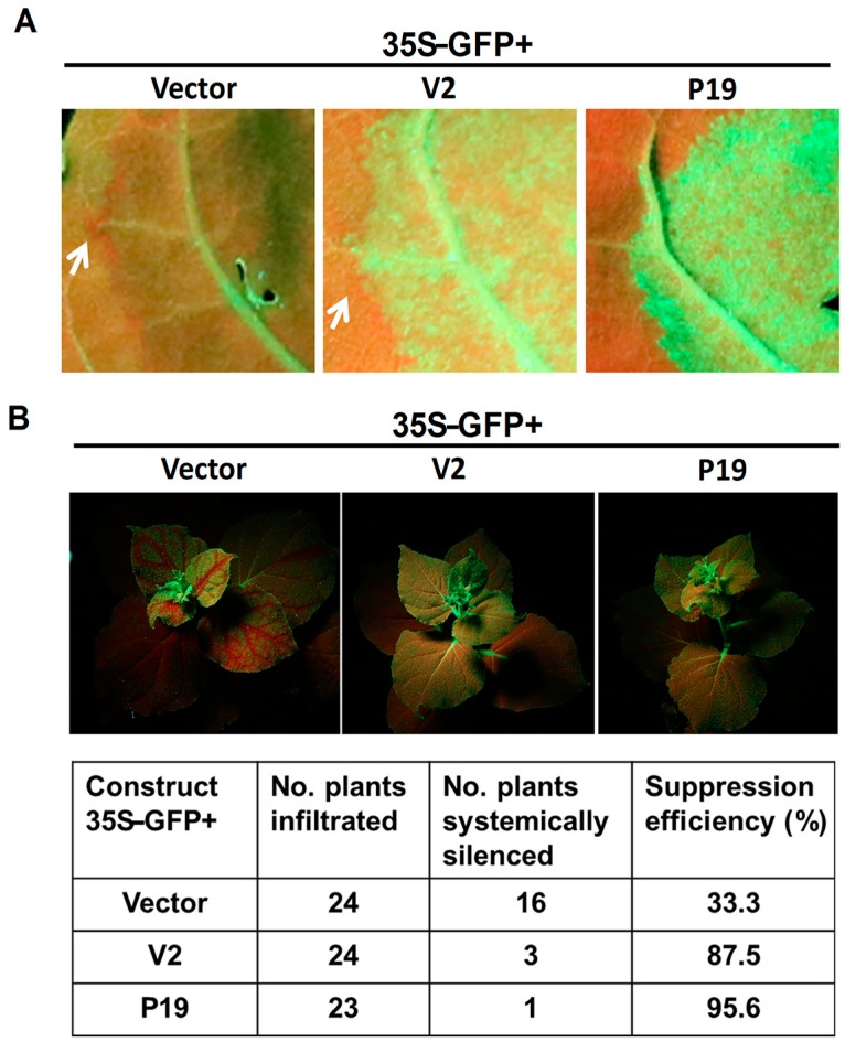
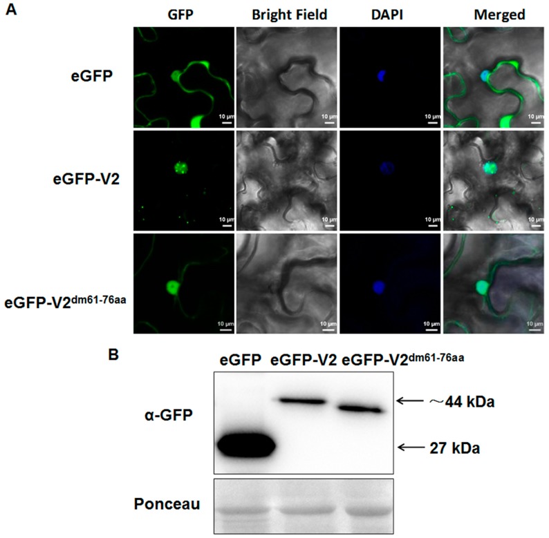
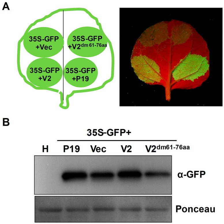
Plant viruses encode virulence factors or RNA silencing suppressors to reprogram plant cellular processes or to fine-tune host RNA silencing-mediated defense responses. In a previous study, Mulberry mosaic dwarf-associated virus (MMDaV), a novel, highly divergent geminivirus, has been identified from a Chinese mulberry tree showing mosaic and dwarfing symptoms, but the functions of its encoded proteins are unknown. In this study, all seven proteins encoded by MMDaV were screened for potential virulence and RNA silencing suppressor activities. We found that V2, RepA, and Rep affect the pathogenicity of a heterologous potato virus X. We showed that V2 could inhibit local RNA silencing and long-distance movement of the RNA silencing signal, but not short-range spread of the green fluorescent protein (GFP) silencing signal in Nicotiana benthamiana 16c plants. In addition, V2 localized to both subnuclear foci and the cytoplasm. Deletion mutagenesis of V2 showed that the basic motif from amino acids 61 to 76 was crucial for V2 to form subnuclear foci and for suppression of RNA silencing. Although the V2 protein encoded by begomoviruses or a curtovirus has been shown to have silencing suppressor activity, this is the first identification of an RNA silencing suppressor from a woody plant-infecting geminivirus.

Keywords: MMDaV; RNA silencing; suppressor; virulence factor.


Conflict of interest statement

The authors declare no conflict of interest. The funding sponsors had no role in the design of the study; in the collection, analyses, or interpretation of data; in the writing of the manuscript, and in the decision to publish the results.

Figures

Figure 1
Figure 1
Genome organization of Mulberry mosaic dwarf-associated geminivirus (MMDaV), showing the open reading frames (ORFs) coded. ORFs encoded on the virion-sense (V) strand and complementary-sense (C) strand are denoted as blue and green colors, respectively. The circle represents the circular, single-stranded DNA of MMDaV. The stem-loop structure that contained the conserved nonanucleotide sequence, TAATATTAC, within the intergenic region (IR) is shown on top of the diagram.
Figure 2
Figure 2
Effects of MMDaV-encoded proteins on potato virus X (PVX) pathogenicity. (A) Symptoms of Nicotiana benthamiana plants inoculated with Agrobacterium cells harboring PVX alone, or recombinant PVX vector expressing individual ORFs of MMDaV. Photographs were taken at 10 days post infiltration (dpi). Experiments were repeated three times, and at least four to six plants were used for each inoculation. (B) Western blot analysis of PVX accumulation in inoculated N. benthamiana plants at 7 to 10 dpi using a monoclonal antibody against PVX CP. Total proteins were extracted from upper non-inoculated leaves as indicated in (A). Two independent plants were used to extract total proteins. H represents total soluble proteins extracted from the healthy N. benthamiana plant, which was used to detect the specificity of the PVX antibody. Ponceau staining of the large subunit of Rubisco served as loading controls.
Figure 3
Figure 3
Effects of MMDaV-encoded proteins on GFP local silencing. (A) N. benthamiana 16c plants were infiltrated with a mixture of Agrobacterium cultures containing 35S-GFP and pCHF3 vectors expressing individual ORFs of MMDaV, respectively. The constructs used for infiltration are indicated. N. benthamiana 16c plants infiltrated with 35S-GFP, plus the empty vector or 35S-GFP plus tomato bushy stunt virus P19 were used as negative or positive controls, respectively. Photographs were taken under UV light with a yellow filter-mounted Canon camera at 5 dpi. Similar results were obtained in three dependent experiments. At least five plants were agroinfiltrated per experiment. (B) Analysis of the GFP mRNA and protein levels in infiltrated leaf patches. 10 μg of total RNA extracted from infiltrated patches at 5 dpi were used in Northern blot analysis. The GFP mRNA was detected using a DIG-labeled GFP-specific probe. Methylene blue staining was used to visualize the loading controls for the mRNA. The expression of the GFP protein was analyzed by Western blot using a monoclonal antibody against GFP. Ponceau staining of the large subunit of Rubisco served as loading controls for the Western blot assay. (C) Suppression of local post-translational gene silencing (PTGS) in wild-type N. benthamiana plants. N. benthamiana plants were infiltrated with a mixture of Agrobacterium cultures containing constructs as indicated in the left panel. Expression of 35S-GFP with the empty vector or 35S-GFP with P19 served as negative or positive controls, respectively. Photographs were taken under UV light at 3 dpi. (D) Western blot analysis of the GFP protein levels in infiltrated N. benthamiana leaf patches using a GFP monoclonal antibody. Total soluble proteins extracted from the healthy N. benthamiana plants were used to detect the specificity of the GFP antibody. Ponceau staining of the large subunit of Rubisco protein are shown as loading controls.
Figure 4
Figure 4
Effect of MMDaV V2 on short- and long-range spread of the silencing signals. (A) MMDaV V2 could not inhibit short-distance spread (10–15 cells) of the GFP silencing signal in N. benthamiana 16c plants. Leaves were infiltrated with a mixture of Agrobacterium cultures containing 35S-GFP and MMDaV V2. Leaves infiltrated with a mixtures of Agrobacterium cultures containing 35S-GFP plus the empty vector or 35S-GFP plus P19, served as negative or positive controls, respectively. Photographs were taken under UV light at 6 dpi. White arrows indicate the red ring, a hallmark of short-distance spread of the mobile RNA silencing signal at the edge of the infiltrated patches. (B) MMDaV V2 could interfere with systemic spread of RNA silencing signal in N. benthamiana 16c plants. The upper panels represent N. benthamiana 16c plants infiltrated with Agrobacterium cells carrying 35S-GFP plus empty vector (with systemic silencing), or 35S-GFP plus MMDaV V2 (with no systemic silencing), or 35S-GFP plus P19 (with no systemic silencing). Photographs were taken under UV light at 20 dpi. The number of N. benthamiana 16c plants systemically silenced at 20 dpi was indicated. N. benthamiana 16c plants that turned red in the major and minor veins of upper young leaves were considered to be systemically silenced.
Figure 5
Figure 5
Deletion of the basic motif of MMDaV V2 fails to localize to subnuclear foci. (A) Subcellular localization of MMDaV V2 and V2 mutant variant in epidermal cells of N. benthamiana. Agrobacterium cells containing 35S-eGFP, 35S-eGFP-V2, 35S-eGFP-V2dm61-76aa was infiltrated into leaves of N. benthamiana, respectively. DAPI staining was used to visualize the nuclei. White bars represent 10 µm. All images were visualized by confocal microscopy (Zeiss LSM880) at 36 to 48 h post-infiltration. Independent infiltration experiments were performed three times, and 5–6 cells were examined in each experiment. (B) Gel blot of total proteins from N. benthamiana leaves infiltrated with constructs represented in (A) using anti-GFP antibody. Ponceau staining serves as a loading control.
Figure 6
Figure 6
Deletion of the basic motif of MMDaV V2 influences its RNA silencing activity. (A) Suppression of RNA silencing in N. benthamiana plants. Leaf patches were infiltrated with a mixture of Agrobacterium cultures containing constructs represented in the left panel. Photographs were taken under UV light at 3 dpi. Three independent infiltration experiments were carried out, and four plants were used per experiment. (B) Western blot analysis of the GFP protein levels in infiltrated leaf patches using a monoclonal antibody against GFP. Total proteins extracted from the healthy N. benthamiana plant (H) were used to detect the specificity of the GFP antibody. Ponceau staining of the large subunit of Rubisco was used as loading controls.

Similar articles

Cited by

References

    1. Calil I.P., Fontes E.P.B. Plant immunity against viruses: Antiviral immune receptors in focus. Ann. Bot. 2017;119:711–723. doi: 10.1093/aob/mcw200. - DOI - PMC - PubMed
    1. Zhao J.H., Hua C.L., Fang Y.Y., Guo H.S. The dual edge of RNA silencing suppressors in the virus-host interactions. Curr. Opin. Virol. 2016;17:39–44. doi: 10.1016/j.coviro.2015.12.002. - DOI - PubMed
    1. Pumplin N., Voinnet O. RNA silencing suppression by plant pathogens: Defence, counter-defence and counter-counter-defence. Nat. Rev. Microbiol. 2013;11:745–760. doi: 10.1038/nrmicro3120. - DOI - PubMed
    1. Alcaide-Loridan C., Jupin I. Ubiquitin and plant viruses, let’s play together! Plant Physiol. 2012;160:72–82. doi: 10.1104/pp.112.201905. - DOI - PMC - PubMed
    1. Leary A.Y., Sanguankiattichai N., Duggan C., Tumtas Y., Pandey P., Segretin M.E., Linares J.S., Savage Z.D., Yow R.J., Bozkurt T.O. Modulation of plant autophagy during pathogen attack. J. Exp. Bot. 2018;69:1325–1333. doi: 10.1093/jxb/erx425. - DOI - PubMed

Publication types

LinkOut - more resources