. 2018 Oct:19:166-178.
doi: 10.1016/j.redox.2018.08.013. Epub 2018 Aug 24.

Cinnamic aldehyde inhibits vascular smooth muscle cell proliferation and neointimal hyperplasia in Zucker Diabetic Fatty rats

Affiliations

Cinnamic aldehyde inhibits vascular smooth muscle cell proliferation and neointimal hyperplasia in Zucker Diabetic Fatty rats

Nicholas E Buglak et al. Redox Biol. 2018 Oct.

Abstract

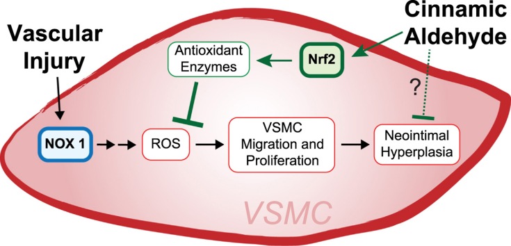
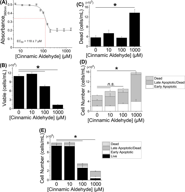
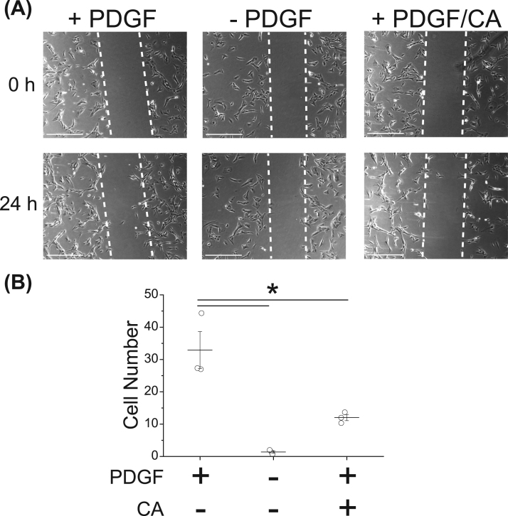
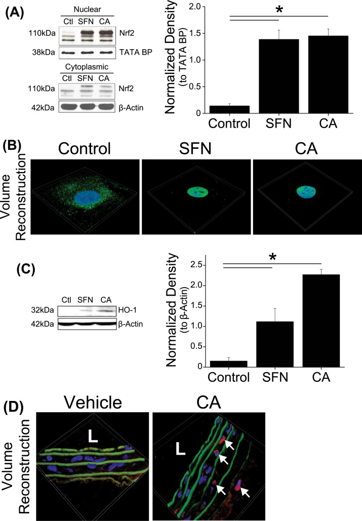
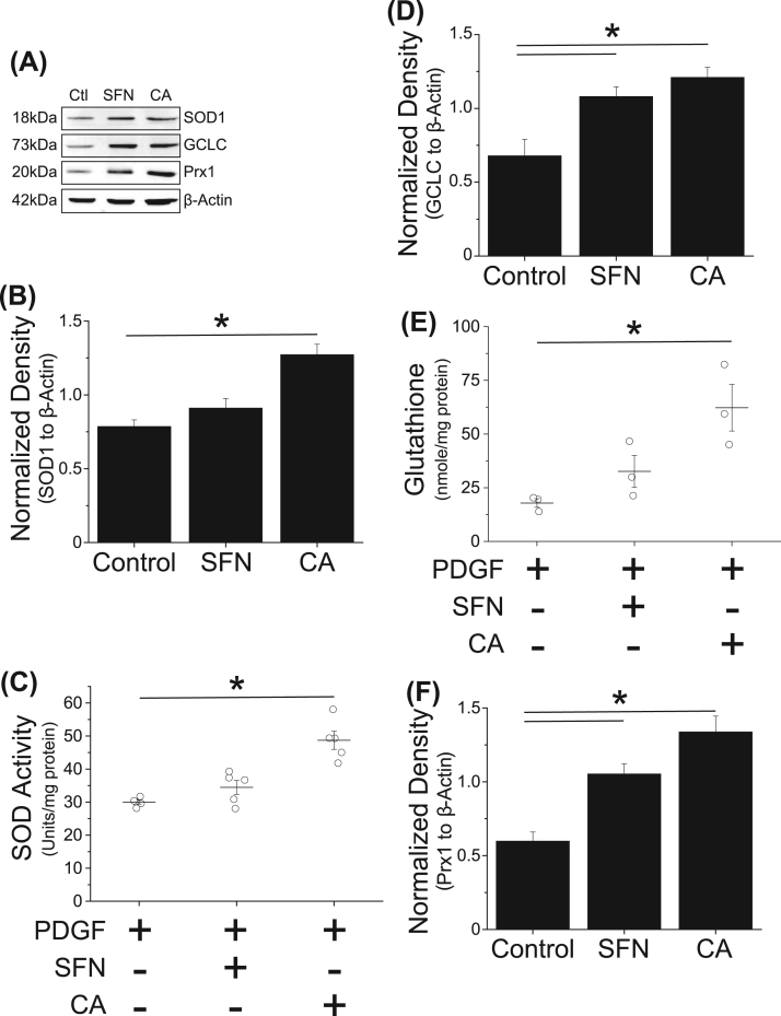
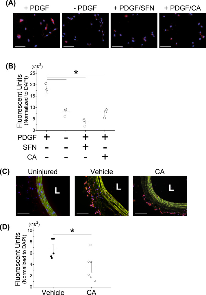
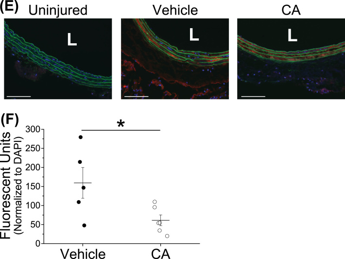
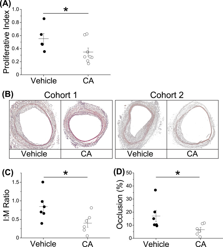
Atherosclerosis remains the number one cause of death and disability worldwide. Atherosclerosis is treated by revascularization procedures to restore blood flow to distal tissue, but these procedures often fail due to restenosis secondary to neointimal hyperplasia. Diabetes mellitus is a metabolic disorder that accelerates both atherosclerosis development and onset of restenosis. Strategies to inhibit restenosis aim at reducing neointimal hyperplasia by inhibiting vascular smooth muscle cell (VSMC) proliferation and migration. Since increased production of reactive oxygen species promotes VSMC proliferation and migration, redox intervention to maintain vascular wall redox homeostasis holds the potential to inhibit arterial restenosis. Cinnamic aldehyde (CA) is an electrophilic Nrf2 activator that has shown therapeutic promise in diabetic rodent models. Nrf2 is a transcription factor that regulates the antioxidant response. Therefore, we hypothesized that CA would activate Nrf2 and would inhibit neointimal hyperplasia after carotid artery balloon injury in the Zucker Diabetic Fatty (ZDF) rat. In primary ZDF VSMC, CA inhibited cell growth by MTT with an EC50 of 118 ± 7 μM. At a therapeutic dose of 100 μM, CA inhibited proliferation of ZDF VSMC in vitro and reduced the proliferative index within the injured artery in vivo, as well as migration of ZDF VSMC in vitro. CA activated the Nrf2 pathway in both ZDF VSMC and injured carotid arteries while also increasing antioxidant defenses and reducing markers of redox dysfunction. Additionally, we noted a significant reduction of neutrophils (69%) and macrophages (78%) within the injured carotid arteries after CA treatment. Lastly, CA inhibited neointimal hyperplasia evidenced by a 53% reduction in the intima:media ratio and a 61% reduction in vessel occlusion compared to arteries treated with vehicle alone. Overall CA was capable of activating Nrf2, and inhibiting neointimal hyperplasia after balloon injury in a rat model of diabetic restenosis.

Keywords: Cinnamic aldehyde; Diabetes; Neointimal hyperplasia; Nrf2; Restenosis; Vascular smooth muscle cells.


Figures

fx1
Graphical abstract
Fig. 1
Fig. 1
100 µM cinnamic aldehyde (CA) reduced PDGF-BB-stimulated ZDF vascular smooth muscle cell (VSMC) number without inducing apoptosis. ZDF VSMC in 25 mM glucose were synchronized overnight and then stimulated with 25 ng/mL PDGF-BB for 24 h with or without varying concentrations of CA. (A) Metabolic activity analyzed by MTT of ZDF VSMC treated with CA dose range (EC50 = 118 ± 7 μM) (n = 3 independent experiment with 8 replicates). (B) Quantification of total viable ZDF VSMC after treatment with increasing doses of CA. (C) Quantification of total dead ZDF VSMC after CA treatment. (D) Quantification of early apoptotic, late apoptotic, and dead ZDF VSMC after treatment with increasing doses of CA. Significant increase shown is only in the late apoptotic cell population. No significant change in other subpopulations. (E) Live subpopulation of ZDF VSMC included with subpopulations from (D) with significant decrease in total cell number at 100 µM and 1000 µM CA shown. (B-E) *p < 0.05 compared to control (0 µM CA); one-way ANOVA with post hoc Tukey's correction for multiple comparisons. Data presented as means ± SEM (n = 3 independent experiments in triplicate).
Fig. 2
Fig. 2
Cinnamic aldehyde (CA) inhibited PDGF-BB-induced ZDF vascular smooth muscle cell (VSMC) migration. ZDF VSMC in 25 mM glucose were synchronized overnight and then stimulated with 25 ng/mL PDGF-BB for 24 h with or without varying concentrations of CA. (A) Phase contrast images of PDGF-treated (+), PDGF-starved (-), and 100 µM CA treated ZDF VSMC at 0 and 24 h. Scale bar = 500 µm. (B) Quantification of migratory ZDF VSMC within the scratch area as indicated by the dashed lines in (A). *p < 0.05 compared to PDGF-treated control; one-way ANOVA with Tukey's correction. Data presented as means ± SEM (n = 3 independent experiments in triplicate).
Fig. 3
Fig. 3
Cinnamic aldehyde (CA) activated the Nrf2 pathway in ZDF vascular smooth muscle cells (VSMC). (A-C) ZDF VSMC in 25 mM glucose were synchronized overnight and then stimulated with 25 ng/mL PDGF-BB with or without varying concentrations of CA. (A) Immunoblot of nuclear and cytoplasmic ZDF VSMC fractions treated with 100 µM CA or 4 µM sulforaphane (SFN) for 6 h probed for Nrf2 with TATA Binding Protein (TATA BP) or β-Actin in the nuclear and cytoplasmic fraction respectively. Plot of nuclear fraction Nrf2 band density normalized to TATA BP. (B) Confocal images of subcellular Nrf2 localization after CA or SFN treatment for 24 h. Green = Nrf2; Blue = Nuclei. (C) Representative immunoblot of whole cell lysates treated with CA or SFN for 4 h probed for heme oxygenase 1 (HO-1). Plot of HO-1 band density normalized to β-Actin (n = 3). (D) Hyperglycemic ZDF rats weighing 350–500 g underwent the carotid balloon injury and were treated with periadventitial application of Pluronic vehicle (100 µL) or CA in Pluronic (100 µM in 100 µL) at the time of surgery. Carotids were harvested three days after surgery. Confocal images of three-day injured carotid arteries stained for Nrf2. Red = Nrf2; Blue = DAPI; Green = elastic lamina; L = lumen. Arrows indicate nuclear co-localization. *p < 0.05 compared to control (Ctl); one-way ANOVA with Tukey's correction. Data presented as means ± SEM (n = 3 independent experiments in triplicate; n = 3 ZDF VSMC or tissue cross-sections for confocal images with representative image shown).
Fig. 4
Fig. 4
Cinnamic aldehyde (CA) increased antioxidant levels in ZDF vascular smooth muscle cells (VSMC). ZDF VSMC in 25 mM glucose were synchronized overnight and then stimulated with 25 ng/mL PDGF-BB with or without 100 µM CA or 4 µM sulforaphane (SFN) for 24 h. (A) Representative immunoblot of whole cell lysates probed for superoxide dismutase 1 (SOD1), glutamate-cysteine ligase catalytic subunit (GCLC), peroxiredoxin 1 (Prx1), and β-Actin. (B) Plot of SOD1 band density normalized to β-Actin (n = 3). (C) Total SOD activity in treated ZDF VSMC (n = 5 with technical duplicates). (D) Plot of GCLC band density normalized to β-Actin (n = 3). (E) Intracellular glutathione levels in treated ZDF VSMC (n = 3 with technical duplicates). (F) Plot of Prx1 band density normalized to β-Actin (n = 3). (B-F) *p < 0.05 compared to control (Ctl); Data analyzed with one-way ANOVA with Tukey's correction. Data presented as means ± SEM.
Fig. 5
Fig. 5
Cinnamic aldehyde (CA) reduced levels of redox markers in ZDF vascular smooth muscle cells (VSMC) and injured carotid arteries. (A-B) ZDF VSMC in 25 mM glucose were synchronized overnight then treated for 24 h with or without stimulation by 25 ng/mL PDGF-BB in the presence of either CA (100 µM) or sulforaphane (SFN) (4 µM). (A) Representative images of ZDF VSMC stained with dihydroethidium (DHE). Scale bar = 100 µm; Red = DHE; Blue = DAPI. (B) Quantification of fluorescence from images in (A) normalized to cell nuclear count (DAPI). (C-F) Hyperglycemic ZDF rats weighing 350–500 g underwent the carotid balloon injury and were treated with periadventitial application of Pluronic vehicle (100 µL) or CA in Pluronic (100 µM in 100 µL) at the time of surgery. Carotids were harvested three days after surgery. (C) DHE staining of uninjured or three-day injured carotid arteries treated with 100 µM CA or vehicle alone. Scale bar = 100 µm; Red = DHE; Blue = DAPI; Green = elastic lamina; L = lumen. (D) Quantification of fluorescence from images in (C) normalized to cell nuclear count. (E) Immunofluorescent images for 3-nitrotyrosine staining of three-day injured carotid arteries. Scale bar = 100 µm; Red = 3-nitrotyrosine; Blue = DAPI; Green = elastic lamina; L = lumen. (F) Quantification of fluorescence from images in (E). (B) *p < 0.05 compared to PDGF-alone control. Data presented as means ± SEM (n = 3 independent experiments in triplicate). (D, F) *p < 0.05 compared to vehicle alone. One-way ANOVA with post hoc Tukey's correction for multiple comparison. Each point indicates the average of three arterial cross-sections per rat (n = 5 for vehicle, n = 6 for CA) with means ± SEM for each group overlaid.
Fig. 5
Fig. 5
Cinnamic aldehyde (CA) reduced levels of redox markers in ZDF vascular smooth muscle cells (VSMC) and injured carotid arteries. (A-B) ZDF VSMC in 25 mM glucose were synchronized overnight then treated for 24 h with or without stimulation by 25 ng/mL PDGF-BB in the presence of either CA (100 µM) or sulforaphane (SFN) (4 µM). (A) Representative images of ZDF VSMC stained with dihydroethidium (DHE). Scale bar = 100 µm; Red = DHE; Blue = DAPI. (B) Quantification of fluorescence from images in (A) normalized to cell nuclear count (DAPI). (C-F) Hyperglycemic ZDF rats weighing 350–500 g underwent the carotid balloon injury and were treated with periadventitial application of Pluronic vehicle (100 µL) or CA in Pluronic (100 µM in 100 µL) at the time of surgery. Carotids were harvested three days after surgery. (C) DHE staining of uninjured or three-day injured carotid arteries treated with 100 µM CA or vehicle alone. Scale bar = 100 µm; Red = DHE; Blue = DAPI; Green = elastic lamina; L = lumen. (D) Quantification of fluorescence from images in (C) normalized to cell nuclear count. (E) Immunofluorescent images for 3-nitrotyrosine staining of three-day injured carotid arteries. Scale bar = 100 µm; Red = 3-nitrotyrosine; Blue = DAPI; Green = elastic lamina; L = lumen. (F) Quantification of fluorescence from images in (E). (B) *p < 0.05 compared to PDGF-alone control. Data presented as means ± SEM (n = 3 independent experiments in triplicate). (D, F) *p < 0.05 compared to vehicle alone. One-way ANOVA with post hoc Tukey's correction for multiple comparison. Each point indicates the average of three arterial cross-sections per rat (n = 5 for vehicle, n = 6 for CA) with means ± SEM for each group overlaid.
Fig. 6
Fig. 6
Cinnamic aldehyde (CA) reduced immune cell invasion into the injured carotid artery. Hyperglycemic ZDF rats weighing 350–500 g underwent the carotid balloon injury and were treated with periadventitial application of Pluronic vehicle (100 µL) or CA in Pluronic (100 µM in 100 µL) at the time of surgery. Carotids were harvested three days or two weeks after surgery. (A) Myeloperoxidase (MPO) staining of uninjured and three-day injured carotid arteries treated with 100 µM CA or vehicle alone. Scale bar = 100 µm; Red = MPO; Blue = DAPI; Green = elastic lamina; L = lumen. (B) Quantification of MPO fluorescence from images in (A) normalized to cell nuclei (DAPI). (C) CD68 staining of three-day injured carotid arteries. Scale bar = 100 µm; Red = CD68 Blue = DAPI; Green = elastic lamina; L = lumen. (D) Quantification of CD68 staining from images in (C). (E) Quantification of CD68 fluorescence from two-week injured arteries of cohort 1. (F) Quantification of CD68 fluorescence from two-week injured arteries of cohort 2. *p < 0.05 compared to vehicle; Student's t-test. Each point indicates the average of three arterial cross-sections per rat (three-day injured: n = 5 for vehicle, n = 6 for CA; two-week injured: n = 3) with means ± SEM for each group overlaid.
Fig. 7
Fig. 7
Cinnamic aldehyde (CA) inhibited cellular proliferation in injured carotid arteries and the development of neointimal hyperplasia. Hyperglycemic ZDF rats weighing 350–500 g underwent the carotid balloon injury and were treated with periadventitial application of Pluronic vehicle (100 µL) or CA in Pluronic (100 µM in 100 µL) at the time of surgery. Carotids were harvested three days or two weeks after surgery. (A) Quantification of vessel proliferative index of three-day injured arteries by BrdU immunohistochemistry. (B) H&E stained arterial cross-sections of two-week injured rats from cohort 1 and cohort 2. (C) Quantification of the intima: media (I:M) ratio of carotid arteries from both cohorts in (B). (D) Quantification of percent vessel occlusion of carotid arteries from both cohorts in (B). *p < 0.05 compared to vehicle; Student's t-test. Each point indicates the average of three arterial cross-sections per rat (three-day injured: n = 5 for vehicle, n = 8 for CA; two-week injured: n = 6) with means ± SEM for each group overlaid.

Similar articles

Cited by

References

    1. Writing Group M., Mozaffarian D., Benjamin E.J., Go A.S., Arnett D.K., Blaha M.J. Executive summary: heart disease and stroke statistics–2016 update: a report from the American heart association. Circulation. 2016;133:447–454. - PubMed
    1. International Diabetes Federation Diabetes Atlas, Eighth edition, 2017.
    1. Matheus A.S., Tannus L.R., Cobas R.A., Palma C.C., Negrato C.A., Gomes M.B. Impact of diabetes on cardiovascular disease: an update. Int. J. Hypertens. 2013;2013:653789. - PMC - PubMed
    1. Lexis C.P., Rahel B.M., Meeder J.G., Zijlstra F., van der Horst I.C. The role of glucose lowering agents on restenosis after percutaneous coronary intervention in patients with diabetes mellitus. Cardiovasc. Diabetol. 2009;8:41. - PMC - PubMed
    1. DeRubertis B.G., Pierce M., Ryer E.J., Trocciola S., Kent K.C., Faries P.L. Reduced primary patency rate in diabetic patients after percutaneous intervention results from more frequent presentation with limb-threatening ischemia. J. Vasc. Surg. 2008;47:101–108. - PubMed

Publication types

MeSH terms