

Irisin promotes cardiac progenitor cell-induced myocardial repair and functional improvement in infarcted heart
- PMID: 30171682
- PMCID: PMC6510276
- DOI: 10.1002/jcp.27037
Irisin promotes cardiac progenitor cell-induced myocardial repair and functional improvement in infarcted heart
Abstract
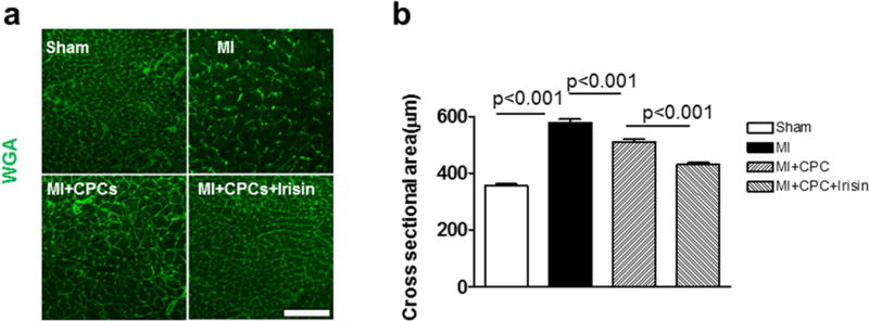
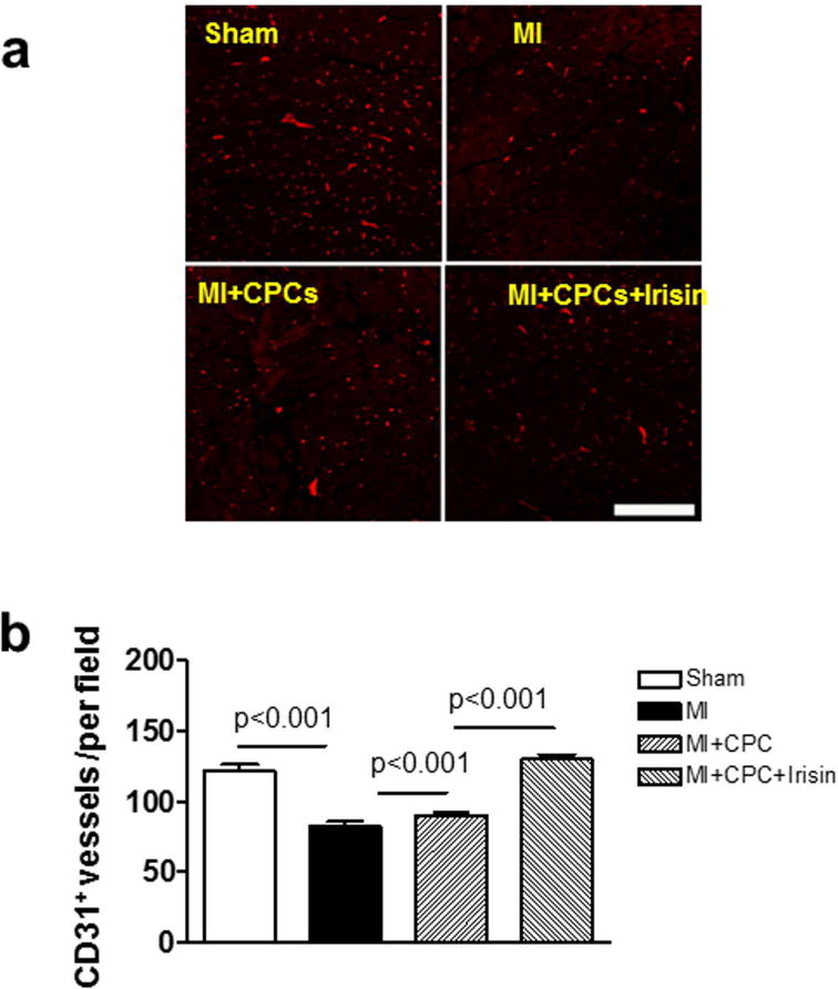
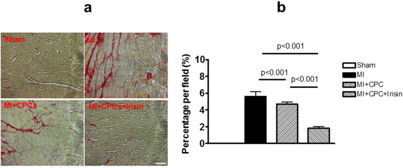
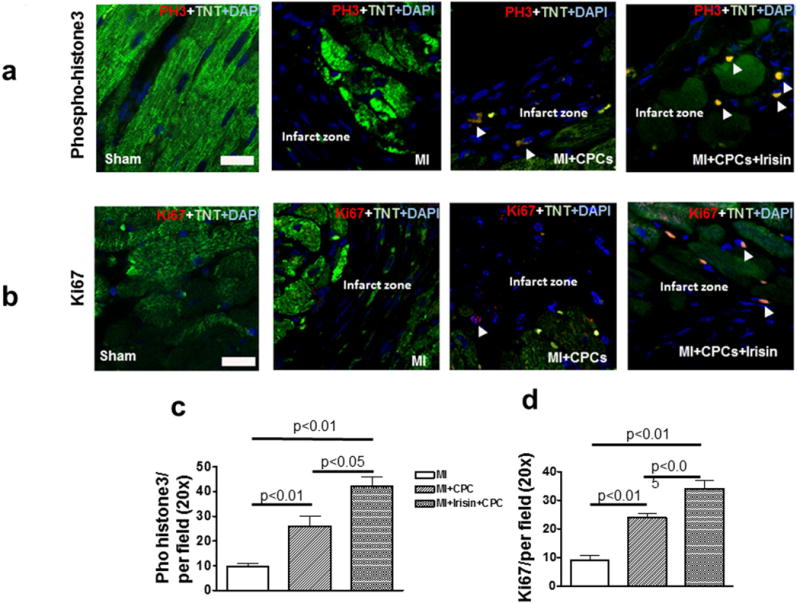
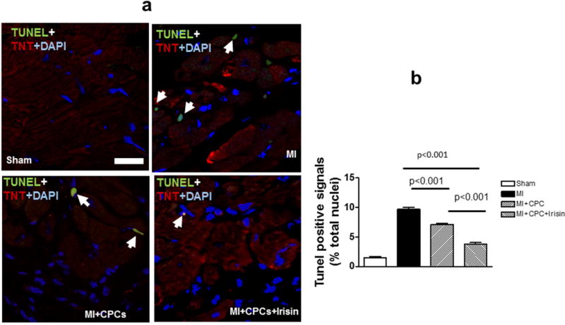
Irisin, a newly identified hormone and cardiokine, is critical for modulating body metabolism. New evidence indicates that irisin protects the heart against myocardial ischemic injury. However, whether irisin enhances cardiac progenitor cell (CPC)-induced cardiac repair remains unknown. This study examines the effect of irisin on CPC-induced cardiac repair when these cells are introduced into the infarcted myocardium. Nkx2.5+ CPC stable cells were isolated from mouse embryonic stem cells. Nkx2.5 + CPCs (0.5 × 10 6 ) were reintroduced into the infarcted myocardium using PEGlylated fibrin delivery. The mouse myocardial infarction model was created by permanent ligation of the left anterior descending (LAD) artery. Nkx2.5 + CPCs were pretreated with irisin at a concentration of 5 ng/ml in vitro for 24 hr before transplantation. Myocardial functions were evaluated by echocardiographic measurement. Eight weeks after engraftment, Nkx2.5 + CPCs improved ventricular function as evident by an increase in ejection fraction and fractional shortening. These findings are concomitant with the suppression of cardiac hypertrophy and attenuation of myocardial interstitial fibrosis. Transplantation of Nkx2.5 + CPCs promoted cardiac regeneration and neovascularization, which were increased with the pretreatment of Nkx2.5 + CPCs with irisin. Furthermore, irisin treatment promoted myocyte proliferation as indicated by proliferative markers Ki67 and phosphorylated histone 3 and decreased apoptosis. Additionally, irisin resulted in a marked reduction of histone deacetylase 4 and increased p38 acetylation in cultured CPCs. These results indicate that irisin promoted Nkx2.5 + CPC-induced cardiac regeneration and functional improvement and that irisin serves as a novel therapeutic approach for stem cells in cardiac repair.
Keywords: cardiac progenitor cell; irisin; myocardial infarction; ventricular function.
© 2018 Wiley Periodicals, Inc.
Conflict of interest statement
The authors declare that they have no competing interests.
Figures


Similar articles
-
Apoptosis-Resistant Cardiac Progenitor Cells Modified With Apurinic/Apyrimidinic Endonuclease/Redox Factor 1 Gene Overexpression Regulate Cardiac Repair After Myocardial Infarction.Stem Cells Transl Med. 2016 Aug;5(8):1067-78. doi: 10.5966/sctm.2015-0281. Epub 2016 Jun 22. Stem Cells Transl Med. 2016. PMID: 27334489 Free PMC article.
-
Expandable human cardiovascular progenitors from stem cells for regenerating mouse heart after myocardial infarction.Cardiovasc Res. 2020 Mar 1;116(3):545-553. doi: 10.1093/cvr/cvz181. Cardiovasc Res. 2020. PMID: 31287499 Free PMC article.
-
Specific inhibition of HDAC4 in cardiac progenitor cells enhances myocardial repairs.Am J Physiol Cell Physiol. 2014 Aug 15;307(4):C358-72. doi: 10.1152/ajpcell.00187.2013. Epub 2014 Jun 18. Am J Physiol Cell Physiol. 2014. PMID: 24944198 Free PMC article.
-
Stem cells in the infarcted heart.J Cardiovasc Transl Res. 2010 Feb;3(1):73-8. doi: 10.1007/s12265-009-9151-4. Epub 2009 Nov 20. J Cardiovasc Transl Res. 2010. PMID: 20560035 Review.
-
New Insights into the Role of Exosomes in the Heart After Myocardial Infarction.J Cardiovasc Transl Res. 2019 Feb;12(1):18-27. doi: 10.1007/s12265-018-9831-z. Epub 2018 Sep 2. J Cardiovasc Transl Res. 2019. PMID: 30173401 Review.
Cited by
-
Exercise-induced myokine FNDC5/irisin functions in cardiovascular protection and intracerebral retrieval of synaptic plasticity.Cell Biosci. 2019 Apr 3;9:32. doi: 10.1186/s13578-019-0294-y. eCollection 2019. Cell Biosci. 2019. PMID: 30984367 Free PMC article.
-
Betulin Alleviates Myocardial Ischemia-Reperfusion Injury in Rats via Regulating the Siti1/NLRP3/NF-κB Signaling Pathway.Inflammation. 2021 Jun;44(3):1096-1107. doi: 10.1007/s10753-020-01405-8. Epub 2021 Jan 4. Inflammation. 2021. PMID: 33392937
-
Integrin-Ligand Interactions in Inflammation, Cancer, and Metabolic Disease: Insights Into the Multifaceted Roles of an Emerging Ligand Irisin.Front Cell Dev Biol. 2020 Oct 26;8:588066. doi: 10.3389/fcell.2020.588066. eCollection 2020. Front Cell Dev Biol. 2020. PMID: 33195249 Free PMC article. Review.
-
Irisin: linking metabolism with heart failure.Am J Transl Res. 2020 Oct 15;12(10):6003-6014. eCollection 2020. Am J Transl Res. 2020. PMID: 33194010 Free PMC article. Review.
-
The Neuroprotective Effect of Irisin in Ischemic Stroke.Front Aging Neurosci. 2020 Dec 22;12:588958. doi: 10.3389/fnagi.2020.588958. eCollection 2020. Front Aging Neurosci. 2020. PMID: 33414714 Free PMC article. Review.
References
-
- Aydin S, Aydin S, Kobat MA, Kalayci M, Eren MN, Yilmaz M, Kuloglu T, Gul E, Secen O, Alatas OD, Baydas A. Decreased saliva/serum irisin concentrations in the acute myocardial infarction promising for being a new candidate biomarker for diagnosis of this pathology. Peptides. 2014;56:141–5. - PubMed
-
- Anastasilakis CD, Mantzoros CS. Circulating irisin levels are lower in patients with either stable coronary artery disease (CAD) or myocardial infarction (MI) versus healthy controls, whereas follistatin and activin A levels are higher and can discriminate MI from CAD with similar to CK-MB accuracy. Metabolism. 2017;73:1–8. - PubMed
Publication types
MeSH terms
Substances
Grants and funding
LinkOut - more resources
Full Text Sources
Other Literature Sources
Medical
