Role of P53-Senescence Induction in Suppression of LNCaP Prostate Cancer Growth by Cardiotonic Compound Bufalin
- PMID: 30166403
- PMCID: PMC6668018
- DOI: 10.1158/1535-7163.MCT-17-1296
Role of P53-Senescence Induction in Suppression of LNCaP Prostate Cancer Growth by Cardiotonic Compound Bufalin
Abstract
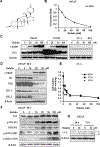
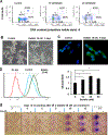
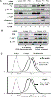
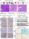
Bufalin is a major cardiotonic compound in the traditional Chinese medicine, Chansu, prepared from toad skin secretions. Cell culture studies have suggested an anticancer potential involving multiple cellular processes, including differentiation, apoptosis, senescence, and angiogenesis. In prostate cancer cell models, P53-dependent and independent caspase-mediated apoptosis and androgen receptor (AR) antagonism have been described for bufalin at micromolar concentrations. Because a human pharmacokinetic study indicated that single nanomolar bufalin was safely achievable in the peripheral circulation, we evaluated its cellular activity within range with the AR-positive and P53 wild-type human LNCaP prostate cancer cells in vitro Our data show that bufalin induced caspase-mediated apoptosis at 20 nmol/L or higher concentration with concomitant suppression of AR protein and its best-known target, PSA and steroid receptor coactivator 1 and 3 (SRC-1, SRC-3). Bufalin exposure induced protein abundance of P53 (not mRNA) and P21CIP1 (CDKN1A), G2 arrest, and increased senescence-like phenotype (SA-galactosidase). Small RNAi knocking down of P53 attenuated bufalin-induced senescence, whereas knocking down of P21CIP1 exacerbated bufalin-induced caspase-mediated apoptosis. In vivo, daily intraperitoneal injection of bufalin (1.5 mg/kg body weight) for 9 weeks delayed LNCaP subcutaneous xenograft tumor growth in NSG SCID mice with a 67% decrease of final weight without affecting body weight. Tumors from bufalin-treated mice exhibited increased phospho-P53 and SA-galactosidase without detectable caspase-mediated apoptosis or suppression of AR and PSA. Our data suggest potential applications of bufalin in therapy of prostate cancer in patients or chemo-interception of prostate precancerous lesions, engaging a selective activation of P53 senescence. Mol Cancer Ther; 17(11); 2341-52. ©2018 AACR.
©2018 American Association for Cancer Research.
Conflict of interest statement
Figures


Similar articles
-
Persistent p21Cip1 induction mediates G(1) cell cycle arrest by methylseleninic acid in DU145 prostate cancer cells.Curr Cancer Drug Targets. 2010 May;10(3):307-18. doi: 10.2174/156800910791190238. Curr Cancer Drug Targets. 2010. PMID: 20370687
-
A natural androgen receptor antagonist induces cellular senescence in prostate cancer cells.Mol Endocrinol. 2014 Nov;28(11):1831-40. doi: 10.1210/me.2014-1170. Epub 2014 Sep 9. Mol Endocrinol. 2014. PMID: 25203674 Free PMC article.
-
Phenylbutyl isoselenocyanate induces reactive oxygen species to inhibit androgen receptor and to initiate p53-mediated apoptosis in LNCaP prostate cancer cells.Mol Carcinog. 2018 Aug;57(8):1055-1066. doi: 10.1002/mc.22825. Epub 2018 May 2. Mol Carcinog. 2018. PMID: 29668110
-
Senescence and aging: the critical roles of p53.Oncogene. 2013 Oct 24;32(43):5129-43. doi: 10.1038/onc.2012.640. Epub 2013 Feb 18. Oncogene. 2013. PMID: 23416979 Review.
-
Bufalin for an innovative therapeutic approach against cancer.Pharmacol Res. 2022 Oct;184:106442. doi: 10.1016/j.phrs.2022.106442. Epub 2022 Sep 9. Pharmacol Res. 2022. PMID: 36096424 Review.
Cited by
-
Multi-omics profiling of PC-3 cells reveals bufadienolides-induced lipid metabolic remodeling by regulating long-chain lipids synthesis and hydrolysis.Metabolomics. 2023 Jan 16;19(2):6. doi: 10.1007/s11306-022-01968-7. Metabolomics. 2023. PMID: 36645548
-
Bufalin stimulates antitumor immune response by driving tumor-infiltrating macrophage toward M1 phenotype in hepatocellular carcinoma.J Immunother Cancer. 2022 May;10(5):e004297. doi: 10.1136/jitc-2021-004297. J Immunother Cancer. 2022. PMID: 35618286 Free PMC article.
-
The multifaceted therapeutic value of targeting steroid receptor coactivator-1 in tumorigenesis.Cell Biosci. 2024 Mar 29;14(1):41. doi: 10.1186/s13578-024-01222-8. Cell Biosci. 2024. PMID: 38553750 Free PMC article. Review.
-
Bufalin inhibits hepatitis B virus-associated hepatocellular carcinoma development through androgen receptor dephosphorylation and cell cycle-related kinase degradation.Cell Oncol (Dordr). 2020 Dec;43(6):1129-1145. doi: 10.1007/s13402-020-00546-0. Epub 2020 Jul 4. Cell Oncol (Dordr). 2020. PMID: 32623699
-
Amphibian-Derived Natural Anticancer Peptides and Proteins: Mechanism of Action, Application Strategies, and Prospects.Int J Mol Sci. 2023 Sep 12;24(18):13985. doi: 10.3390/ijms241813985. Int J Mol Sci. 2023. PMID: 37762285 Free PMC article. Review.
References
-
- Scher HI, Fizazi K, Saad F, Taplin ME, Sternberg CN, Miller K, de Wit R, Mulders P, Chi KN, Shore ND et al.: Increased survival with enzalutamide in prostate cancer after chemotherapy. N Engl J Med 2012, 367(13):1187–1197. - PubMed
-
- Petrylak DP, Tangen CM, Hussain MH, Lara PN, Jones JA Jr, Taplin ME, Burch PA, Berry D, Moinpour C, Kohli M et al.: Docetaxel and estramustine compared with mitoxantrone and prednisone for advanced refractory prostate cancer. N Engl J Med 2004, 351(15):1513–1520. - PubMed
-
- de Bono JS, Oudard S, Ozguroglu M, Hansen S, Machiels JP, Kocak I, Gravis G, Bodrogi I, Mackenzie MJ, Shen L et al.: Prednisone plus cabazitaxel or mitoxantrone for metastatic castration-resistant prostate cancer progressing after docetaxel treatment: a randomised open-label trial. Lancet 2010, 376(9747):1147–1154. - PubMed
-
- Yin S, Jiang P, Ye M, Hu H, Lu J, Jiang C: A Critical Assessment of Anti-cancer Activities of Bufadienolides. Horizons in Cancer Research 2013, 52:63–88.
Publication types
MeSH terms
Substances
Grants and funding
LinkOut - more resources
Full Text Sources
Other Literature Sources
Medical
Research Materials
Miscellaneous

