Protective Effects of 2,3,5,4'-Tetrahydroxystilbene-2- O-β-d-glucoside on Ovariectomy Induced Osteoporosis Mouse Model
- PMID: 30154383
- PMCID: PMC6163345
- DOI: 10.3390/ijms19092554
Protective Effects of 2,3,5,4'-Tetrahydroxystilbene-2- O-β-d-glucoside on Ovariectomy Induced Osteoporosis Mouse Model
Abstract
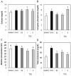
2,3,5,4'-Tetrahydroxystilbene-2-O-β-d-glucoside (TSG), an active polyphenolic component of Polygonum multiflorum, exhibits many pharmacological activities including antioxidant, anti-inflammation, and anti-aging effects. A previous study demonstrated that TSG protected MC3T3-E1 cells from hydrogen peroxide (H₂O₂) induced cell damage and the inhibition of osteoblastic differentiation. However, no studies have investigated the prevention of ovariectomy-induced bone loss in mice. Therefore, we investigated the effects of TSG on bone loss in ovariectomized mice (OVX). Treatment with TSG (1 and 3 μg/g; i.p.) for six weeks positively affected body weight, uterine weight, organ weight, bone length, and weight change because of estrogen deficiency. The levels of the serum biochemical markers of calcium (Ca), inorganic phosphorus (IP), alkaline phosphatase (ALP), and total cholesterol (TCHO) decreased in the TSG-treated mice when compared with the OVX mice. Additionally, the serum bone alkaline phosphatase (BALP) levels in the TSG-treated OVX mice were significantly increased compared with the OVX mice, while the tartrate-resistant acid phosphatase (TRAP) activity was significantly reduced. Furthermore, the OVX mice treated with TSG showed a significantly reduced bone loss compared to the untreated OVX mice upon micro-computed tomography (CT) analysis. Consequently, bone destruction in osteoporotic mice as a result of ovariectomy was inhibited by the administration of TSG. These findings indicate that TSG effectively prevents bone loss in OVX mice; therefore, it can be considered as a potential therapeutic for the treatment of postmenopausal osteoporosis.
Keywords: 2,3,5,4′-tetrahydroxystilbene-2-O-β-d-glucoside (TSG); bone loss; menopause; osteoporosis; ovariectomy.
Conflict of interest statement
The authors declare no conflict of interest.
Figures


Similar articles
-
Protective effects of 2,3,5,4-tetrahydroxystilbene-2-o-β-D-glucoside against osteoporosis: Current knowledge and proposed mechanisms.Int J Rheum Dis. 2018 Aug;21(8):1504-1513. doi: 10.1111/1756-185X.13357. Int J Rheum Dis. 2018. PMID: 30146742
-
Antiosteoporotic activity of echinacoside in ovariectomized rats.Phytomedicine. 2013 Apr 15;20(6):549-57. doi: 10.1016/j.phymed.2013.01.001. Epub 2013 Feb 18. Phytomedicine. 2013. PMID: 23428402
-
Protective Effect of Acteoside on Ovariectomy-Induced Bone Loss in Mice.Int J Mol Sci. 2019 Jun 18;20(12):2974. doi: 10.3390/ijms20122974. Int J Mol Sci. 2019. PMID: 31216684 Free PMC article.
-
Biological Effects of Tetrahydroxystilbene Glucoside: An Active Component of a Rhizome Extracted from Polygonum multiflorum.Oxid Med Cell Longev. 2018 Nov 4;2018:3641960. doi: 10.1155/2018/3641960. eCollection 2018. Oxid Med Cell Longev. 2018. PMID: 30524653 Free PMC article. Review.
-
Current pharmacological developments in 2,3,4',5-tetrahydroxystilbene 2-O-β-D-glucoside (TSG).Eur J Pharmacol. 2017 Sep 15;811:21-29. doi: 10.1016/j.ejphar.2017.05.037. Epub 2017 May 23. Eur J Pharmacol. 2017. PMID: 28545778 Review.
Cited by
-
A quinoxaline-based compound ameliorates bone loss in ovariectomized mice.Exp Biol Med (Maywood). 2021 Dec;246(23):2502-2510. doi: 10.1177/15353702211032133. Epub 2021 Jul 25. Exp Biol Med (Maywood). 2021. PMID: 34308655 Free PMC article.
-
Icaritin ameliorates RANKL-mediated osteoclastogenesis and ovariectomy-induced osteoporosis.Aging (Albany NY). 2023 Oct 3;15(19):10213-10236. doi: 10.18632/aging.205068. Epub 2023 Oct 3. Aging (Albany NY). 2023. PMID: 37793008 Free PMC article.
-
Restorative effect of modified dioscorea pills on the structure of hippocampal neurovascular unit in an animal model of chronic cerebral hypoperfusion.Heliyon. 2019 Apr 28;5(4):e01567. doi: 10.1016/j.heliyon.2019.e01567. eCollection 2019 Apr. Heliyon. 2019. PMID: 31183430 Free PMC article.
-
Exploring the role and mechanism of Fuzi decoction in the treatment of osteoporosis by integrating network pharmacology and experimental verification.J Orthop Surg Res. 2023 Jul 18;18(1):508. doi: 10.1186/s13018-023-03842-1. J Orthop Surg Res. 2023. PMID: 37464262 Free PMC article.
-
Menaquinone 4 Reduces Bone Loss in Ovariectomized Mice through Dual Regulation of Bone Remodeling.Nutrients. 2021 Jul 27;13(8):2570. doi: 10.3390/nu13082570. Nutrients. 2021. PMID: 34444729 Free PMC article.
References
MeSH terms
Substances
LinkOut - more resources
Full Text Sources
Other Literature Sources
Medical
Miscellaneous

